250 mg of Clinical

This forum made possible through the generous support of SDN members, donors, and sponsors. Thank you.

sevoflurane

Ride
20+ Year Member
Joined
Jul 16, 2003
Messages
6,030
Reaction score
3,808
Lot’s of doom and gloom here on this board lately. While there is certainly cause for concern, I thought I’d lighten things up and start a thread that stears us away from business and more towards the clinical side of things… kinda like the old days.

It’s always fun to do clinical cases here for all to learn. If you have any interesting anesthesia material you’d like to post, go for it. This board is meant to be constructive. Hopefully there is some good thought provoking material some of you out there might have for us.

If you know the answer from the get go, let the med studs have a crack at it first.
There are never wrong answers as one can learn from what is not right.


I’ll start with 2 different clinical posts.

Members don't see this ad.
 
Last edited:
Case #1:

photo_zps5da02e52.jpg

RU lobectomy for pancoast tumor.

1) What is a pancoast tumor and what clinical signs can you see with it?
2) What monitors and how would you position this patient?
3) What strategies can you employ in order to improve outcome?
4) What are the structures labeled 1-7
 
Last edited:
Case #2.

ScreenShot2014-02-08at50441PM_zps5f3bfb4d.png


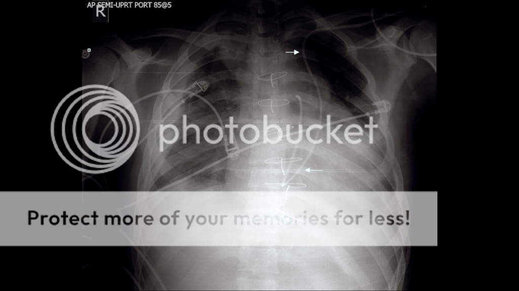
This one is just an X-ray.

1) What do you see that may be a bit off?

This Xray has followup images.
 
Members don't see this ad :)
Case #1:


This was a case from 3-4 years ago. RU lobectomy for pancoat tumor.

1) What is a pancoast tumor and what clinical signs can you see with it.
2) What monitors and how would you position this patient.
3) What strategies can you employ in order to improve outcome?
4) What are the structures labeled 1-7


1. Pancoast tumor is a neoplasm at the apex of the lung that can disrupt the sympathetic ganglion and some other nerves. Usually of NSCLC nature they may have some association with paraneoplastic syndromes. Horners syndrome, ptosis, miosis and anhydrosis, scapular pain, arm pain. Of course the typical weight loss, night sweats and fevers as well.

2. I did a CT surgery rotation during my surgery rotation and these pts were usually positioned on their side with bean bags holding them in place, with their arms secured in front. Im not sure if this would not be the case for a RUL lobectomy. They require a double lumen ETT as well.

cant orient myself at all in that picture! lol
 
Case #2.


This one is just an X-ray.

1) What do you see that may be a bit off?

This Xray has followup images.

This looks like a childs xray? IDK i see sternotomy wires, so maybe not a child? or maybe a child who had a congenital issue and underwent ct surgery at a young age, but I dont know if they use sternotomy wires on kids. Swan ganz on the wrong side of the heart? or on the right side but the heart chambers are on the wrong side? Im just babbling now, lol, but maybe it is the correct placement but this is a child that has situs inversus?
 
  • Like
Reactions: 1 user
cant orient myself at all in that picture! lol

-Moo-Moo, my bad (anybody know what that's from? it involves aliens and cows).
The orientation is cephalic to the top of the screen caudal to the bottom.

Great answer btw. I will hold off on commenting for now. :watching:
 
Last edited:
This looks like a childs xray? IDK i see sternotomy wires, so maybe not a child? or maybe a child who had a congenital issue and underwent ct surgery at a young age, but I dont know if they use sternotomy wires on kids. Swan ganz on the wrong side of the heart? or on the right side but the heart chambers are on the wrong side? Im just babbling now, lol, but maybe it is the correct placement but this is a child that has situs inversus?

I havent completed peds if this is a child, Ill just go through my ABC's with this image:

AP film-probably portable xray where pt left shoulder is internally rotated, lower body rotated the left laterally? unevenly exposed

a: the trachea is mostly midline, the carina is so high this cant be an adult? lol
b: bones look intact with a decent breath taken
c: cant really find the aortic notch, there's some silhuoetting on the left heard border or maybe all of that is the left heart? right heart border is present. things get skewed with portable xrays
d. i can make out right diaphragatic recess but not on the left
e. ecg leads in place, swan ganz doing something. IDK if the pt is rotated strangely (sternotomy is completely misaligned with vertebre) or the mass on the left side of the chest is just a big heart taking up extra space, or both.
 
Last edited:
I think it would be good to have a formal title for these clinical threads, that way people can go through and search them out a lot easier. I took me a bit to find the thread about different types of local not working. Here's my idea for the title 'Clinical Thread: 4-5 word description of case'
eg Clinical Case: Pancoast Tumor or Clinical Case: Line Xray abnormality
Something so that when people remember the bare bones of the thread it will be easier to find, b/c these threads are top notch
just my 2c
 
Case #1:

photo_zps5da02e52.jpg

This was a case from 3-4 years ago. RU lobectomy for pancoat tumor.

1) What is a pancoast tumor and what clinical signs can you see with it.
2) What monitors and how would you position this patient.
3) What strategies can you employ in order to improve outcome?
4) What are the structures labeled 1-7

1) @IkeBoy18 covered most of it. I'd also add that there would be a cough with no clear etiology (until you imaged it).
2) Aline and central venous access. Dual lumen tube. Left lateral decub. TEE potentially.
3) Unless contraindicated (previous manipulation of spine with residual hardware, spina bifida, coagulopathy/thrombocytopenia come to mind briefly) thoracic epidural, lower TV when on 1 lung ventilation (maintain O2 and CO2) use of PEEP, preop consider split lung function tests depending on how much resecting the surgeon thinks they will need)
4) Not even a clue.
 
Last edited:
Case #2.

ScreenShot2014-02-08at50441PM_zps5f3bfb4d.png


This one is just an X-ray.

1) What do you see that may be a bit off?

This Xray has followup images.

Abdomen: No remarkable gas patterns. Domes of diaphragm do not appear to have any obvious malformation.
Thorax (bones of the, orientation and film quality included): AP film, over penetrated. Rotation to right. No boney malformations noted on a quick scan.
Mediastinum: Sternotomy wires. Difficult to see on my screen, but I don't think trachea is deviated. Some straight radio opaque line in trachea (again, difficult to see), I thnk it is an ETT. Right heart border visible. Line on L subclavian to PA?
Lung (single): Hazy R infiltrate in R middle and RLL. Looks more alveolar than interstitial. L lung fields difficult to eval on my screen. I'm thinking most of what I'm seeing is heart, because L costophrenic angle is sharp and clear, making me think that LLL doesn't have a significant infiltrate, but it could just be a segment that is clean versus the rest. But I can't tell. Also tough to say it ins't partly because of penetration.
Lungs (compared to one another): Difficult to say much.


Does this kid get a lot of pulmonary infections?


Overall impression: I wonder if this is situs inversus. Swan coming in on left, takes a turn like it is in through atria and vent and into vessel.

Be gentle, I haven't had to think for a while. I'm just getting warmed back up.
 
Hey... no worries. Thanks for playing.
I like how you guys think broadly. To me it means you have a "system" you use in order to evaluate an X-ray. You can apply that to any imaging technology and pretty much all of medicine. Great way to evaluate patients. :thumbup:

You mentioned Situs Inversus... Great thought, but no dice. I see what you mean though. That PA cath looks weird. So let's see if we can glean a little from your answer:

Below is a CXR of Situs Inversus in a child. I want you to notice 2 things. Look at the heart (1) and look at the long bones and the growing plates/epiphyseal plate as well as the angle of the ribs (2). Compare them to the CXR in the original case.

DextrocardiaInversus.jpg


See why it's not Situs Inversus in a child? Nice stab. :pompous:


Yeah. It is clearly not SI when compared to that film. I see the age difference and the clear inversion of the heart. Maybe I'm just imagining it, but that heart looks a little shifted. That PA catheter is what is really throwing me off. I couldn't come up with a good explanation for it on the left side. I thought of maybe a L sided SVC or maybe it hitched a ride on an accessory vein, but I just can't come up with anything. SI was a dumb guess, and now I regret it.
 
Members don't see this ad :)
1) Line on L subclavian to PA?

2) Does this kid get a lot of pulmonary infections?

1) Great question. I'll hold off.

2) Nothing too significant from what I can tell. Heart looks big although it's an AP film.
 
Does that happen to be kid with tet or some other type of single ventricle? Maybe somewhere in midst of BT shunting or fontan completion?
 
Does that happen to be kid with tet or some other type of single ventricle? Maybe somewhere in midst of BT shunting or fontan completion?


The X-ray in the original post looks to be an adult, although certainly not the 70 y/o version.

Any stabs on anatomy on case #1? :snaphappy:

I like the lung ventilation strategy that Physio Doc proposed. If there were no deterrents, I'd let the CO2 rise a little if it helped the surgical field and it was appropriate to do so with comorbid conditions. Right shift of the O2 dissociation curve does move it to the right... Right? That means easier unloading of O2.

Hb-O2-dissociation-curve.jpg
 
Last edited:
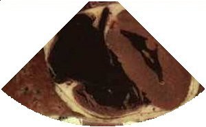
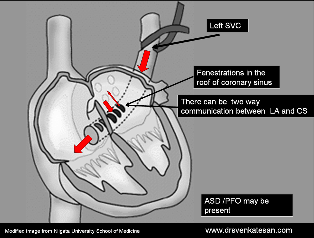
Defects related to case #2 lead to a dilated coronary sinus and filling of the CS prior to that of the RA on a bubble study initiated in the left extremity.

vh_cs.jpg
 
For case #1:

  1. Azygous Vein
  2. Svc
  3. Phrenic
  4. R. IMV
  5. R. IMA
  6. R. Upper lobe
  7. R. Middle lobe
 
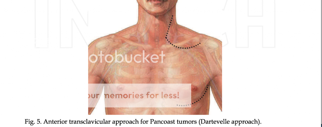
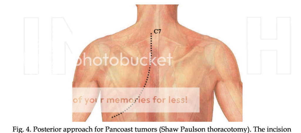
Someone mentioned lateral position for the Pancoast Tumor case. True. There is, however, more than one approach:

F1large_zps624e5a49.jpg


Physical findings (i.e. horners syndrome) along with neurovascular findings on MRI/CT will help the surgeon determine which approach to take.
 
17e68b5e-2c52-46fe-bc5c-f8f2d7b9b227_zps9a2e4611.png


Ouch....

e45fe1a5-409f-46d1-9781-feb3359d837c_zps746ca8e3.png


This approach is just as painful depending on how many ribs are taken. Removing several large sections of thoracic ribs might buy you a large bioprosthetic patch.

Paravertebral catheters or Thoracic epidurals are key. These patients can have tremendous pain post-op. As perioperative anesthesiologists, we can place these before incision and reduce our intraoperative narcotics and possibly chronic pain as well as recurrence (at least in breast surgery). Furthermore, removing an upper lung in a patient that already has pulmonary issues may render them susceptible to tachypnea, hypercapnea and other respiratory complications.

Then there is OLV...
 
case 2: Funny, I just looked up this paper 2 days ago because we have an adult pt in ICU right now with one of these. My pt's x-ray shows CVC going in right side, crossing an innominate, and down the persistent left SVC. Very cool.
http://www.cardiovascularultrasound.com/content/6/1/50

Nailed it... and super cool you are taking care of one of these patients. I think someone mentioned it earlier, so koodos to that person as well.

So embryologically, the left superior vena cava fails to regress.

Here's a CT angio that demonstrates the L SVC.


TEE bubble study is nice to see as well. As mentioned, you will see air enter the Coronary sinus BEFORE the RA when injecting air-saline mixture into the LUE. Usually, due to the increase in blood flow through the CS, the CS will be dilated. You can place calipers and measure the CS to check for dilation if you are thinking you might have a persistent left SVC.

Here is a pic that demonstrates anatomic features of the PLSVC. The PLSVC in continuity with the CS. Various other congenital heart defects may be present.

coornary-sinus-unroofed.png
 
For case #1, as far as monitors... usual stuff. A-line, a couple of big bore IV's.
If your patient is sick and requires a CVL, I would probably not place a subclavian on the same side of the tumor if you are going to do a posterior approach (def. not anterior approach cuz you'll be in the way of the surgical field). If you are going to place one, it's probably better to place it on the opposite side.
 
Let's get some more clinical threads up on SDN.
IMO, the clinical aspects of SDN is where the rubber meets the road. It's also why the anesthesia forum has been so popular in the past.
 
  • Like
Reactions: 1 users
I agree, thanks for posting these. :thumbup:

No prob... you posted an epic one a little while back. Started it off with an EKG if I remember correctly. That was a good one.
 
  • Like
Reactions: 1 user
Say at the end of the pancoast tumor case, the surgeon tracks down some exparel mixed with some .5% marcaine and infiltrates the phrenic nerve.

That's kinda weird. Why would he do that?
 
Say at the end of the pancoast tumor case, the surgeon tracks down some exparel mixed with some .5% marcaine and infiltrates the phrenic nerve.

That's kinda weird. Why would he do that?
Never seen that done, but I'd guess it's to reduce referred pain? C3-5 wouldn't be covered by a thoracic epidural.
 
  • Like
Reactions: 1 user
Very good. There is evidence in the literature that this indeed is helpful for referred pain after thoracic surgery.

http://www.ncbi.nlm.nih.gov/pubmed/11473840 N=90

"Phrenic nerve block with 2% lidocaine should be performed in all patients undergoing a major thoracic surgery procedure. These results strongly support the hypothesis that irritation of the pericardium and/or mediastinal-diaphragmatic pleural surfaces results in pain that is referred to the shoulder via the phrenic nerve."

http://www.ncbi.nlm.nih.gov/pubmed/11473840 N-48

"Ipsilateral shoulder pain after thoracotomy is common and may be severe, even in the presence of a functioning thoracic epidural. We have shown that infiltration of the phrenic nerve with local anesthetic significantly and safely reduces this shoulder pain, potentially allowing the ideal goal of a pain-free thoracotomy."
 
Any OTHER reasons to infiltrate the phrenic nerve?
 
south park, 1st season. classic.
SPcowinquiry.jpg

SPcarlsfault.jpg


that, unfortunately, is all i have to contribute to this otherwise excellent clinical thread.

Very nice. Probably the best response in the thread. :D :thumbup:
 
Re: Persistent left SVC

Residents- say you are doing your echo prior to mitral valve surgery and discover a persistent left svc. Do you have any suggestions for the surgeon on what may need to be done differently for the case?
 
Just guessing, so the operative lung does less work on POD 1? I'm not sure if that'd be desirable or undesirable. Educate me :)


:smug: You are on the right track. :thumbup:

Removing a lobe, particularly an upper lobe like in the pancoast tumor case, leaves a large air space where the lobe previously existed. This space can lead to PAL.
Persistent Air Leak (PAL) is a complication of thoracic surgery. Increased chest tube duration as well as patient discomfort lead to longer hospital stays and overall health care cost. More worrisome however, is the increase risk of infections/empyema, bronchopleural fistulas and thrombi-embolic disease from being tied down to the CT. Indeed, PAL has been proven to lead to increased morbidity post lobectomy.

So what can we do to decrease that space and decrease PAL and improve outcome? We can attempt to raise the ipsilateral hemidiaphragm to reduce the space and it’s associated complications via the prophylactic techniques listed below:

Prophylactic space reduction techniques: A simple technique to transiently reduce the functional size of pleural cavity is to anesthetize the phrenic nerve. This is recommended only for patients with near normal preoperative lung function. The phrenic neurovascular pedicle is grasped gently as it courses above the hilum with a babcock clamp and 1 to 2 cc of 0.5% bupivacaine without adrenalin are injected as a wheal around pedicle but not through pericardium.The nerve is not injected directly, and crushing the pedicle is not advocated. The elevated hemidiaphragm will promote early pleural-pleural apposition and will reduce the amount of free space within the hemithorax. Another technique to consider for space reduction after a VATS resection may be pleural tenting. Dense pleural adhesions may compromise the ability to create an intact tent. Hemothorax may be a possible complication after pleural tenting. Like other intra operative adjuncts, pleural tenting should be reserved for patients undergoing and upper lobectomy with a troublesome air leak.”

So injecting the phrenic has 2 potential upsides:
1)Reduction in pain
2)Decreases the chance of PAL

Thoracic epidurals, phrenic nerve infiltration and OLV strategies are areas where we may be able to improve outcomes. As mentioned above, you need to pick your patients for phrenic nerve infiltration as those with moderate to severe respiratory dysfunction may not tolerate temporary ipsilateral hemidiaphramatic paralysis.
 
Last edited:
  • Like
Reactions: 1 user
Sevo, that's awesome. I just learned something. Thanks.

No takers to my question? Hint-you wouldn't have to do this for a cabg or avr. But you'd have to do it to open the heart without the field becoming flooded...
 
Residents- say you are doing your echo prior to mitral valve surgery and discover a persistent left svc. Do you have any suggestions for the surgeon on what may need to be done differently for the case?

Do you have to clamp the PLSVC?
 
Sevo, that's awesome. I just learned something. Thanks.

No takers to my question? Hint-you wouldn't have to do this for a cabg or avr. But you'd have to do it to open the heart without the field becoming flooded...

Glad to help HB. Thanks for chiming in with your very relevant question. :thumbup:

Med studs and residents... where does the retrograde cannula go again? ;)

I'll let HB give guidance to his question.

Ohh... and HB, I hope you've been getting on top of the epic champagne pow we've bee seeing lately. Caught some deep stuff after a 61" dump a couple weeks back.
You're a Mammoth rider... yes?
 
Alrighty then. So- usually for intracardiac procedures, i.e. mitrals, tricuspids, ASDs etc, in order to obtain a bloodless field we cannulate both cavae. One in the SVC, one in the IVC. For AVR and CABG, a single 2-stage cannula is fine.

When we talk about persistent left SVC, we're usually talking about having bilateral SVCs. A small percentage of PLSVCs completely lack a right (normal) SVC, but this is unusual (though I've seen it).

So in addition to the usual bicaval cannulation, you generally need a *third* cannula, to drain the LSVC flow. How this is accomplished varies from shop to shop but the easiest way is for the surgeon to take a regular straight venous cannula and insert it into the coronary sinus as they would a retrograde cardioplegia catheter.

Another way would be to temporarily clamp the PLSVC, IF there is a good-sized innominate vein to bring left-sided blood to the right SVC. This is not always the case, and in addition, the azygous/hemiazygous system is often aberrant in these folks as well, and you could get flow into the coronary sinus through such a system.

Again for CABG or AVR, your single 2-stage cannula will drain as it usually does even in the presence of a PLSVC, so no need to change cannulation strategy there. However-

Retrograde cardioplegia is generally not given if there is a PLSVC. You could in theory clamp the PLSVC, but again there can be runoff down the azygous system. Generally only antegrade plege is given.

Sevo- I was a Mammoth guy when I lived in SoCal, but now the nearest ski resort is 2500 miles away from me. Traded the snow for surf- if only I could work less and surf more! But yeah- been following the bonanza they finally got after one of the crappiest winters ever so far. Good for them.

Maybe next year I'll hit up the CPB conference in Aspen to get my fix...
 
Sevo: thanks for the great little nugget about phrenic nerve infiltration. I hadn't heard of that technique.

(However, don't go mixing Exparel and regular bupi!)
 
Top