Good learning case.

This forum made possible through the generous support of SDN members, donors, and sponsors. Thank you.

sevoflurane

Ride
20+ Year Member
Joined
Jul 16, 2003
Messages
6,025
Reaction score
3,784
65 y/o male h/o DM, COPD, bilateral iliac stents, endo AAA repair, presents to the primary care physician with vague epigastric pain radiating to the back. CXR reveals something very similar to this:

PA-chest1.jpg


(this xray is off of the internet... not the patient I took care of)

What next?

Members don't see this ad.
 
Last edited:
  • Like
Reactions: 1 user
Was that aneurysm purely abdominal, or also thoracic? Because that's one big aorta otherwise (unless it's a very strange hiatal hernia :) ). I would be concerned about aortic aneurysm/dissection, and I would probably consult his CT surgeon about this. Contrast CT next?
 
Members don't see this ad :)
Was that aneurysm purely abdominal, or also thoracic? Because that's one big aorta otherwise (unless it's a very strange hiatal hernia :) ). I would be concerned about aortic aneurysm/dissection, and I would probably consult his CT surgeon about this. Contrast CT next?

Yes sir.

This is what you get:

Descending thoracic aorta aneurysm. 6-7cm in width. 6.5 cm in length. Starts just distal to the arch, but still confined to the thoracic aorta.

What next?
 
Last edited:
TEVAR with GA, a-line, cordis, TEE, perfusionist and bypass on standby. lots of vasoactive drips ready
 
TEVAR with GA, a-line, cordis, TEE, perfusionist and bypass on standby. lots of vasoactive drips ready

He has bilateral iliac stents and an endo AAA graft. Can we still do a TEVAR?
 
i have seen a patient get a TAVR in a similar circumstance so I think it can be possible. they actually stented the iliac so they would be able to do the TAVR and the pt had a AAA repair in the past as well.
 
i have seen a patient get a TAVR in a similar circumstance so I think it can be possible. they actually stented the iliac so they would be able to do the TAVR and the pt had a AAA repair in the past as well.

You are correct. It can be done. So with this history, you really need to look at the anatomy itself. If the iliacs and the abdominal aorta are very torturous you run the risk of snagging the previously deployed grafts and causing serious problems. Seen it before and it can get ugly. This specific patient was not a candidate.

So if TEVAR is not possible what are our options? How do we do this case?
 
Last edited:
in my very limited resident experience with thoracic aneurysms would say open thoracic repair. on our side: smooth induction with lots of narcotics, versed, splash of etomidate, paralytic. a-line x 2, cordis, PAC, TEE. Case needs bypass and likely DHCA if there is arch involvement. so EEG or SED and cerebral oximetry.
 
Members don't see this ad :)
TEVAR with GA, a-line, cordis, TEE, perfusionist and bypass on standby. lots of vasoactive drips ready
I agree with the lines +/- spinal drain.
Awake fiberoptic intub and Rt sided double lumen tube with CT surg on standby. (Though they will already likely be at bedside). The cxr looks like you've got some major tracheal deviation right above the carina.
 
I agree with the lines +/- spinal drain.
Awake fiberoptic intub and Rt sided double lumen tube with CT surg on standby. (Though they will already likely be at bedside). The cxr looks like you've got some major tracheal deviation right above the carina.

Why awake FOB versus intubation following standard/cardiac/vascular induction with neuromuscular blockade?

Is there a reason you want a right versus left-sided tube?

Have you ever done an awake FOB with a DLT (not fun at all)?

If CT is only on stand-by, who is doing the procedure? In every hospital I have ever been, this would be their bailiwick, not Vascular.

That tracheal deviation does not look that bad.

Agree with Sevo, these are super fun cases.
 
Can you do traditional bypass when you are clamping the thoracic aorta?
 
Open repair is my opinion. Lines as described. Asleep airway management of choice. That tracheal deviation would make me downsize my DBL TUBE so as to pass it carefully. I don't see the advantage of doing it wake. However, I would be cognizant of tracheal collapse with muscle relaxants. Therefore, I would attempt to pass the tube without muscle relaxants and good topicalization. I like the LTA atomizer for this. Mask the pt, topicalize, mask again all while deepening the anesthetic carefully. Unless his anatomy isn't favorable for airway management.
The problem I see here is that you can't easily crash on bypass. This is a difficult case for sure and careful gentle planning and approach is best ( no **** genius).
 
Why awake FOB versus intubation following standard/cardiac/vascular induction with neuromuscular blockade?

Is there a reason you want a right versus left-sided tube?

Have you ever done an awake FOB with a DLT (not fun at all)?

If CT is only on stand-by, who is doing the procedure? In every hospital I have ever been, this would be their bailiwick, not Vascular.

That tracheal deviation does not look that bad.

Agree with Sevo, these are super fun cases.

With this mass effect on the trachea, I worry about tracheal collapse post-induction necessitating a sub-sternal trach.
I think it would be difficult to direct the tube to the left side in this case.
I've never put a DLT with FOB. I was thinking more along the lines of a tube exchange.
Also, if that deviation doesn't look bad, what does a bad deviation look like??
 
  • Like
Reactions: 1 user
We normally used bronchial blocker, since we'd keep pt intubated and wanted to avoid exchange at end of case.

Also vascular did these with high cross clamp only and no bypass.
 
Pent sux tube. The airway will be fine.
 
Fem-fem lines awake. Aline, right cordis +\- pulm cath, tee, 2 great iv lines. fem fem bypass ready to go if needed.

Cardiac induction. SL ET then bronch. Depending on tracheal anatomy put dl or blocker or even mainstem sl. If you get into any airway trouble, fully heparinize and start fem fem bypass.

Open chest, put left atrial line, cool, +\- heparin, atrial fem bypass, repair TAA, wean off bypass, remove cardiac lines close chest, send to icu.

Other options for bypass: simple bypass, dhca if cant access the arch, or partial CPB bypass.
 
Last edited:
  • Like
Reactions: 1 user
Thanks for posting this case. I haven't seen a thoracic aneurysm in the wild since residency, just not part of my practice. These threads help hold off the brain rot, help me not lose everything I don't think about often.
 
  • Like
Reactions: 1 user
Thanks for posting this case. I haven't seen a thoracic aneurysm in the wild since residency, just not part of my practice. These threads help hold off the brain rot, help me not lose everything I don't think about often.
+1. I actually did one of these at 3 AM in residency. Still remember it fondly.
 
Yep. Chest pain in a drunk guy. Dissecting big aneurysm of the descending thoracic aorta.

Of course, I had been up and running for 21 hours when he came in. :)
 
With this mass effect on the trachea, I worry about tracheal collapse post-induction necessitating a sub-sternal trach.
I think it would be difficult to direct the tube to the left side in this case.
I've never put a DLT with FOB. I was thinking more along the lines of a tube exchange.
Also, if that deviation doesn't look bad, what does a bad deviation look like??

We have a CT of his chest, so it would be interesting to see what the actual path of his airway looks like, but that CXR just looks like a relatively small degree of deviation. The path just below the cords continues straight, and it appears to maintain width, while pushed slightly off to the right side, before coming back midline just above the carina. The fact that the trachea returns to midline may make proper placement of a left DLT more challenging, more so than the slight deviation itself. A few months ago, we had a guy here whose trachea made a greater than ninety degree turn off to the right just a few cm below the cords, almost crossed the clavicle, then turned again back toward midline. That was bad. Another thing to consider, would lateral mass effect from an aortic aneurysm necessarily lead to collapse of a cartilaginous structure with neuromuscular blockade? This is a real concern with sizeable anterior mediastinal masses, but there, the direction of force is different in a supine patient.

By the presentation, it does not seem as though we are crashing back to the OR to deal with an acute dissection, so I would first review the CT with the radiologist and/or surgeon, looking specifically at airway anatomy for evidence of compression, and other effects of an enlarged aorta on adjacent structures (esophagus). I would perform a directed history and physical, looking for any other cardiac history (given several risk factors), as well as any positional dyspnea, reflux, or other symptoms. He would get an arterial line, RIJ MAC, good PIVs, and a lumbar drain. I would also discuss with the surgeons if evoked potential monitoring would add anything to assessment of spinal perfusion, given the location of his thoracic aneurysm and previous AAA stent. I see nothing in his history so far that would be a strong indication for PAC. Provided no positional symptoms, no significant airway compromise on CT, and normal airway exam, he would receive a gentle vasculopath induction with neuromuscular blockade, with careful placement of a slightly smaller DLT (37 or 39), which would be guided into place via FOB down the bronchial lumen first. Assuming no GI pathology, I would also place a TEE probe.

Is the surgeon willing to do this procedure quickly, with a high cross-clamp and no bypass, or are we thinking atrial-femoral bypass with either DHCA or ascending aortic cannulation as well, to keep the brain going? Also, where in his iliacs are his stents (and how recently were they placed), and what is the risk of femoral cannulation with iliac artery stents in place (may be a stupid question, but could the cannula snag part of a stent?)?
 
  • Like
Reactions: 1 users
My guess is we have to go with open repair given his prior AAA and iliac stents (unless these are widely patent and they can get enough access to do a TEVAR). The other question is how proximal the aneurysm extends, and whether there is sufficient landing area to place an endovascular graft (does he need a left SC/LCC bypass?). The risk of SCI is definitely a real issue here. If it involves the arch, we typically do DHCA with antegrade cerebral perfusion via R axillary cannulation. If not, left heart bypass is an option (left atrium to distal to the cross clamp) to provide distal perfusion and hopefully protect the spinal cord and kidneys. I'd do a GA with a left sided double lumen tube (I'd just pass a fiberoptic bronchoscope through the tube shortly after getting it into the trachea and guide it down the left bronchus--as above), bilateral radial a-lines, MAC, and a lot of blood. Platelets for after circ arrest (if necessary), and a lumbar drain. I'd probably avoid the AFOI because of the hypertension and tachycardia that it would likely produce (in a patient with a giant aortic aneurysm). Good case.
 
  • Like
Reactions: 1 user
I'd probably avoid the AFOI because of the hypertension and tachycardia that it would likely produce (in a patient with a giant aortic aneurysm). Good case.

I wouldn't do AFOI either -- but if i did, I'd use remi
 
Thanks for posting this case. I haven't seen a thoracic aneurysm in the wild since residency, just not part of my practice. These threads help hold off the brain rot, help me not lose everything I don't think about often.

You bet. Giving back to this forum is something I've tried to do for a while now as it previously gave so much to me... and still does. :thumbup:

I wouldn't do AFOI either -- but if i did, I'd use remi

Well, I wouldn't do an AFOI either -- but if I did, I'd use Dos Equis. Stay thisty my friends...
 
  • Like
Reactions: 3 users
You guys are a smart bunch :bookworm:. A lot of great answers and posts. Always impressed by the caliber of responders here.
For me, this is a super fun case because it encompasses nearly every procedure known to the anesthesiologist and the physiology and plumbing can be quite interesting.

Lines/monitors/setup for this case:

Left sided DLT: No need for AFOI + standard induction. A-line in the dependent extremity, double stick right IJ (Mac + triple lumen), Swan, 14G PIV, Cerebral Ox, TEE, lumbar drain with pressure monitor. As Noy pointed out, this is a case that really needs to be discussed with the CT surgeons prior to bringing the patient back to the OR. Prepare for massive transfusion protocol (if you are not a major center, make sure you have appropriate products the day before).

A couple of questions to carry on the discussion:

  1. Is this most likely a median sternotomy or a typical lateral thoracotomy approach?
  2. What is partial bypass and how does it work? Do we need an oxygenator? How do we manage anticoagulation? How do we monitor upper and lower perfusion pressure? What should the perfusion pressure be?
  3. Can we place a thoracic epidural if we are performing partial bypass? Can we place TEA catheter with a lumbar drain in place?
  4. What do we do with a lumbar drain? How do we manage it?
 
Last edited:
  • Like
Reactions: 1 user
Is the aortic valve involved at all?

Valve is good. Based on the CT, the thought is that a crossclamp will be able to be placed distal to the arch and it's vessels.
 
Not a big fan of the "no bypass" technique with previous endo AAA:


75a9b994-34e8-4da4-98e3-b85331d8cf4c_zps333356a6.png
 
  1. Is this most likely a median sternotomy or a typical lateral thoracotomy approach?
Usually lateral as this gives best access to the descending TAA. Median if it is ascending and involving the aortic valve.
  1. What is partial bypass and how does it work? Do we need an oxygenator? How do we manage anticoagulation? How do we monitor upper and lower perfusion pressure? What should the perfusion pressure be?
There are different types of bypass you can do which i was eluding to earlier:
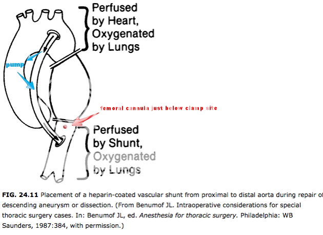
1) simple shunt - basically a heparinized tubing from LV or acending aorta to descending aorta.
2) partial bypass - left atria or pulm vein or even ascending aorta to the left femoral artery. Generally heparinize coated centrifugal pump requires no systemic heparin or low dose 100u/kg. no need for oxygenator as the blood is already oxygenated. Heat exchanger is not even needed.
3) DHCA: fem-fem bypass that you esentially cool the body to 20 degrees, drain entire blood supply, work on aorta quick, rewarm body unless need cardiac surgery too (av repair) then it gets more complicated.

Some posts are confusing these points. In 1 and 2 the heart is not stopped. It keeps pumping and thats why you still have cerebral flow. The TAA is then clamped above and below. However the afterload is super high immediately after cross clamp and this can be difficult to manage. In partial bypass u need to measure both right radial and femoral artery pressure. The perfusionist and anesthesiologists work in a team because the anesthesiologist is managing upper blood flow, and the perfusionist is managing the lower blood flow.
The Femoral flow is confusing too... Its not only providing anterade flow to that leg but the rerograde flow to the decending aorta below the clamp which should help perfuse spinal cord and gut. The radial artery will give a traditional arterial pulse wave, whereas the femoral will give you a flat line. MAP should be around 70
 
  • Like
Reactions: 1 user
Great thread and great responses.

I have been lucky, all the TAAs I've dealt with since leaving training have been amenable to TEVAR.

But I know I'll see another one of these one day. They're great cases-once the patient is safely in the ICU.
 
You guys are a smart bunch :bookworm:. A lot of great answers and posts. Always impressed by the caliber of responders here.
For me, this is a super fun case because it encompasses nearly every procedure known to the anesthesiologist and the physiology and plumbing can be quite interesting.

Lines/monitors/setup for this case:

Left sided DLT: No need for AFOI + standard induction. A-line in the dependent extremity, double stick right IJ (Mac + triple lumen), Swan, 14G PIV, Cerebral Ox, TEE, lumbar drain with pressure monitor. As Noy pointed out, this is a case that really needs to be discussed with the CT surgeons prior to bringing the patient back to the OR. Prepare for massive transfusion protocol (if you are not a major center, make sure you have appropriate products the day before).

A couple of questions to carry on the discussion:

  1. Is this most likely a median sternotomy or a typical lateral thoracotomy approach?
  2. What is partial bypass and how does it work? Do we need an oxygenator? How do we manage anticoagulation? How do we monitor upper and lower perfusion pressure? What should the perfusion pressure be?
  3. Can we place a thoracic epidural if we are performing partial bypass? Can we place TEA catheter with a lumbar drain in place?
  4. What do we do with a lumbar drain? How do we manage it?
Why the PAC? Shouldnt the TEE be enough? Agree with everything else.
 
Why the PAC? Shouldnt the TEE be enough? Agree with everything else.

Culture of our institution, post-op management, surgeon preference, sudden increase in afterload, continuous cardiac output and SVO2 during partial bypass AND one lung ventilation. We don't have CRNA's running around. This is a solo gig. It's nice to look up and get some quick info in the middle of a potential crisis. TEE does take time away from drawing drugs up, giving products, etc. But yeah, you could def. do it w/o a PAC.
 
Last edited:
  • Like
Reactions: 1 user
  1. Is this most likely a median sternotomy or a typical lateral thoracotomy approach?
Usually lateral as this gives best access to the descending TAA. Median if it is ascending and involving the aortic valve.
  1. What is partial bypass and how does it work? Do we need an oxygenator? How do we manage anticoagulation? How do we monitor upper and lower perfusion pressure? What should the perfusion pressure be?
There are different types of bypass you can do which i was eluding to earlier:
1) simple shunt - basically a heparinized tubing from LV or acending aorta to descending aorta.
2) partial bypass - left atria or pulm vein or even ascending aorta to the left femoral artery. Generally heparinize coated centrifugal pump requires no systemic heparin or low dose 100u/kg. no need for oxygenator as the blood is already oxygenated. Heat exchanger is not even needed.
3) DHCA: fem-fem bypass that you esentially cool the body to 20 degrees, drain entire blood supply, work on aorta quick, rewarm body unless need cardiac surgery too (av repair) then it gets more complicated.

Some posts are confusing these points. In 1 and 2 the heart is not stopped. It keeps pumping and thats why you still have cerebral flow. The TAA is then clamped above and below. However the afterload is super high immediately after cross clamp and this can be difficult to manage. In partial bypass u need to measure both right radial and femoral artery pressure. The perfusionist and anesthesiologists work in a team because the anesthesiologist is managing upper blood flow, and the perfusionist is managing the lower blood flow.
The Femoral flow is confusing too... Its not only providing anterade flow to that leg but the rerograde flow to the decending aorta below the clamp which should help perfuse spinal cord and gut. The radial artery will give a traditional arterial pulse wave, whereas the femoral will give you a flat line. MAP should be around 70


Very nice. Great response RxBoy. Kudos man!

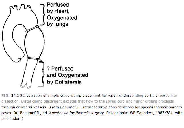
We elected not to poke a huge hole in the ascending aorta (like the picture below) and instead placed the cannula in the relatively low pressure LEFT atrium. The distal end of the cannula was then anastamosed to the femoral artery below the crossclamp. The right lung was able to oxygenate the entire patient w/o any issues whatsoever. SVO2 stayed in the 70-75% range. No need for an oxygenator or heat exchanger. ACT in the 275-300 range.

Here is a graphic for those interested. Our cannulation site (red addition to the graphic representation) was just below the crossclamp, so the partial bypass was very physiologic with mostly anterograde flow and little to no retrograde flow.

c5047884-537a-4b41-a463-75ddf70b8730_zpsea2b37d0.png
 
Last edited:
You mentioned communication to perfusion. You are absolutely right!

So before we went lateral we placed a right femoral arterial line so that perfusion could transduce their lower extremity pressures. They stole about 2 l/m of blood flow from the left atrium which then left us roughly the same amount to perfuse the upper extremity and brain. It actually helped to decrease the afterload after placement of the proximal crossclamp thereby decreasing wall tension on the LV. Deep transgastric views showed that contractility was essentially unchanged after application of the x-clamp.
 
Last edited:
Upper extremity pressures were 90-120 systolic. Femoral MAPs around 55-65mmhg.
 
Who wants to try and tackle questions #3 and #4 above? Spinal cord protection is one of the other big aspects of this case.
 
I did similar case last night. Thoracic dissection from valve to diaphragm. TEE was classic. DHCA with antegrade cerebral perfusion, stented the descending thoracic aorta, tube graft from valve to arch, left arch alone (no compromise of arch vessels), tissue valve, bypass RCA (it was dissected as well). Young guy, like 45. Per family, he would never want to be on coumadin so he could keep working construction, so he got a tissue valve. Went well. Awake, extubated, no neuro deficits today.
 
Top