High Yield Anatomy/Neuroanatomy thread

This forum made possible through the generous support of SDN members, donors, and sponsors. Thank you.

sswang00

MS4
10+ Year Member
Joined
Feb 20, 2010
Messages
144
Reaction score
5
Anatomy is one of those subjects most of us don't want to devote too much time to because it isn't as high yield as some of the other subjects on the Step 1.
So how about we quiz each other with high yield anatomy facts? I'll get the ball rolling.

Question: A woman comes in with an ovarian mass and is scheduled for surgery to remove the tumor. During the surgey, which ligament should be ligated to prevent excess bleeding?

Members don't see this ad.
 
Last edited:
  • Like
Reactions: 1 user
Suspensory ligament?

Woman tripped down the stairs wearing high heels, inverting her ankle. Which ligament is most likely injured?
 
Suspensory ligament?

Woman tripped down the stairs wearing high heels, inverting her ankle. Which ligament is most likely injured?

Yup, you got it.
Ant __Fibular ligament? I forgot the whole name. But i know its on the lateral part of the ankle and its connected to the fibula.

Question: Surgeon performs thyroidectomy. What nerve is damaged and what are the associated symptoms if there was an injury while ligating:
1) Superior laryngeal artery
2) Inferior laryngeal artery
 
Question: Surgeon performs thyroidectomy. What nerve is damaged and what are the associated symptoms if there was an injury while ligating:
1) Superior laryngeal artery
2) Inferior laryngeal artery

Recurrent laryngeal n - pt can present w/ hoarseness after surgery.
Sup. laryngeal n. since sup laryngeal n innervates the cricothyroid muscle, if you nick the nerve then the pt will have problem with the vocal ligament?
Inf. laryngreal n. Same story, affects the muscles of the larynx.
So I'm guessing there's problem with phonation? I can never remember the actions of the laryngeal muscles. Sorry.

Q: In horseshoe kidney, what structure prevents the ascent of the kidney?
 
Last edited:
Members don't see this ad :)
I got about 6-7 lower limb anatomy questions on my test. I wasnt ready for that but I wasn't unprepared either.

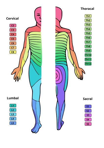
The only one that really got me asked me what nerve root innervates sensation to the lateral thigh. ANd im like oh lateral femoral cutaneous and im like hmm is that L2 or L3, and I couldn't decide.. So i just put L2 down and moved along.. And after the test I went to look at that one, and it happens that the nerve is innervated by L2 AND L3, so I'm just hoping that it was an experimental question.
 
Q: In horseshoe kidney, what structure prevents the ascent of the kidney?

inferior mesenteric artery?

Q: Fusion of the caudal portion of the kidneys during embronic development would result in which congenital malformation?
a. bicornuate uterus
b. horseshoe kidney
c. cryptorchidism
d. hypospadias
e. renal agenesis
 
Last edited:
Yup, you got it.
Ant __Fibular ligament? I forgot the whole name. But i know its on the lateral part of the ankle and its connected to the fibula.

Question: Surgeon performs thyroidectomy. What nerve is damaged and what are the associated symptoms if there was an injury while ligating:
1) Superior laryngeal artery
2) Inferior laryngeal artery

what are the answers to this? superior and inferior laryngeal nerve?
 
what are the answers to this? superior and inferior laryngeal nerve?
Ligation of inf.thyroid artery during thyroidectomy can cause Unilateral damage to recurrent laryngeal nerve(hoarse voice, movement of vocal fold on affected side towards the midline), or bilateral damage to recurrent laryngeal nerve(dyspnea cuz both vocal folds move towards the midline and close the air passage).

Ligation of superior thyroid artery during thyroidectomy can cause damage to superior laryngeal nerve(weak voice with loss of projection, vocal cord on affected side appears flaccid).
 
My plan was to basically only due arm and leg anatomy.. knowing that going into the test anatomy will be my weak point. Any body else go in with this strategy??

Would FA be enough for anatomy??
 
Patient presents with right sided ataxia, left sided nystagmus, right sided loss of pain and temperature in the face, left sided loss of pain and temperature in the body and right sided ptosis. Where is the lesion?

A. Right Lateral Pons
B. Left Medial Midbrain
C. Right Lateral Medulla
D. Spinal Cord at T-1
E. Right Post-central Gyrus
 
A. Right lateral pons

?

left 12th rib broken/pierced - what structure is damaged
 
Last edited:
I was going for lateral medullary syndrome with PICA but I forgot to add a few symptoms like dysphagia and hoarseness. Lateral pontine syndrome (AICA) would've also had facial paralysis though.

Oh and to your question I'm thinking spleen?
 
Members don't see this ad :)
I got about 6-7 lower limb anatomy questions on my test. I wasnt ready for that but I wasn't unprepared either.

The only one that really got me asked me what nerve root innervates sensation to the lateral thigh. ANd im like oh lateral femoral cutaneous and im like hmm is that L2 or L3, and I couldn't decide.. So i just put L2 down and moved along.. And after the test I went to look at that one, and it happens that the nerve is innervated by L2 AND L3, so I'm just hoping that it was an experimental question.
424px-Dermatoms.svg.png


psychforme saves the day once again
 
I was going for lateral medullary syndrome with PICA but I forgot to add a few symptoms like dysphagia and hoarseness. Lateral pontine syndrome (AICA) would've also had facial paralysis though.

Oh and to your question I'm thinking spleen?

haha yeah, the CN IX, X deficiences like losing gag reflex, hoarseness are needed


what is the hypothalamic nucleus for hunger ?
 
lol yea they all look the same.
I just try and remember it like this:

-Tibial ("TIP") helps you stand on your "tippie toes" so it innervates the sole of the foot= s1/2
-Peroneal ("PED") helps you Evert your foot so your pointing it laterally..makes sense in innervate lateral leg

-Then for medial leg&thigh region, I try to think of the KNEE being in the middle of your leg. Knee jerk reflex = L2,3,4 = Femoral Nerve.
So thigh is L2 and medial leg is L3,4 (saphenous branch off femoral)
 
Last edited:
hey..thanks! i was having a hard time remembering those stuff :)
lol yea they all look the same.
I just try and remember it like this:

-Tibial ("TIP") helps you stand on your "tippie toes" so it innervates the sole of the foot= s1/2
-Peroneal ("PED") helps you Evert your foot so your pointing it laterally..makes sense in innervate lateral leg

-Then for medial leg&thigh region, I try to think of the KNEE being in the middle of your leg. Knee jerk reflex = L2,3,4 = Femoral Nerve.
So thigh is L2 and medial leg is L3,4 (saphenous branch off femoral)
 
is l3 not also thigh?


oh and peroneal = fibular, which is your lateral leg bone...only makes sense that it innerv. lateral
 
lol yea they all look the same.
I just try and remember it like this:

-Tibial ("TIP") helps you stand on your "tippie toes" so it innervates the sole of the foot= s1/2
-Peroneal ("PED") helps you Evert your foot so your pointing it laterally..makes sense in innervate lateral leg

-Then for medial leg&thigh region, I try to think of the KNEE being in the middle of your leg. Knee jerk reflex = L2,3,4 = Femoral Nerve.
So thigh is L2 and medial leg is L3,4 (saphenous branch off femoral)

Is this info in FA? Haven't studied anatomy yet.. Gonna start that by the end of this week. How should I approach anatomy?? Anatomy hasalways been my weakness...
 
Is this info in FA? Haven't studied anatomy yet.. Gonna start that by the end of this week. How should I approach anatomy?? Anatomy hasalways been my weakness...
yes it is, in the musculoskeletal section of the 2010 at least, not sure about earlier versions
 
Name the
a. Spinal level
b. Anatomical landmark
c. space that you are looking to penetrate into (epidural/subdural/subarachnoid)

1. taking a CSF sample
2. Injecting anesthetics into a woman prior to labor
 
okay well i don't know the answer to 2 but I think 1 is

a. L4-L5
b. iliac crest plane
c. skin, ligaments, epidural space, dura, sub dural space, arachnoid and your there

? and no idea for #2
 
Is there much straight anatomy tested (ie what branches off of what.. Etc)? Or is most of it clinical Anatomy?
 
After going through msot of UW, all of the Anatomy questions encountered are related to a certain disease or therapeutic intervention.
 
IMG, you still haven't taken your test yet? dude, you are going to MURDER THAT thing
 
What is the reason why infants have a positive Babinski reflex, and at what age does it dissapear?
 
What is the reason why infants have a positive Babinski reflex, and at what age does it dissapear?


Because the Corticospinal tracts are not fully myelinated till age 1-2. Once this happens, the hyperexcitability of the reflex arc diminishes.

My question:

Lesion to what part of the brain produces speech from an individual lacking all emotion (expressive dysparody)?
 
Expressive dysprosody - Lesion to Broca's Area in the
Non-Dominant Hemisphere (Inferior Frontal Lobe )

How about a man who wakes up in the middle of the night and believes someone has "pranked him" by placing an arm in his bed, to his left. Where is the lesion?​
 
Last edited:
Expressive dysprosody - Lesion to Broca's Area in the
Non-Dominant Hemisphere (Inferior Frontal Lobe )

How about a man who wakes up in the middle of the night and believes someone has "pranked him" by placing an arm in his bed, to his right. Where is the lesion?​

hemineglect - lesion of right(nondominant) parietal ?

while on the topic what constitutes Gertsmann syndrome?​
 
Expressive dysprosody - Lesion to Broca's Area in the
Non-Dominant Hemisphere (Inferior Frontal Lobe )

How about a man who wakes up in the middle of the night and believes someone has "pranked him" by placing an arm in his bed, to his right. Where is the lesion?​


hey Knucles,

I get that this is inferior parietal lobe (non-dominant) hemisphere. How do you find out if this person is right or left dominant?

Should we assume that people are right handed i.e. left dominant.​
 
yes i think its safe to assume that, also i believe i read that even some proportion left handed people are left hemisphere dominant, i stand corrected though
 
I thought for neglect syndromes, it is if it is right arm neglect, then left parietal lesion regardless of dominant side. Are you guys saying that this, like speech, follows dominant sides?
 
Not being aware of half of the world (hemi-neglect) is hallmark of a non-dominant parietal lobe lesion. Since I didn't mention that the person was left handed, you're to assume they're right handed and most likely left dominant. Hence, left parietal lobe. (On the other hand, most left handed people are also left dominant. I wouldn't worry about this unless they specifically mention the dominance is not left.)

Gerstmann syndrome is a lesion of the dominant (left) hemisphere parietal lobe. Symptoms include being unable to distinguish between right and left.
Agraphia (not being able to write)
Acalculia (unable to perform simple arithmetic)
Inagnosia (unable to identify fingers)
Reading ability is intact


So in conclusion:

Hemineglect = Nondominant Parietal lobe lesion
Gerstmann syndrome = Dominant Parietal lobe lesion

So with hemi-neglect is the neglect always directed towards the non-dominant arm (bc of damage to nondominant lobe)... ie right handed person, right parietal damage --> left hemi-neglect??.... will left parietal damage lead to right hemi-neglect OR will it lead to Gerstmann Syndrome??
 
So with hemi-neglect is the neglect always directed towards the non-dominant arm (bc of damage to nondominant lobe)... ie right handed person, right parietal damage --> left hemi-neglect??.... will left parietal damage lead to right hemi-neglect OR will it lead to Gerstmann Syndrome??
Gerstmann.

I guess I messed up my question earlier. It seems the pranked arm should have been on his left. Since hemispatial neglect has to be on the left side, correlating to the non-dominant right lobe.)


See wiki:

Hemispatial neglect results most commonly from brain injury to the right cerebral hemisphere, causing visual neglect of the left-hand side of space. Right-sided spatial neglect is rare because there is redundant processing of the right space by both the left and right cerebral hemispheres, whereas in most left-dominant brains the left space is only processed by the right cerebral hemisphere. Although most strikingly affecting visual perception ('visual neglect'), neglect in other forms of perception can also be found, either alone, or in combination with visual neglect.
For example, a stroke affecting the right parietal lobe of the brain can lead to neglect for the left side of the visual field, causing a patient with neglect to behave as if the left side of sensory space is nonexistent; although they can still turn left. In an extreme case, a patient with neglect might fail to eat the food on the left half of their plate, even though they complain of being hungry. If someone with neglect is asked to draw a clock, their drawing might show only the numbers 12 and 1 to 6, the other side being distorted or left blank. Neglect patients may also ignore the contralesional side of their body, shaving or adding make-up only to the non-neglected side.
Neglect may also present as a delusional form, where the patient denies ownership of a limb or an entire side of the body. Since this delusion often occurs alone without the accompaniment of other delusions, it is often labeled as a monothematic delusion.
Wow thanks for the awesome reply:thumbup:

Where can you find this info from?? I dont think its in FA.. is it in HY (I didnt find it in the Cerebral Cortex Chapter 22)?? BRS?
 
Wow thanks for the awesome reply:thumbup:

Where can you find this info from?? I dont think its in FA.. is it in HY (I didnt find it in the Cerebral Cortex Chapter 22)?? BRS?

Yes it is in FA and like 80% of the things, its tucked in between stuff so you dont notice the subtlety (that's a word right? All these medical words have replaced English as a second language.)

I would however suggest to you to give HY Neuro a quick read, I went through all of it in 8 hours and it is one of the best books I have seen. Heck, I would say you can skip neuro section of FA (except Path and Pharm) and just read HY Neuro instead.
 
AV malformations?

My question: What is the anterior drawer sign for?

Test for ACL. Flex knee and apply pressure on the back of the knee to test for movement of the tibia.

If the tibia displaces excessively forward indicated a lesion in the ACL.
If the tibia displaces excessively backwards indicated a lesion in the PCL.
 
Test for ACL. Flex knee and apply pressure on the back of the knee to test for movement of the tibia.

If the tibia displaces excessively forward indicated a lesion in the ACL.
If the tibia displaces excessively backwards indicated a lesion in the PCL.

:thumbup:
 
Top