This is a friend and not a patient. Here is text from an email he sent me:
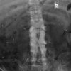
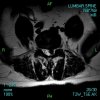
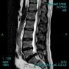
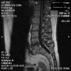
It's not really funny. The tumor is huge filling more that 3/4 of the canal at the C3-C4 level. He believes it's a meningioma. He's rather amazed that my only complaint has been my "arthritic" hand symptoms which have not increased since they began 10 months ago. He did show me that my right knee reflex is hyper reactive though surprisingly to him my arm is not. By comparison my left leg is slightly numb compared to the right leg: all the way down into the foot. I've know the left shin has been numb for several years and always assumed it had to do with round house kicks from karate and bone on bone blocks I would use.
Meanwhile, I broke my right foot in judo 4 weeks ago. LOL. It's improving now. But, the neurosurgeon broke my heart when he said I have to stop judo NOW. He was pretty impressed I didn't paralyze myself landing on my neck which I've done several times.
Now I have to find someone who's an expert at intra-canal tumors in the cervical spine. >From what I was told, nobody is an expert because they're very rare in that location. Well, I have the opinion from the one neurosurgeon who is chief of neurosurgery at our local hospital and he wants to go in the old way through the back of the spine, opening a large section and hoping to "pop" the tumor out....... I'm trying to get another opinion from someone at AAA Medical Center, a teaching hospital. When I called their neurosurgery department and said cervical canal tumor she gave me a specific physician's name. He does most of the cervical spine work there, so that's kind of heartening to hear. He does do minimally invasive surgery though (something I should have asked the doctor yesterday) I don't know if he would do it in this particular case. I guess I'll find out.
The doctor yesterday feels my hand muscles are somewhat atrophied but is not sure if that might just be normal appearance for me: I really don't know.
I have caught you up to date. Hopefully you are doing well. BTW, I don't see much difference between 4mg and 8mg of zanaflex.


")