- Joined
- Jul 6, 2004
- Messages
- 1,977
- Reaction score
- 563
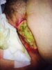
I saw a pt with surgeon who was treated with RT in 1998 for unknown primary "poorly diff epithelial malignancy". He had groin dissection, 7 nodes taken 2 w disease and ECE. He had adjuvant RT, 50 to the groin with conedown to 60 Gy to I guess what they considered the high risk region. He says they took a week break due to dermatitis after 15 fractions. Don't really have much more than that. He had lymphedema on the ipsilateral side that developed a few years later, some tightness but tolerable.
He noticed a small blister in the groin in January 2015 that over the course of the next 6 months slow increased in depth. No other symptoms, no infectious like sxs. Surgery saw him and were concerned about RT necrosis and sent him for 40 tx of hyperbaric O2. On HBO the wound got particularly worse and the size expanded from 2 x 2 to 9.5 x 6 cm by the end of treatment. They debrided the tissue but could only go so far, surgeon was concerned about exposure of femoral vessels. Pathology doesn't show any recurrent disease. They have him packing the wound, the surgeon discussed possible need of an amputation before (sounded extreme but the wound is quite large). They'll see plastics next wk.
Is this consistent with RT necrosis almost 20 years out, Is it unusual to see such enlargement while on HBO. Would you avoid HBO again given that it increased in size. I think the wound has stabilized but it is quite impressive.
Last edited: