The death of the PAC

This forum made possible through the generous support of SDN members, donors, and sponsors. Thank you.
D

deleted171991

An article from the excellent Paul Marik, in the Annals of Intensive Care: "Obituary: pulmonary artery catheter 1970 to 2013" (via ScanCrit).
Conclusions
There is no evidence that the use of the PAC has improved patient outcomes. There are, however, convincing data that the hemodynamic parameters obtained from the PAC are inaccurate, are incorrectly interpreted and that these data frequently lead to excessive and inappropriate therapeutic interventions that maybe harmful. In addition, the data suggest that the hemodynamic parameters obtained from the PAC have little utility in managing critically ill patients in the ICU and operating room. These data therefore suggest that the PAC has a limited role in the ICU and operating room and challenge those experts who believe that the 'pulmonary artery catheter is still a valuable tool for hemodynamic monitoring'. The PAC, however, has a role in the diagnosis and operative management of patients with pulmonary hypertension and acute right ventricular failure.

Food for thought. I personally hope that, in the future, all these invasive monitors will go the way of the bloodletting.

Members don't see this ad.
 
Last edited by a moderator:
I agree. It really irks me when I'm rotating through the TBICU, which is run by the trauma surgeons and whenever they have a patient on pressor, they want to float a swan. Whenever they see something hazy on the chest X-ray, they want to do a bronch. Not only does it waste my time since I have to do the procedures, it rarely changes management and places the patient at risk for potentially life threatening complication.
 
Members don't see this ad :)
Every heart I do in PP gets one. If nurse calls surgeon about hypotension or any issue it's "What's the CO?"
 
  • Like
Reactions: 1 user
Marik argues that the CO, as measured by the PAC, can be up to 80% off the real CO, as measured by the direct Fick method. It tends to underestimate CO especially in patients with a degree of TR, which represent 70% of the elderly, leading to overtreatment. There is a huge intermeasurement variability, because of all the factors that can affect PA thermodilution, from respiration/PPV to the location of the probe.

This, while the transpulmonary thermodilution methods are way more precise, with an inter-measurement variability of less than 7%.

This is the important stuff:

Potential benefits of the PAC
The benefits of the PAC are somewhat difficult to define. In some patients the diagnosis between non-cardiogenic and cardiogenic pulmonary edema is difficult to make. In such circumstance the PCWP has been used to make this distinction. However, with advancements in echocardiography, catheterization of the pulmonary artery for this purpose is seldom required. It would appear that the role of the PAC is limited to diagnosing patients with pulmonary hypertension and managing these patients in the perioperative period (see below), as well as the diagnosis of intracardiac shunts (echocardiography may be better) and amniotic fluid embolism.
Indications for the use of the PAC
Doppler echocardiography is frequently used to calculate the pulmonary artery systolic pressure (sPAP). However, the sPAP as determined by Doppler echocardiography is an inaccurate estimate of sPAP, and this technology is not considered a reliable method for the diagnosis and management of pulmonary arterial hypertension (PAH). Pulmonary artery catheterization is therefore required to confirm the diagnosis of PAH, classify PAH, assess its severity and to test the vasoreactivity of the pulmonary circulation. Pulmonary artery catheterization has been recommended in patients with significant PAH (sPAP > 50 mmHg and/or RV enlargement) undergoing a major surgical intervention. While this recommendation is not supported by high quality evidence, it would appear to be logical as the PA pressure is the only reliable hemodynamic parameter derived from the PAC and its use may allow for the rational titration of vasoactive agents.

Over 30 randomized controlled trials have studied perioperative hemodynamic optimization in a variety of settings, using various goals and techniques of hemodynamic optimization. This approach has been demonstrated to reduce the risk of complications and mortality in elective non-cardiac surgery patients. The initial preemptive hemodynamic studies used the PAC and targeted ‘supranormal’ goals while more recent studies have ‘optimized’ CO using esophageal Doppler or dynamic indices of fluid responsiveness. Meta-analyses of these studies have demonstrated that both approaches reduce surgical mortality and morbidity. In addition, these meta-analyses have demonstrated that the PAC has been largely replaced by less invasive hemodynamic monitoring techniques.
 
Last edited by a moderator:
Every heart I do in PP gets one. If nurse calls surgeon about hypotension or any issue it's "What's the CO?"
This reminds me of the dinosaur eye surgeons who want retrobulbar block or even GA for their cataract patients.
 
Still use them for all my liver transplants. Best indicator for impending RV failure post reperfusion.
 
It tends to underestimate CO especially in patients with a degree of TR, which represent 70% of the elderly, leading to overtreatment.

100% of patients with a PA catheter have a PA catheter stenting open their tricuspid valve.
 
  • Like
Reactions: 1 users
Still use them for all my liver transplants. Best indicator for impending RV failure post reperfusion.

Interesting, even in the otherwise healthy HCC guys who are only eligible because of exception points? Where I trained we would drop a TEE if we had any concerns of their cardiac status leading up to or following reperfusion, and even that was rare. Then again our surgeons were some of the best in the world and all things being considered, patients ended up being as stable as you could expect for undergoing a liver transplant.
 
I could argue that the thermodilution is more accurate than the Fick equation using O2, the Fick Equation using C02, flowtrac, PICCO or any other measurement of CO in certain situations. All of them are just a way to measure a physiologic number of interest to hopefully help our patients.

I do agree that a swan is not indicated in every cardiac surgery.

But if I am titrating ionotropic drugs and other therapies to augment a patients CO….I am going to go with a swan above any other measure in the OR. Just know its limitations like any other tool we use to measure a number and know how to fact check your tools…..if you SWAN is telling you the patient has a CI of 1.5 and you get a MVO2 and it is 70 be suspicious of your swan.

And I don't see how TEE has any benefit over a swan when all you care about is CO…Echo has it own inaccuracies and assumptions when calculating areas and gradients.

FFP what do you use when you are titrating ionotropes?
 
Interesting, even in the otherwise healthy HCC guys who are only eligible because of exception points? Where I trained we would drop a TEE if we had any concerns of their cardiac status leading up to or following reperfusion, and even that was rare. Then again our surgeons were some of the best in the world and all things being considered, patients ended up being as stable as you could expect for undergoing a liver transplant.


Preop cardiac status is important and part of it but I'm also concerned about about other factors that may increase acid load on reperfusion and put even a healthy heart into RV failure. It's worth mentioning that I don't get that many healthy recipients. Most of our MELD score's are >30 and I find myself transfusing platelets as I place lines.
 
So if you believe Marik, the PA catheter produces inaccurate, non-reproducible, and dubiously interpreted data...

...and bedside echo solves all of those problems.:uhno:
 
Members don't see this ad :)
So if you believe Marik, the PA catheter produces inaccurate, non-reproducible, and dubiously interpreted data...
Actually that would explain why the outcomes are worse with a PAC (and that's not just Marik). First do no harm.

The PAC makes a lot of assumptions/approximations related to the left heart that are usually not true, and now it seems that even the CO is unreliable, but I can see why it's great for PA pressure measurement.

I trust Marik because the guy wrote a beautiful (even if slightly biased) evidence-based ICU (hand)book, already in the 2nd edition.
 
Last edited by a moderator:
I did my residency at a place that took the same stance as the OP. Prior to starting my fellowship, I had never floated a PAC in the ICU, and only did them for open heart cases in the OR (along with the occasional liver). Then I did my fellowship, when I actually learned how to use a PAC and interpret it for diagnostic decision making.

All my experiences together did teach me that the PAC is generally an unreliable monitor and should not be used routinely. However, there are VERY select circumstances in which a PAC can give you useful information. While I agree that the TEE is the most accurate monitor to assess cardiac function, volume status, etc, it is also not a continuous monitor. Leaving a TEE probe on in the esophagus will have it's own set of complications and add costs.

Bottom line: although their readings may not be the best, PAC/CVP monitors are the only continuous monitors we have. Although I would not use them routinely, some times you just have to work with what you've got.
 
Bottom line: although their readings may not be the best, PAC/CVP monitors are the only continuous monitors we have. Although I would not use them routinely, some times you just have to work with what you've got.
Continuously unreliable? :)

Since you mentioned CVP: that's another monitor which has been proven to be unreliable, except for the low values, which correlate pretty well with hypovolemia.
 
Last edited by a moderator:
Continuously unreliable? :)

Since you mentioned CVP: that's another monitor which has been proven to be unreliable, except for the low values, which correlate pretty well with hypovolemia.

With the CVP, as with any continuous monitor, I put more stock into the trends rather than the absolute values. Even if a CVP of 16 may not necessarily indicate intravascular hypervolemia, seeing it suddenly rise would almost certainly indicate some form of myocardial dysfunction.
 
INotrope. Not IONotrope.

I agree in principle that the PAC is overused, and often misused.

I don't use a swan for routine CABG or AVR with a normal ventricle.

I do find it useful for mitral surgery, or anytime I suspect either ventricle may struggle.

I do not ever use a "regular" swan with bolus thermodilution. Always the CCO swan. The most useful number it gives you IMO is the real-time mixed venous, which is about the only thing I can't get by echo in the OR.

3D measurement of the LVOT area makes cardiac output by TEE pretty frigging accurate I think.

So yeah. I don't use swans very often, but I think they have their place.

I have a huge issue, though, when the ICU starts INotropes for an index of 1.9 when the patient otherwise looks great and is not acidotic. That is just flat-out dumb, and an instance of how the monitor can be misused and lead to badness if the INotropes lead to things like dysrhythmias.
 
All hearts get PAC where I am. Also, pretty much all hearts have an echo probe in as well. And we get paged at all hours to come do an echo if there are questions, even if there is a PAC in place
 
With the CVP, as with any continuous monitor, I put more stock into the trends rather than the absolute values. Even if a CVP of 16 may not necessarily indicate intravascular hypervolemia, seeing it suddenly rise would almost certainly indicate some form of myocardial dysfunction.
wrong
 
  • Like
Reactions: 2 users
We still place a PAC in just about every heart. I think it compliments TEE.... although if I had to pick I'd pick TEE>>>>>>>>>PAC.
CVP is a terrible monitor for volume management. I find respiratory variation on a pulse ox more useful.
 
There are a ton of things between your left heart and your CVP monitor. Any of them will influence the readings, most importantly lung/intrathoracic pressures. All a CVP will tell you is what the pressure in the thoracic vena cava is, but you'll have no idea how to interpret that correctly. Trends don't matter either, because of the same reason. To quote Marik's book, and the studies he mentions, the pooled correlation coefficient between CVP and measured blood volume was 0.16, between baseline CVP and a change in stroke index/cardiac index was 0.18, between delta-CVP and stroke index/cardiac index was 0.11.

His opinion in 2009 was that:
Paul E Marik said:
The CVP is a relic from the past and should never be measured in modern critical care medicine (except in acute cor pulmonale). The CVP and PCWP are no more useful than "the phases of the moon" in evaluating a patient's volume status.

If the CVP increases, what does that mean? Venoconstriction? Increased venous return? TR? Right ventricular dysfunction? Pulmonary artery vasoconstriction? Intrapulmonary problems (ventilation - most frequently, circulation)? Etc. There is a plethora of things that can influence the value, even more than with the Swan.

If you want to find out how the LV is doing, put an echo probe on/in the patient.

P.S. I trained in a place where every single cardiac surgery patient would get a Swan, and we treated numbers to keep the surgeon happy.
 
Last edited by a moderator:
  • Like
Reactions: 1 user
There are a ton of things between your left heart and your CVP monitor. Any of them will influence the readings, most importantly lung/intrathoracic pressures. All a CVP will tell you is what the reading in the thoracic vena cava is, but you'll have no idea how to interpret that correctly. Trends don't matter either, because of the same reason. To quote Marik's book, and the studies he mentions, the pooled correlation coefficient between CVP and measured blood volume was 0.16, between baseline CVP and a change in stroke index/cardiac index was 0.18, between delta-CVP and stroke index/cardiac index was 0.11.

His opinion in 2009 was that:


If the CVP increases, what does that mean? Venoconstriction? Increased venous return? TR? Right ventricular dysfunction? Pulmonary artery vasoconstriction? Intrapulmonary problems (ventilation - most frequently, circulation)? Etc. There is a plethora of things that can influence the value, even more than with the Swan.

If you want to find out how the LV is doing, put an echo probe on/in the patient.

:thumbup: Thank you for clarifying. I had thought that trends had some value and was unaware of the literature you quoted.

I do agree that the Echo is the superior assessment of cardiac function. Perhaps soon when hand held ultrasounds become more available, it will be more feasible to obtain them quickly and routinely.
 
I was also trained with the idea that trends matter. But then I also had attendings who would not put a CVP monitor in for a hepatectomy, regardless of the surgeon's tantrums.
 
Echo>>>>PAC... but I still like seeing PA pressures when I'm doing a million things at once... especially after protamine. Maybe I'm just old fashioned. ;)

8d5ff300-170d-461c-bfd4-9f4509625503_zps1386357f.jpg


I think the PAC still holds some value in certain scenarios.
 
  • Like
Reactions: 1 user
But that makes perfect sense, sevo! Protamine can induce pulmonary vasoconstriction, and the PAC is in the right place to measure that. Plus you have the time correlation, so you know that it's most probably from protamine.
 
Yeah. They are like health IT: 10 years behind everybody else and highly-resistant to change.
 
Part of the reason surgeons like pa cath data is because the numbers can be easily communicated over the phone by the ICU nurse. If one could easily transmit real time echo images to their smart phones they wouldn't need a swan.

We still put in PA cath for every pump case.
 
I trust Marik because the guy wrote a beautiful (even if slightly biased) evidence-based ICU (hand)book, already in the 2nd edition.

:thumbup:

One of my SICU attendings in medical school printed out a half dozen chapters from the book for the residents and students. Phenomenal, phenomenal book.
 
PAC are valuable tool to directly measure right sided pressures…TEE can give you an estimate. PAC can measure CO just as well as TEE…but has the advantages of being able to measure a MVO2, it can stay in when your patient is transferred to CVICU (please don't bring up htee bc it has a long way to go and good luck in a post op heart getting reasonable TTE images), and it is a quick value to obtain when in house or at home. In my opinion, the best studies that Marik quoted in an attempt to make his point about the PAC inaccuracies compared thermodilution PAC to a direct aortic flow probe (unfortunately we do not have those available for patients) in animal models. Also, the only alternative he offers to replace the PAC is a tranpulmonary thermodilution device (PICCO). Many studies have compared transpulmonary thermodilution to PAC thermodilution and have shown the two be comparable without significant variability when measuring CO. For those of you on this forum who do hearts in practice, what tool do you use to measure CO when titrating ionotropes….and what is used post op.
 
So I should skip studying PAC for the orals? Ill save those brain cells for Halothane and Mapleson circuit facts.
 
I just ordered it off Amazon because of this thread.
Just don't forget it was written in 2009, so some things might not be perfectly up-to-date. If I were to buy it now, I would probably order the Kindle edition (the paperback is slightly heavy and definitely not a handbook). I still prefer to read it while relaxing in the bathtub - it reads like a novel.

Being a single-author book, it has strong opinions, some not evidence-based. I enjoy the author's passion, but I can see some people disliking it, or the design and print quality of the book (no graphics, tight text, cheap thick paper).

For those who haven't read the article in the first post of this thread, I will quote the abstract below, because the book is written in the same tone.
The birth of the intermittent injectate-based conventional pulmonary artery catheter (fondly nicknamed PAC) was proudly announced in the New England Journal of Medicine in 1970 by his parents HJ Swan and William Ganz. PAC grew rapidly, reaching manhood in 1986 where, in the US, he was shown to influence the management of over 40% of all ICU patients. His reputation, however, was tarnished in 1996 when Connors and colleagues suggested that he harmed patients. This was followed by randomized controlled trials demonstrating he was of little use. Furthermore, reports surfaced suggesting that he was unreliable and inaccurate. It also became clear that he was poorly understood and misinterpreted. Pretty soon after that, a posse of rivals (bedside echocardiography, pulse contour technology) moved into the neighborhood and claimed they could assess cardiac output more easily, less invasively and no less reliably. To make matter worse, dynamic assessment of fluid responsiveness (pulse pressure variation, stroke volume variation and leg raising) made a mockery of his ‘wedge’ pressure. While a handful of die-hard followers continued to promote his mission, the last few years of his existence were spent as a castaway until his death in 2013. His cousin (the continuous cardiac output PAC) continues to eke a living mostly in cardiac surgery patients who need central access anyway. This paper reviews the rise and fall of the conventional PAC.

Some ICU people are fans of the Evidence-Based Practice of Critical Care book (again the Kindle edition is better, because that one has is almost baby Miller size, though lighter), which is your usual "collection of disparate chapters by many authors" type of book. It's from the same period as Dr. Marik's, possibly more objective, definitely not as enjoyable (for me).
 
Last edited by a moderator:
nexfin-patients.jpg

The Future is here. You MUST see this device in action. UNBELIEVABLE! A true leap in technology

The nexfin has been one of the most unreliable monitors I have ever seen. The only time I've seen it accurately give out readings is in completely healthy patients. In sicker patients I have never seen it correlate with the PAC, Echo, arterial line, SVV measured by Aline, or even the NIBP.

The only patients Ive seen it work reliably in are those that don't need cardiac output or svv measurements.
 
So I should skip studying PAC for the orals? Ill save those brain cells for Halothane and Mapleson circuit facts.

I wouldn't go that far. ANYTHING is fair game in the oral boards. And they find a way to ask about every major subspecialty, including cardiac and ICU. Besides, even Marik admitted that the PAC has some use in specific situations (pulm HTN, RV failure, etc).
 
The nexfin has been one of the most unreliable monitors I have ever seen. The only time I've seen it accurately give out readings is in completely healthy patients. In sicker patients I have never seen it correlate with the PAC, Echo, arterial line, SVV measured by Aline, or even the NIBP.

The only patients Ive seen it work reliably in are those that don't need cardiac output or svv measurements.


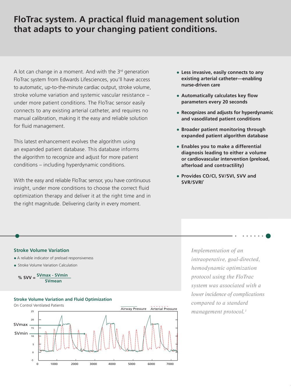
How about the Flotrac and other monitors using the arterial waveform? These monitors are quite useful for C.O. and correlate well with TEE.
I have no experience with the Nexfin myself except a demo by the rep on an ASA1 person. On the other hand, the monitors which utilize the arterial waveform to obtain C.O. seem to be quite useful in Cardiac patients especially for trending of C.O.
 
The accuracy of the Pulse algorithm may be compromised under the following circumstances:(i)aortic valve regurgitation,(ii)post-aortic reconstruction,(iii)intra-aortic balloon pump,(iv)highly damped peripheral arterial lines,(v)severe peripheral arterial vasoconstriction,(vi)inaccurate sodium and hemoglobin measurements,(vii)arrhythmias,(viii)intra- or extracardiac shunts.
 
A272
October 16, 2012
3:00:00 PM - 4:30:00 PM
Room 103A
Influence of Systemic Vascular Resistance on the Accuracy of Cardiac Output and Tracking Changes in Vigileo-Flotrac System
Koichi Suehiro, M.D., Katsuaki Tanaka, M.D.,Ph.D., Tomoharu Funao, M.D.,Ph.D., Takashi Mori, M.D.,Ph.D., Kiyonobu Nishikawa, M.D.,Ph.D.
Osaka City University Graduate School of Medicine, Osaka, Japan
[Introduction]

The Vigileo-FloTrac system (Edwards Lifescience, Irvine, CA) is a less invasive method to obtain continuous cardiac output (CO) using pulse contour analysis. Previous study has suggested that acute changes in peripheral vascular resistance might decrease the ability of Vigileo-FloTrac system to measure accurate cardiac output [1]. The aim of this study was to examine the ability of Vigileo-FloTrac system to measure CO and track CO changes induced by increased vasomotor tone in low and high systemic vascular resistance states.

[Subjects and Methods]

After obtaining approval from the ethics committee of our institution and informed consent from all patients, 40 patients scheduled for cardiac surgery were enrolled in this study. Study protocol was performed under stable hemodynamic condition. Hemodynamic variables including CO measured with Vigileo-FloTrac system (APCO), CO measured by pulmonary artery catheter thermodilution method (ICO), and systemic vascular resistance index (SVRI) was recorded before (T1) and after (T2) phenylephrine administration. As a statistical analysis, we used Bland and Altman analysis to compare ICO and APCO. The percentage error was considered clinically acceptable when it was below 30%. We defined the percentage increase of ICO and APCO between T1 and T2 as ΔICO and ΔAPCO, respectively. We used the 4 quadrant plots to compare the concordance rate of ΔICO and ΔAPCO. The concordance rate was considered as acceptable when it was over 60%, and good when it was over 90%. We divided the patients into 3 groups according to the SVRI value at point T1, and defined the patients with low SVRI (below 1200 dyne s/cm5/m2), normal SVRI (1200 to 2500 dyne s/cm5/m2), and high SVRI (over 2500 dyne s/cm5/m2), as L group, N group, and H group, respectively. We compared the ability of Vigileo-FloTrac system by using the Bland and Altman method and the 4 quadrant plots in L group, N group, and H group patients. For all analyses, a P value <0.05 was considered as statistically significant.

[Results]

A total of 155 paired data were collected. The numbers of data were 44 in the L group, 63 in the N group, and 48 in the H group. The mean bias between ICO and APCO was 1.85, 0.52, and -1.34 L/min, in the L, N, and H group patients, respectively. The percentage error was 46.3, 26.4, and 61.4%, in the L, N, and H group, respectively. The concordance rate between ΔAPCO and ΔICO was 67.5%, 28.8%, and 7.7% in the L, N and H group patients, respectively.

[Conclusions]

The ability of new third-generation Vigileo-FloTrac system to measure CO and track changes in CO induced by phenylephrine administration was not clinically acceptable. It was much influenced with the systemic vascular resistance. The Vigileo-FloTrac system has the reliable ability to measure CO only in the normal peripheral resistance state, and not in the low and high systemic vascular resistance state. And the trending ability of the Vigileo-FloTrac system against ICO was clinically acceptable only in the low peripheral vascular state. Consequently, we should be aware that the change in systemic vascular resistance may affect the accuracy of the Vigileo-FloTrac system for measuring CO in conducting goal-directed therapy.

[References]
 
I agree. It really irks me when I'm rotating through the TBICU, which is run by the trauma surgeons and whenever they have a patient on pressor, they want to float a swan. Whenever they see something hazy on the chest X-ray, they want to do a bronch. Not only does it waste my time since I have to do the procedures, it rarely changes management and places the patient at risk for potentially life threatening complication.

This sounds eerily similar to where I trained, where the trauma surgeon run TBICU had me float swans in anything that stood still long enough.

I have PTSD from trying to place CVLs into these head and neck burn patients and float the Swan so I could literally flood them until they fell off their Starling curves...
 
Just imagine the patients' PTSD. Not because of you, but most patients are hyperalgic in the ICU and remember vividly their painful experiences.
 
Top