Adjuvant Prostate Patient w Apparent Bulky Nodes

This forum made possible through the generous support of SDN members, donors, and sponsors. Thank you.

Haybrant

1K Member
15+ Year Member
Joined
Jul 6, 2004
Messages
1,977
Reaction score
563
57 yo guy high anxiety diagnosed 3 months ago with very high risk prostate cancer Gleason 4+5 in all 12 cores PSA 10 ng/ml. Bone scan was negative/CTAP did not show any adenopathy. He did get referred beforehand to speak about RT. I basically told him look you're young but we dont have evidence of survival benefit with trimodality therapy, your chance of needing RT after surgery is essentially assured. He was not hearing any of it, mind was already made up that he was getting it taken out. We wanted the robot

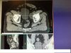
Underwent RRP on 10/16/17 with pathologic T3bN0M0 3.5 cm adenocarcinoma Gleason 4+5 with + margin, + SVI, + ECE, 0/5 lymph nodes positive for disease, had persistent positive PSA of 0.43 ng/ml. Referred back by Urology on on the same day, got started on ADT right away (been on ADT for a month). I sim'd him and now Im seeing fairly bulky external iliac adenopathy 4cm on the left, 3 cm on the right, no other gross nodes evident, no clear distant disease. Picture attached. There is room from bowel but obvious fem heads would be an issue if trying to boost anything here. Havent attempted to boost a guy like this before; What to do now? Escalate cytotoxic systemic therapy hope for shrinkage?

Members don't see this ad.
 

Attachments

  • Prostate nodal_2.jpg
    Prostate nodal_2.jpg
    100.4 KB · Views: 124
Last edited:
Members don't see this ad :)
I hope so. Bilat lymphoceles tho? What would you do, contrast CT?
Yeah, I generally contrast my sims if they are high risk postop, just in case. I think it might help here. Certainly could be bilateral if the surgeon did a good dissection. It's only been a few months out from surgery
 
Could be lymphoceles, for sure. There is no way to tell them from mets. I see these a lot after lymphadenectomy for endometrial cancer.
 
  • Like
Reactions: 1 user
I think it is reasonable to sim in 2-3 months after starting AD as the lesions may shrink and make planning easier- would try to get 70 Gy to the gross nodes. If dont, change in size, most likely lymphocele. Would definitely advocate zytiga, or taxane after radiation. (I know no data in post-op, but absolutely reasonable to extrapolate)
 
Last edited:
I would probably try to get a postop MRI to: 1) evaluate for gross disease in the postop bed, 2) should be able to distinguish lymphocele from enlarged lymph node.
 
  • Like
Reactions: 1 users
Agree with those comments above about a lymphocele. I see this quite a bit after pelvic LND.

His Pre-op PSA is 10, so presumably even though he has de differentiated high gleason disease, his cancer does secrete PSA. So in my mind the likelihood he has bulky lymph nodes with a very low PSA is low, so I think these are lymphoceles. Would get a diagnostic MRI or CT though to evaluate.
 
Lymphoceles for sure. Inadequate LN dissection with 5 nodes. Just plain bad medicine.
 
LND for prostate Ca is not therapeutic. How many nodes need to be sampled to adequately stage patients? Nobody knows, IMO.

Lymphoceles for sure. Inadequate LN dissection with 5 nodes. Just plain bad medicine.
 
If they weren't there pre-op, I wouldn't expect a PSA of 0.43 to give you bulky LNs like that. Agree with lymphoceles.

You can do contrast enhanced scan to confirm lymphocele if you want. MRI is reasonable as well, although the likelihood it shows you gross disease is near zero.

I'd treat him however you would standard post-op. I'd be interested in knowing if anybody else is getting F18 PET or similar imaging in this case of immediate post-op salvage. Try and find metastatic disease (you know this guy is going to develop it at some point) before he goes through 7 weeks of RT? Obviously now that he's gotten hormones it likely won't matter, but maybe before he started hormones.

As a question for others in this thread:
1) If you were treating this patient definitively, would you have treated the lymph nodes electively? Do you routinely treat the lymph nodes in high-risk Prostate?
2) Would you treat the lymph nodes electively in the post-op setting?
 
With gleason 9, seminal vesicle involvement, there is greater than 50% chance of lymph node involvement. It really seems reasonable to treat them given limited side effects of imrt. We can argue if someone with 10-15% chance needs ln treatment, but when the chance of involvement is 50% + and the toxicity is low, I cant see leaving them out.
 
Members don't see this ad :)
With gleason 9, seminal vesicle involvement, there is greater than 50% chance of lymph node involvement. It really seems reasonable to treat them given limited side effects of imrt. We can argue if someone with 10-15% chance needs ln treatment, but when the chance of involvement is 50% + and the toxicity is low, I cant see leaving them out.
I would treat, but obviously it's a controversial issue with not a lot of data.
 
Excellent location. Would take nodes to at least 70Gy, could even go higher. Do this commonly in Gyn cancers. Wouldn't worry about it.
 
I've had very bad luck with using contrast MRI and diagnostic CT to tell a metastatic node from a lymphocele. "Cystic structure, cannot rule out postoperative change" is a common Radiology phrase, in my neck of the wood.
 
Last edited:
I've had very bad luck with using contrast MRI and diagnostic CT to tell a metastatic node from a lymphocele. "Cystic structure, cannot rule out postoperative change" is a common Radiology phrase, in my neck of the wood.
Metastatic nodes aren't cystic; CYA by radiology
 
No, in cervical cancer and H&N cancer mets are very often cystic. They look like a tiny enhancing rim around a radiolucent cavity.
What I'm trying to say, lymphocele vs. nodal met determnation by a RadOnc unfortunately turns out to be a classic case of using clinical judgement.

Sure but I haven't seen prostate cancer nodes be cystic, which is the question here.

Lymphocele only makes clinical sense if patient has had surgery, which shouldn't happen for cervical cancer (ideally).
 
  • Like
Reactions: 1 user
Lymphocele didnt occur to me because I thought urologists did not do much of a lymph node dissection, especially with robot.
I have seen lymphoceles at this exact location in gyn.
 
Last edited:
  • Like
Reactions: 1 user
From what i understand you cant get many LN's with Robot bc of complications with access - So all these types of patients are inadequately dissected.

my radiologists looked at it and said its for sure lymphocele to the point they said don't even get a contrast scan; would you though? Ive never seen a lymphocele in a prostate pt and ive seen probably close to 500-600+ such scans I would guess. But I agree that scan in September was negative and his psa isn't exactly high.
 
I think if the radiologist says for sure it's a lymphocele, you trust them (if you deem them trustworthy), document in your note your discussion (including name of physician you discussed it with if possible) and go about your life.

I think Robotic Pelvic LND depends on skill of operator. I've seen quite a few post-prostates after RALP and I've routinely seen 20+ LNs harvested. It may be an issue with pathologic sectioning thickness as well rather than just the surgeons.
 
Another idea: to call the robotisist and ask if he/she remembers where the nodes where taken from. Lymphocele must correspond to the surgical bed.
 
  • Like
Reactions: 1 user
routinely see pathetic lymph nodes counts -I thought I saw some literature about the lymph node counts decreased with the robot.
 
routinely see pathetic lymph nodes counts -I thought I saw some literature about the lymph node counts decreased with the robot.

we had a big robot guy come to speak once and they also say you cant really access many lymph nodes. For sure he couldn't access this external iliac area, but the radiology thought maybe its where the trocars went? I guess I can discuss w the surgeon to find out, I didn't realize lymphceles only arise where nodes were dissected?
 
Hm trocars... never thought about it, don't know the answer to this question.
 
@DoctwoB as a urologist who stops by this forum from time to time, can you give your input regarding RALP and lymph node dissection, and how adequacy of LN dissection compares to the days of RRP?
 
Those are probably lymphoceles.
I'd still treat pelvis though pN0 (0/5) is simply inadequate. On the other hand he may be already metastatic out of the pelvis, but right now, giving large volume RT is his best together with ADT for a long lasting remission.

With a persistent PSA of 0.43 ng/ml post-surgery, we would have probably done PSMA-PET-CT here before starting ADT.
 
It is a lymphocele. Compare Hounsfield units between that and the bladder, and I bet it will be pretty similar. I've seen lymphoceles develop post prostatectomy with selective lymph node sampling.
 
sent the image to the surgeon he said that's the exact area they sample in so we're good. He thanked me for sharing the cool image

I presume I should contour the entire lymphocele.
 
sent the image to the surgeon he said that's the exact area they sample in so we're good. He thanked me for sharing the cool image
I presume I should contour the entire lymphocele.

If you are going to treat the pelvis, then the lymphatic CTV is displaced / enlarged by the lymphocele, since they need to be included in the CTV.
I have had patients sent back to the surgeon to drain big lymphoceles, as I felt that treating such a big volume would result in a greater volume of bowel being treated too.
It's your call I guess. From the way they look on the axial image, there's not that much bowel around them, but I am not sure how much high they reach.
Radiologists can drain lymphoceles too, but I'd surely first ask the surgeon.

Having those those lymphoceles drained or waiting for them get smaller may delay the beginning of your RT.
You cound sonsider giving ADT and starting RT a few motnhs later. He does have persistent PSA elevation and although it's not higher than the "magical" 0.7 ng/ml as per RTOG9601, he may benefit from ADT.
 
i definitely have him on adt, ya the location of these aren't too bad so will treat through it. thanks all
 
To weigh in from the surgical standpoint, those definitely look like lymphoceles to me and I would absolutely not treat those specifically. Prostate cancer node mets are pretty much never cystic, and the location and apperance of these collections is classic for lymphoceles. Now given his high risk pT3b disease with persistent PSA and + margins, adjuvant radiation is certainly indicated, and if you feel that pelvic LN radiation is indicated due to the high risk of occult LN mets I wouldn't argue with you, but I wouldn't let these lymphoceles affect that decision, if anything radiating them may delay healing/resorption (or maybe could actually help as a form of sclerotherapy? you tell me.)

To provide some background, while the extent of lymph node dissection does vary between institutions and surgeons, what is typically done at every place I've been is an external illiac and obturator Lymph node dissection. This can be done about equally well from a robotic and open procedure. These aren't huge packets, and the lymph node count can be highly variable and pathologist dependent. From good looking packets in the OR I've seen as few as 1 or as many as 20 nodes. Keep in mind that this is almost purely for diagnostic purposes, the therapeutic role of lymph node dissection in clinical N0 disease is debated and minimal at best, so it's hardly worth shaming or blaming a surgeon for a low node count.

Where the robot and open surgeries differ is that it is easier to extract hypogastric or common illiac nodes with an open surgery. Usually this doesn't matter as we don't go after these nodes anyways, the exception being if there is a clinically suspicious node on pre-op scan or we find something concerning intraoperatively. It can be done robotically, but is much slower and more tedious then doing it open. The other key difference between the two concerns lymphoceles. I'd bet that if we scanned all our open prostates post op, a large % of them would have lymphoceles because some lymph almost always leaks and the fluid is contained extraperitoneally. Robotically, the dissection is usually intraperitoneal, and so lymphoceles are relatively rare as the fluid would drain into the peritoneum. Of course a delayed lymph leak could still cause a lymphocele after the peritoneum re-epithliealizes, but it is much less common. Regardless, they very rarely cause a problem unless they are big enough to cause a mass effect or get secondarily infected.

thanks doctwoB, this thread started with me concerned about new external iliac nodes but I was corrected that these likely represent lymphoceles which was confirmed by my radiologist/urologist and good friends at SDN! I don't have any plan to "cover them" meaning to go to a higher curative dose to these regions. But we "cover" them in our prophylactic regions as they are at risk lymphatic regions part of our whole pelvis initial treatment field. If the lymphocele is quite large that it is going to lead to significant increase to OAR's then a decision has to be made whether covering the entire region would be beneficial
 
thanks doctwoB, this thread started with me concerned about new external iliac nodes but I was corrected that these likely represent lymphoceles which was confirmed by my radiologist/urologist and good friends at SDN! I don't have any plan to "cover them" meaning to go to a higher curative dose to these regions. But we "cover" them in our prophylactic regions as they are at risk lymphatic regions part of our whole pelvis initial treatment field. If the lymphocele is quite large that it is going to lead to significant increase to OAR's then a decision has to be made whether covering the entire region would be beneficial

Can I ask why on the bolded? I know it's controversial in seemingly every high-risk situation, but none of the 3 major adjuvant trials did whole pelvis. IMO you have a positive margin that is likely driving your 0.43.
 
You said nodes were negative by CT preop. Doubt those nodes "blew up" in just such a short time. He's still node-negative IMHO at this point unless you did like an Axumin PET. My point is, statistically speaking, in a known N0 patient (which he was) who is quickly re-imaged, the pre-test prob of N1 is low and you thus need a rather specific test to "rule in" the nodal positivity.

Add Zytiga in this case; I certainly would. Apalutamide may be what we (rad onc "we") use soon. Certainly N1=Stage IV and thus the indication may already be there... *IF* we call this N1. But Zytiga is valid per STAMPEDE even if N0.
 
You said nodes were negative by CT preop. Doubt those nodes "blew up" in just such a short time. He's still node-negative IMHO at this point unless you did like an Axumin PET. My point is, statistically speaking, in a known N0 patient (which he was) who is quickly re-imaged, the pre-test prob of N1 is low and you thus need a rather specific test to "rule in" the nodal positivity.

The question is if you can safely say that pN0 (0/5) is "enough" to call this patient a "true" pN0.
I would in some cases. But I wouldn't do it in a patient with a advanced local disease, persistant PSA elevation postop and a Gleason score of 9. Chances are high that he would have been pN1 had he received an adequate lymphadenectomy.
But then again, I am just speculating. I would have to read the surgery report and have a look at the pathology report to see which nodes were resected, how much lymphatic tissue was taken out, etc... The pN0 (0/5) could also have been the work of a "lazy" pathologist who didn't want to look hard enough for nodes in the specimen he got.

Add Zytiga in this case; I certainly would. Apalutamide may be what we (rad onc "we") use soon. Certainly N1=Stage IV and thus the indication may already be there... *IF* we call this N1. But Zytiga is valid per STAMPEDE even if N0.
Would you get Zytiga approved in this indication in the US?
We wouldn't get it in Europe. Stampede included very few if any patients with this scenario (post op XRT), most patients had distant metastasis and were treated without XRT. And those who weren't metastatic actually got primary XRT + ADT +/- Zytiga.
 
Can I ask why on the bolded? I know it's controversial in seemingly every high-risk situation, but none of the 3 major adjuvant trials did whole pelvis. IMO you have a positive margin that is likely driving your 0.43.
Perhaps. It depends on how big the positive margin was (the pathologists report should state that). This guy had a PSA of 10 ng/ml before surgery and all cores involved, which means that the tumor infiltrated more or less the whole organ. If his prostate wasn't tiny this means that his PSA density was rather low for a rather big tumor. This is understandable bearing in mind that he had a Gleason score 9 tumor which is poorly differentiated.
So a persistant elevation of 0.43 ng/ml is in my opinion a bit too high "only" because of a positive margin. There is a considerable tumor mass driving this PSA.
If we speculate that his prostate was around 40 cm3 and 20cm3 was tumor at a PSA of 10 ng/ml, then a PSA of 0.43 ng/ml would correspond to about 1 cm3 of tumor. That's quite a big volume for "just" a positive margin.

So he may be metastatic already... The question is: only in the pelvic nodes and can one salvage that with regional irradiation or already micrometastatic in distant sites?


You’re presuming that all aspects of prostate tissue and cancer produce psa at similar rates. That 0.4 could represent a big chunk of benign tissue left behind from poor surgical technique. Or a small area of residual high grade disease at the positive margin. Or a node. Or undetectably small bone met. No way to know for sure right away.
 
You’re presuming that all aspects of prostate tissue and cancer produce psa at similar rates. That 0.4 could represent a big chunk of benign tissue left behind from poor surgical technique. Or a small area of residual high grade disease at the positive margin. Or a node. Or undetectably small bone met. No way to know for sure right away.

urologists love speculating about PSA production and where it comes from and differential rates of PSA's. Anyway, the last part of this is correct we don't know where its coming from and we are hedging that with only 5 nodes dissected there is still high chance he has subclinical nodal disease, look up the chance on the nomogram its very high. We could be wrong and he'll have bony disease the day he stops ADT or sooner, but gotta do the best you can with what you have
 
  • Like
Reactions: 1 users
You’re presuming that all aspects of prostate tissue and cancer produce psa at similar rates. That 0.4 could represent a big chunk of benign tissue left behind from poor surgical technique. Or a small area of residual high grade disease at the positive margin. Or a node. Or undetectably small bone met. No way to know for sure right away.
Indeed. I presume that pretty much the entire prostate was infiltrated by a gleason score 9 tumor, since all cores were positive. The pathology report should state how much of the prostate was infiltrated by what kind of a disease.
A "big chunk of residual disease" that produces a PSA of 0.4 ng/ml would be quite a big chunk, I hope this did not happen. There are series reporting persistant PSA levels post-prostatectomy because of benign tissue left behind (usually in the anastomosis area), but not at level sof 0.4 ng/ml. That's too high. And the margin would have to be quite big for a persistant PSA of 0.43 ng/ml.
But hey, I don't have the pathology report, that's why I am just guessing. To me, it seems like a rather big tumor with a rather low PSA pre-OP and a too high PSA post-OP. Way too high for just a small burden of residual disease.
 
urologists love speculating about PSA production and where it comes from and differential rates of PSA's. Anyway, the last part of this is correct we don't know where its coming from and we are hedging that with only 5 nodes dissected there is still high chance he has subclinical nodal disease, look up the chance on the nomogram its very high. We could be wrong and he'll have bony disease the day he stops ADT or sooner, but gotta do the best you can with what you have

I don’t disagree that the risk of undetected nodal disease is high, but you’re weighing too much on the node count. He could have had 20 negative nodes and I would still be concerned that he had disease in the unsampled internal or common iliac nodes.
 
Would you get Zytiga approved in this indication in the US?

Yeah. Just say, "I'm really concerned about the nodes," i.e., you're worried the patient has N1 disease which means he has Stage IV disease. I see a ton of people making the case this guy has N1 disease REGARDLESS of the data, so just stage him that way for Pete's sake.

At the end of the day I don't know why all us angels are dancing on the head of this pin. We know postop radiation and anti-androgen therapy improves outcomes here. An LHRH and Zytiga is the best sockdolager of anti-androgen therapy we have now. Use it. Boost that juicy nodal-looking thing or not, it prob won't affect this guy's outcome. Applying radiation in some fashion, which will be HIGHLY disparate from rad onc to rad onc but at the end of the day still seems to "do its thang," plus the drug therapy, will.
 
Random whole pelvis question - when using IMRT for whole pelvis do you guys go to L5/S1 or higher than that? I was looking at RTOG 0924 protocol and they list L4/L5 (http://rpc.mdanderson.org/rpc/credentialing/files/0924.pdf) then a lot of other places is L5/S1 which I know they did in the 3D days. RTOG atlas shows L5/S1. Thanks
I think a lot of people nowadays just look for where the external/internal illiacs branch off from the commons. More accurate IMO to use the CT than traditional bony landmarks.
 
Top