- Joined
- Jun 10, 2015
- Messages
- 36
- Reaction score
- 4
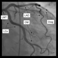
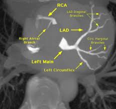
I got a question wrong on UWqbank about LAD vs LCX so I was looking through more pics on google. After going through them, I was thinking if the question asks about LAD I should always go for the upper right artery on CT since I have seen it on most of the CT on upper right, but then on some google pics it's the one on the bottom. How can we tell them apart for sure?