Inappropriate SRS fractionation

This forum made possible through the generous support of SDN members, donors, and sponsors. Thank you.

Gfunk6

And to think . . . I hesitated
Moderator Emeritus
Lifetime Donor
20+ Year Member
Joined
Apr 16, 2004
Messages
4,660
Reaction score
5,075
I'm curious to what your experience has been with this in the community. For brain mets 1.5 cm and less not near critical structures, I always treat single fraction. Usually 18-20 Gy x1 Rx to the 70-90% IDL. The only time I fractionate is if the size is > 1.5 cm or if it is near critical structure.

I understand in the community you sometimes have to compromise based on your hardware availability (e.g. no single fraction SRS if you have 1 cm MLCs). However, I've seen people with state of the art accelerators (e.g. TrueBeam/VersaHD) who treat 5-6 Gy x 5 or 8-9 Gy x 3. These are for unresected brain mets not resection cavity s/p surgery.

Less BED = inferior LC rates. The cynic in me says they do it for money, but curious what others think.

Members don't see this ad.
 
Desire to avoid a concern for increased toxicity? Regardless of how low of a concern that really is? Personally I do send out for GK and believe in single fraction. If you can't do it, don't
 
Last edited:
Desire to avoid a concern for increased toxicity?

As I wrote above, this is a legitimate concern for large mets (increased risk of necrosis) and proximity to optic structures/brainstem. I'm talking about small peripheral mets in 'watershed' regions of the brain that are 1.5 cm or less.
 
Members don't see this ad :)
From the LC point of view for such a small lesion with either 9 Gy x 3 or 6 Gy x 5 would still be around ~90%
(Minniti, IJROBP,2016 Italy ; Marcrom UAB, IJROBP, 2017 UAB) for bigger lesions, so I suspect it to be > 90% for smaller lesions. So, I don't think there is anything to lose control wise. Sometimes, it is easier to stick with what you know and if people have been doing 3 fractions with good results, they may be more comfortable sticking with that regimen. I hope it is not for the money...but don't you make more from a 1 fx SRS than a 3 fx SRT?
 
As a caveat: at my institution we do too much fractionation due to anecdotal concern about necrosis or brainstem toxicity from multiple attendings. Concern over asymptomatic radiographic necrosis, etc. It's an issue, but I want to present my biases before the rest of my post:

We have data from Minniti that shows that 9Gy x 3 gives 91% control in > 2cm disease. Do we really think that 9Gy x 3 to a lesion < 2cm in size will give < 91% control? We know that fractionating in > 2cm results in lower rates of radionecrosis. Will radionecrosis rates be different even in smaller tumors?

Do we have data that single fraction is better than multifraction SRS? This (https://www.redjournal.org/article/S0360-3016(14)01712-X/fulltext) shows no difference despite FSRS being used for larger tumors, post-op cavities, etc. Numerical radionecrosis is 10% for single fraction, 5% for multi, even with the baseline characteristics favoring single fraction. What if baseline characteristics were the same, would it show an advantage to control and necrosis to multifraction?

We know, just recently, that 5Gy x 5 is worse than 6Gy x 5 for intact mets: Fractionated stereotactic radiation therapy for intact brain metastases. - PubMed - NCBI

Is anyone aware of data suggesting 8Gy x 3 is worse than 9Gy x 3?

For the record: BED10 of 27/3 and 18/1 are equivalent. Obviously risk for long-term toxicity based on BED is going to be vastly different between the two. Granted that it's on the extremes (highest fractionated BED versus lowest single fraction BED).

I hate when strong proponents of single fraction insist on doing 15Gy x 1 stubbornly instead of just fractionating.

I honestly prefer the 9Gy x 3 approach to anything over about 1cm in size, unless I can very easily meet V12 < 7-10cc (dural based, vertex, etc.)

I'll certainly look at the plan and see if single fraction is reasonable but I'm not gung-ho about it under any definition. We do 1mm margins to PTV on Linac-based SRS.

I see ROMegatron posted essentially the same thing above.
 
  • Like
Reactions: 3 users
As a caveat: at my institution we do too much fractionation due to anecdotal concern about necrosis or brainstem toxicity from multiple attendings. Concern over asymptomatic radiographic necrosis, etc. It's an issue, but I want to present my biases before the rest of my post:

We have data from Minniti that shows that 9Gy x 3 gives 91% control in > 2cm disease. Do we really think that 9Gy x 3 to a lesion < 2cm in size will give < 91% control? We know that fractionating in > 2cm results in lower rates of radionecrosis. Will radionecrosis rates be different even in smaller tumors?

Do we have data that single fraction is better than multifraction SRS? This (https://www.redjournal.org/article/S0360-3016(14)01712-X/fulltext) shows no difference despite FSRS being used for larger tumors, post-op cavities, etc. Numerical radionecrosis is 10% for single fraction, 5% for multi, even with the baseline characteristics favoring single fraction. What if baseline characteristics were the same, would it show an advantage to control and necrosis to multifraction?

We know, just recently, that 5Gy x 5 is worse than 6Gy x 5 for intact mets: Fractionated stereotactic radiation therapy for intact brain metastases. - PubMed - NCBI

Is anyone aware of data suggesting 8Gy x 3 is worse than 9Gy x 3?

For the record: BED10 of 27/3 and 18/1 are equivalent. Obviously risk for long-term toxicity based on BED is going to be vastly different between the two. Granted that it's on the extremes (highest fractionated BED versus lowest single fraction BED).

I hate when strong proponents of single fraction insist on doing 15Gy x 1 stubbornly instead of just fractionating.

I honestly prefer the 9Gy x 3 approach to anything over about 1cm in size, unless I can very easily meet V12 < 7-10cc (dural based, vertex, etc.)

I'll certainly look at the plan and see if single fraction is reasonable but I'm not gung-ho about it under any definition. We do 1mm margins to PTV on Linac-based SRS.

I see ROMegatron posted essentially the same thing above.

Brother from a different mother
 
I do a lot of 9 Gy x 3 and 6 x 5 ,even for smaller mets. I dont feel comfortable with 0 margin, which is required for single session treatment.
I dont think fusion capabilities at most facilities allows no margin. There often is a delay between the MRI,CT sim and then treatment, because of insurance approval,planning time etc- this also makes me more comfortable adding margin (UCSF actually has a really good paper showing worse control with longer intervals) If there is more than one lesion, I feel a margin is required for rotational error.
Lastly, I dont think there is much, if any, billing difference (I am employed so dont have this knowledge offhand)
For spine SBRT, I believe there is actually retrospective data from Pitt that fractionated treatment has superior control than single dose xrt.

(This is theoretical, but preclinical data also hint 8-9GY in many models is the sweet spot for eliciting immune effect, possibly with cGas/Sting-IL2 from micronuclei/ damaged DNA in cytosol . 12+ Gy apparently release exonucleases in many models, so not as much DNA in cytosol to cause immune response. my take from astro lecture
 
Last edited:
I do a lot of 9 Gy x 3 and 6 x 5 ,even for smaller mets, I dont feel comfortable with 0 margin, which is required for single session treatment.
I dont think even think fusion capabilities at most facilities allows you to have no margin. There often is a delay between the MRI and CT sim and then treatment because of insurance approval etc, which also makes me more comfortable adding margin (UCSF actually has a good paper on this showing worse control with longer intervals) Also, if there is more than one lesion, I feel margin is required for rotational error.
Lastly, I dont think there really is much if any billling difference (I am employed so dont know this things)

Also for SBRT of the spine, I believe there is actually data now that fractionated treatment has superior control than single dose xrt.

Can you link the UCSF paper? I am quite interested. I have always been suspicious of no margin...
 
Can you link the UCSF paper? I am quite interested. I have always been suspicious of no margin...
The paper wasnt on margin, but length of time between mri and stereo, and given that there can often be delay, I like margin to compensate for this. Duke has a randomized paper showing increased toxicity with 1mm and 2-3mm margin for single fracion srs?

Interval From Imaging to Treatment Delivery in the Radiation Surgery Age: How Long Is Too Long? - PubMed - NCBI
 
Last edited:
Can you link the UCSF paper? I am quite interested. I have always been suspicious of no margin...
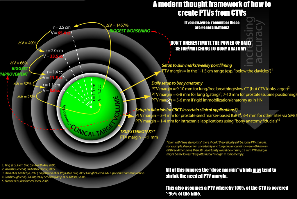
I made this graphic more than a decade ago. "Even with true stereotaxy there should be some margin... if uncertainty were ~0.6cm in all three dimensions, 3D uncertainty would be ~1mm... a 1mm PTV margin might be the lowest 'truly attainable' margin in radiotherapy..."
iRidtAV.png
 
  • Like
Reactions: 1 user
This is beautiful. Do you have a source?

I don't agree with 100% of it, but it's a good starting point for the residents.
source is me (this is why I shoulda done academics, ha)
 
I don't agree with 100% of it

Keep in mind PTV margin = Block margin minus 0.5 cm, give or take. So when I say 1.0 cm PTV margin for bony anatomy (port film) setup for lung, for instance, that's a 1.5 cm block margin, and that was a valid block margin for 3DCRT in the RTOG trials e.g. As was a 1.5 cm block margin for 3D prostate (which equals a 1.0 cm PTV margin etc etc etc).
 
Members don't see this ad :)
This is beautiful. Do you have a source?

I don't agree with 100% of it, but it's a good starting point for the residents.
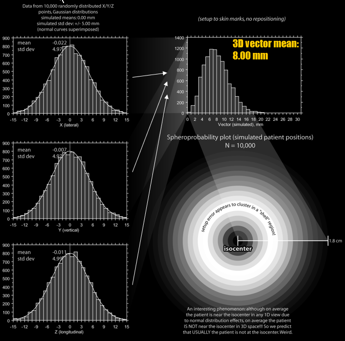
you can run a mathematical sim of the old saying "daily setup error is plus or minus a half cm" and show that PTV margins need to be in the ~1.4-1.5 cm range (which equals the old daily block margins of 2 cm with just daily skin mark setup). Daily bony anatomy setup would get you in the PTV=1 range, possibly. But this is true daily setup error just to skin marks. Every maneuver--daily port films, CBCT, framed stereotaxy--makes it smaller. The setup errors with or without IGRT are always normally distributed unidimensionally yet behave in an "anti-magnetic" fashion about the isocenter. I tried to coin a term "spheroprobability"... it never caught on.
xEC9oHU.png
 
Thanks for the diagram. The truebeam/trilogy machine isocenter is supposed to have a mechanical accuracy of within .75 mm. (Ours was just measured at .69 mm by varian), meaning that the 3d error vector is always over 1mm, so true sub mm accuracy is not really deliverable on a linac? Even the "stereotatic" novalis linac still has a machine isocenter with .6mm tolerance.
 
The truebeam/trilogy machine isocenter is supposed to have a mechanical accuracy of within .75 mm.
Yes. And, I have always wondered what this REALLY meant. Like, the iso is within 0.75mm in one dimension ~95% of the time? All of the time? What's the "average" unidimensional error? Who knows, right? We just say sub-mm accuracy, and in fact, mathematically, it's impossible. If the 1D error is +/- 0.5mm (ie, thats the std dev), something like ~30-40% of all setups will be more than 1mm off in 3D space. 3D is what counts. Who cares what the L/R, or up/down, or in/out... error is, alone.
 
You need to weigh in a lot of factors.
Isocenter accuracy per vendor is one, CBCT accuracy is another (if you are using it to correct positioning), possible patient movement is another (masks not as rigid as stereotactic rings),
And pretreatment inaccuracy also needs to be weighed in: contouring accuracy, CT-MRI-fusion precision.

Some also argue to perform all machine precision tests directly before treatment. Your Linac is probably a bit more precise at 9am treating the first patient than at 3am after having treated 20 patients.

Landmark article
Errors and margins in radiotherapy. - PubMed - NCBI
 
  • Like
Reactions: 1 user
You need to weigh in a lot of factors.
Isocenter accuracy per vendor is one, CBCT accuracy is another (if you are using it to correct positioning), possible patient movement is another (masks not as rigid as stereotactic rings),
And pretreatment inaccuracy also needs to be weighed in: contouring accuracy, CT-MRI-fusion precision.

Some also argue to perform all machine precision tests directly before treatment. Your Linac is probably a bit more precise at 9am treating the first patient than at 3am after having treated 20 patients.

Landmark article
Errors and margins in radiotherapy. - PubMed - NCBI
The "margin recipes" can only be used once you have a complete dataset to draw from. E.g., you would measure the setup error of CBCT each day (how? CBCT once, reposition, CBCT again, measure the error... it's almost never 0/0/0 in x/y/z) across many patients, perhaps in just one disease site, to get a robust set of data (1000+ measurements?). Then you have a mean and SD for that data set. And then, you multiply the mean (systematic error) by 2 or 2.5 and add that to 0.7 times the SD (random error) to derive a PTV (to wholly cover the CTV ~95% of the time). Ideally, and actually commonly it turns out, the unidimensional mean (systematic error) is <1 mm almost every time you do an error-measuring expedition... *with IGRT.* Without IGRT, the systematic error can be very bad... and actually bad is considered to be >1mm. We don't have that data for machine/isocenter, least not that I've ever seen, but it's likely a moot point. At the end of the day, the isocenter will be <1mm "accurate" (and it's constantly checked anyways, for machine QA, or pre-tx SRS QA)... but knowing that it's not unidimensionally sub-0.5mm just means we should probably add 1mm 3D margin always, GK or linac or cyberknife or whatever. However, if someone could tell me the mean unidimensional isocenter error on a machine WAS under half a mm, I wouldn't nec. be compelled to do that... although I still would, as I know I have 1mm contouring errors, CT/MRI fusion errors, at least, as you say. All these things are relatively under-appreciated IMHO. Errors propagate. A zero PTV makes little sense mathematically.
 
  • Like
Reactions: 1 user
While we are on the topic of error, I have seen a couple cases where the enhancement pattern on the planning mri and CT sim were different so that some (1-2mm) enhancement on the CT was clearly outside of the mri volume, despite good fusion and close interval between the two scans.
 
The "margin recipes" can only be used once you have a complete dataset to draw from. E.g., you would measure the setup error of CBCT each day (how? CBCT once, reposition, CBCT again, measure the error... it's almost never 0/0/0 in x/y/z) across many patients, perhaps in just one disease site, to get a robust set of data (1000+ measurements?). Then you have a mean and SD for that data set. And then, you multiply the mean (systematic error) by 2 or 2.5 and add that to 0.7 times the SD (random error) to derive a PTV (to wholly cover the CTV ~95% of the time). Ideally, and actually commonly it turns out, the unidimensional mean (systematic error) is <1 mm almost every time you do an error-measuring expedition... *with IGRT.* Without IGRT, the systematic error can be very bad... and actually bad is considered to be >1mm. We don't have that data for machine/isocenter, least not that I've ever seen, but it's likely a moot point. At the end of the day, the isocenter will be <1mm "accurate" (and it's constantly checked anyways, for machine QA, or pre-tx SRS QA)... but knowing that it's not unidimensionally sub-0.5mm just means we should probably add 1mm 3D margin always, GK or linac or cyberknife or whatever. However, if someone could tell me the mean unidimensional isocenter error on a machine WAS under half a mm, I wouldn't nec. be compelled to do that... although I still would, as I know I have 1mm contouring errors, CT/MRI fusion errors, at least, as you say. All these things are relatively under-appreciated IMHO. Errors propagate. A zero PTV makes little sense mathematically.

This is precisely why I advocated for our physicists to setthe margin for all our SRS.
So they measured and measured and measured and came back with a number. I firmly believe it's their task to give us an estimate for a CTV-PTV-margin in SRS, just as it is our (the physicians) task to set a GTV-CTV-margin.
This is why we came up with 1mm + 1mm = 2mm.
 
  • Like
Reactions: 1 users
While we are on the topic of error, I have seen a couple cases where the enhancement pattern on the planning mri and CT sim were different so that some (1-2mm) enhancement on the CT was clearly outside of the mri volume, despite good fusion and close interval between the two scans.
when I encounter this, I go with the CT. Also, why I always do a contrast-enhanced CT sim for SRS.
 
Spoke to varian and .75 isocenter tolerance is in one dimension, not the 3d error vector, and is due to factors like gantry sag at certain angles due to weight of machine. Action level on winston lutz test is now 1.25 mm. Sub mm accuracy is not possible on linac.
 
  • Like
Reactions: 1 user
Spoke to varian and .75 isocenter tolerance is in one dimension, not the 3d error vector, and is due to factors like gantry sag at certain angles due to weight of machine. Action level on winston lutz test is now 1.25 mm. Sub mm accuracy is not possible on linac.

[3*(0.75mm*0.75mm)]^0.5 = 1.29mm
 
Even with a theoretical machine having no isocenter uncertainties, there are uncertainties related to imaging slice thickness (usually not more than 1 mm), image fusion (including MRI deformation), and patient set up (even with a rigid frame, there is some flex to the frame when locked onto the table). Using CT to target certainly helps, but even with a perfect fusion, MRI may show disease not as well seen on CT.

Whether or not you use a PTV for this depends on whether or not you believe you really need the Rx dose to cover all or most your target, or whether the penumbra (which can be sharp for small lesions treated with gamma knife or cones) is sufficient. Kind of makes SRS sound like voodoo.
 
For what it is worth- i ask to have dose prescribed to GTV. Metrics such as CI and GI are calculated to GTV. We will also analyze dose to gtv + 1mm. Usually if you prescribe 24 GY to GTV, 90% of the gtv +1mm will be getting 21 Gy. When you prescribe to PTV, if you look at CI based on GTV, it can be surprisingly high.

Is there good data for v12 for brain mets?

Even with gamma knife, there are rotational errors with the frame on. At least you can correct it with linac.
 
Penumbra is the radiation oncologist's best friend. Very true for intracranial SRS also.
 
I agree with the penumbra argument.

I used to do 5 x 5 Gy plus 2 mm PTV. Now I do 5 x 6 Gy with no margin (prescribed to 99.5% coverage of GTV).

It takes 2 mm to get from 30 Gy to 25 Gy anyway. This is more or less in all spherical angles (unless there are OARs next to the target) given that I typically use multiple non-coplanar angles in linac SRS.

However, there is a lot of heterogeneity in these details across groups that probably account for much of the heterogeneity in the results.
 
"Whether or not you use a PTV for this depends on whether or not you believe you really need the Rx dose to cover all or most your target, or whether the penumbra (which can be sharp for small lesions treated with gamma knife or cones) is sufficient. Kind of makes SRS sound like voodoo."
Penumbra and systemic treatment-anyone who is alive one year later, almost by definition benefited significantly from systemic therapy, which would have local benefits as well. Regarding instituitonal variation- that is a good point- there is a variety of fall offs and hot spots. For example, UAB planning technique will produce hot spots of 40%. I think virtually all srt should have multiple non coplanar arcs.
 
Sure, but then you lose the advantage of rigid fixation to the skull. Probably a wash I imagine
There is rotation in a fixed frame, and motion is really about the same as within a mask, with outliers for both. Frame based systems didnt really seem to be more accurate than mask based systems (astro lecture by josh Yamada) We just werent catching it in the past for frame based systems with optical guidance and cone beams.
Motion monitoring for cranial frameless stereotactic radiosurgery using video-based three-dimensional optical surface imaging. - PubMed - NCBI
 
Is there good data for v12 for brain mets?
Best one is from Minniti.

Stereotactic radiosurgery for brain metastases: analysis of outcome and risk of brain radionecrosis. - PubMed - NCBI

"For V10 Gy >12.6 cm3 and V12 Gy >10.9 cm3 the risk of radionecrosis was 47%"

"Lesions with V12 Gy >8.5 cm3 carries a risk of radionecrosis >10% and should be considered for hypofractionated stereotactic radiotherapy especially when located in/near eloquent areas."

12 Gy gamma knife radiosurgical volume is a predictor for radiation necrosis in non-AVM intracranial tumors. - PubMed - NCBI

https://www.redjournal.org/article/S0360-3016(09)00856-6/abstract
 
From ICRU 91, "Prescribing, Recording and Reporting of Stereotactic Treatments with Small Photon Beams":

The concept of PTV was introduced in ICRU Report 50 (ICRU, 1993) and restated in ICRU Reports 62, 71, 78, and 83 (ICRU, 1985; 1999; 2004; 2007; 2010). The PTV concept is a tool to ensure with a clinically acceptable probability that the prescribed dose will be delivered to all parts of the CTV despite geometrical uncertainties such as organ motion and setup variations. It is also used for dose prescription and reporting. It surrounds the CTV typically with a margin, which takes into account both the internal and the setup (external) uncertainties.

The setup margin accounts specifically for uncertainties in patient positioning and alignment of the therapeutic beams during the treatment planning and through all treatment sessions. For SRT, this may be due to uncertainties in, for example, external fiducial alignment versus patient skull or, IGRT-related uncertainties, such as, for example, the physician/therapist not consistently identifying the prostate on the CBCT. Although small-field treatments tend to be delivered over fewer sessions and are thus less forgiving to random error, the immobilization and image-guidance protocols tend to be more stringent and thus still permit tight PTV margins.

The PTV is a geometric concept that does not respect anatomy and may encroach on neighboring OAR. Such areas of potential overlap pose a dilemma to the physician who must often make a clinical decision between reducing the probability of delivering the planned dose to the CTV or accepting a higher risk of normal tissue complications. In many cases, the choice will be to incompletely irradiate the PTV. To achieve a compromise treatment plan, one or more sub-volumes of the PTV may need to be created. No matter what PTV subvolumes are created during the planning process, the dose should be reported to the entire, uncompromised PTV. Doing so ensures that reporting adequately reflects the lower probability of adequate dose coverage to the CTV.

In cranial radiosurgery, especially when dedicated devices are used, the total geometric uncertainties are usually quite small. In end-to-end testing, these can be less than 1 mm. In these cases, it is not uncommon for patients to be treated without any expansion from CTV to PTV (for that matter, any expansion from GTV to PTV). As with CTV, there may be rational reasons for which a 0.5 mm to 1 mm uncertainty is ignored in the planning process. However, even if the dose will be prescribed directly to the GTV, it is recommended to evaluate and report the dose to a PTV. It is then possible to compare and evaluate clinical studies. As the penumbra region can be asymmetric, plans providing similar GTV/CTV coverage may variably irradiate the PTV. Knowledge of the minimum dose to the PTV can lead to plan modifications. As an illustration of this, we can consider the ratio of the prescription isodose volume to the target volume [PITY (Shaw et al., 1993)] that is commonly used as an index to evaluate plan quality. Somewhat arbitrarily, a plan, which irradiates more normal tissue than PTV to the prescription dose, can be considered of poor quality.

Factors contributing to the setup uncertainty include methods of patient positioning/immobilization, mechanical uncertainty of the equipment (e.g., sagging of gantry, collimators, and couch), mechanical and software uncertainties in image-guidance (e.g., concordance of imaging and treatment isocenters, image co-registration algorithm uncertainties), dosimetric uncertainties (e.g., output factors, dose calculation algorithm uncertainties), planning image co-registration errors, CT localization errors, and human factors. The importance of these factors will vary from center to center and within a given center from machine to machine, protocol to protocol, and patient to patient.

As the mechanics and quality assurance of radiotherapy treatments improve, target contouring accounts for a larger proportion of the "setup" uncertainty. The impact can be great if significant normal tissue is misidentified as tumor or significant tumor is misidentified as normal tissue. Adding margins to account for all possible variations in target contouring is not a solution to this problem but peer-review and multi-disciplinary treatment planning may reduce mis-targeting. Intra- and inter-observer variations can be limited by well-defined imaging protocols for target definition and training (e.g., atlas, teaching courses). The more protocols that provide detail about the imaging technique, contrast agent (if any), display settings, contouring tools and contouring technique used, the more reproducible the contours should be.

External or setup margins are often derived, at least initially, from the published experience of others but this should not replace local quality assurance protocols including thorough end-to-end testing (see Section 7) and issues such as multimodality localization and/or pre and post treatment imaging. The PTV margin concept was not designed to specifically address the issue of interplay between organ motion and the small beamlets of intensity modulated radiation. It was also not designed to account for the possible effects of tracking a tumor with a field smaller than the CTV. When moving tumors are treated with sub-fields or beamlets smaller than the target volume, the possibility will arise that certain parts of the target or adjacent normal tissue will be under or overdosed (ICRU, 2010). These issues tend not to be solved by increasing the target volume and must be evaluated on a technique-specific basis.
 
Keep in mind that adding a 1mm PTV to a 1cm diameter GTV will increase your treated volume, and your V8/V10/V12Gy by ~30%. The data from the hfSRT series above is from my institution, and almost none of the cases were treated with a PTV margin, and our control rates are as good any comparable published series. There are a number of patients in the series with >10 mets. We do recommend some sort of intrafraction motion monitoring solution for treatment with no PTV though.
 
It's probably one of those things 6Gy x 5 prescribed to a GTV gives enough spill off of 5Gy x 5 to automatically account for PTV margin, to reiterate what Neuronix said above
 
It's probably one of those things 6Gy x 5 prescribed to a GTV gives enough spill off of 5Gy x 5 to automatically account for PTV margin, to reiterate what Neuronix said above

Well, if a 1mm margin was truly necessary as has been posited above, the 30Gy/5fx numbers would not look so good. We are also about to publish over 400 cases worth of SRS QA data, so hopefully we will soon obviate some of the claims of all the linac accuracy naysayers.
 
Well, if a 1mm margin was truly necessary as has been posited above,

Impossible to know, impossible to claim necessity, and impossible to port your results to other practices quite likely. E.g., if you were trained by “generous contourers” and do the same yourself, PTV of 1mm likely not necessary. Or if you have an especially spatially warpy MRI that “oversizes” a lesion. Whole host of unknowns. Or if you Rx to higher IDLs making the absolute dose 1mm from target edge greater than people who prescribe to lower IDLs. All one can say is: you’re doing great. If you had a whole host of zero vs 1mm margin outcomes, and 1mm was no better or instead worse, that’d be more proscriptive. But just zero margin outcomes: less so. It’s possible someone could (try to) replicate the approach and not see same results. My feeling about a 1mm margin is:
1) It doesn’t make control rates worse
2) It has an undetectable effect on toxicities
3) The target volume is still less than 1cc when you add 1mm to a 1cm lesion...
 
  • Like
Reactions: 1 user
Impossible to know, impossible to claim necessity, and impossible to port your results to other practices quite likely. E.g., if you were trained by “generous contourers” and do the same yourself, PTV of 1mm likely not necessary. Or if you have an especially spatially warpy MRI that “oversizes” a lesion. Whole host of unknowns. Or if you Rx to higher IDLs making the absolute dose 1mm from target edge greater than people who prescribe to lower IDLs. All one can say is: you’re doing great. If you had a whole host of zero vs 1mm margin outcomes, and 1mm was no better or instead worse, that’d be more proscriptive. But just zero margin outcomes: less so. It’s possible someone could (try to) replicate the approach and not see same results. My feeling about a 1mm margin is:
1) It doesn’t make control rates worse
2) It has an undetectable effect on toxicities
3) The target volume is still less than 1cc when you add 1mm to a 1cm lesion...

You typically don't prescribe to an IDL with non-cone based linac SRS, you normalize to coverage of the lesion. Smart linac SRS planners don't constrain the hotspot, because the simple act of doing so worsens the moderate isodose falloff.

And, there was a typo in my previous line. Addition of 1mm to 1cm increases volume by ~70%. For a 2cm lesion, it's about 30%. However, the concomitant V12 increase takes you from almost no likelihood of RN to above 20% likelihood (based on Flickinger/Brenemen data)

Anyway, I agree, for small lesions, likelihood of toxicity is low, but my point is that with proper image guidance, and proper QA of your machines, and your fusions, no PTV expansion is necessary. For larger lesions, the risk increase is non-trivial.

Citation for the above claim? The risk of RN treating a 2cm lesion with single fraction SRS is likely not near 0 even if you do no PTV margin. What would you V12 brain be in that situation with and without margin?
 
You typically don't prescribe to an IDL
Even if you think you're not, you do, semantically. This is like saying we didn't have PTVs in the era before CT scanning, 3DCRT, etc. The PTV was always there, just inchoate. The IDL wholly covering the lesion (or "target") is always there whether you have directly ("I like this coverage that I visualize") or indirectly ("I pre-specify this much coverage for my target, regardless what I visualize") caused it to be.
Smart linac SRS planners don't constrain the hotspot
Yes, they would be dumb planners. The hotspot is an ICRU-defined entity which *always* exists outside the target region. Inhomogeneity is what exists inside the target (not a "hot spot"). And by historic definition, in SRS, the "hottest spot" in the plan is 100%. Always. Never less, never more.
And, there was a typo in my previous line. Addition of 1mm to 1cm increases volume by ~70%. For a 2cm lesion, it's about 30%. However, the concomitant V12 increase takes you from almost no likelihood of RN to above 20% likelihood
I don't need any data to know that the RN risk of treating a 2.2 cm lesion is not 20% higher than treating a 2cm lesion. (If it's 1% for 2 cm, you're saying it's 20x greater for 2.2cm?) That don't sound right at all.
no PTV expansion is necessary
I probably agree... for you, in your clinic.
 
Last edited:
Yes, they would be dumb planners. The hotspot is an ICRU-defined entity which *always* exists outside the target region. Inhomogeneity is what exists inside the target (not a "hot spot"). And by historic definition, in SRS, the "hottest spot" in the plan is 100%. Always. Never less, never more.

Sigh. Not this semantics argument again. Everyone knows what emt409 means, man, just let it go. Hotspot colloquially means max heterogeneity. No good IMRT plan is putting anything more than a few voxels of 100% of the dose outside the PTV. The technical definition is antiquated.

Historic definition for SRS is based on GKSRS, and IMO doesn't apply to Linac based. Max heterogeneity can be convered to IDL prescription and vice-versa, and colloquial use of "hotspot" to describe areas receiving higher than prescription dose (and by what percentage they are) is not unreasonable.
 
Citation for the above claim? The risk of RN treating a 2cm lesion with single fraction SRS is likely not near 0 even if you do no PTV margin. What would you V12 brain be in that situation with and without margin?

upload_2018-6-28_16-42-21.png


I put the above together for a talk a while ago. The below is a regression of data from one of our groups's previous works.

upload_2018-6-28_16-44-10.png


With fairly surprising consistency, for a well planned linac SRS treatment (1-9 mets in this series) V12 is roughly 2 x the treated volume + 2.5cc. The relationship is nearly identical for gamma knife (I have that in a diff plot somewhere)

V12 above 10 to 12 cc is one of most commonly cited predictor of RN (Flickingker, Korytko, Blonigen/Brenemen, Minniti, etc).

For a 2cc target, GTV before expansion is 4.2 cc, corresponding to about 11cc of V12, a touch less for a single target, a touch more if distributed in multiple volumes. Adding 1mm expansion, PTV is now 5.6cc, corresponding to 13.6cc of V12, above the threshold. Flickinger quotes RN rates far higher for that threshold, we tend to see rates more in line with the Minniti series.

@scarbrtj , with respect to the IDL rx'ing, that is just incorrect. If the plans were identically heterogeneous for given target sizes, you could make a case for that being true for a single target, but especially in situations with multiple targets, the paradigm ceases to function.

The notion idea of rx'ing SRS to an optimal IDL is justified from Ian Paddick GI paper which showed optimal GI's as a function of IDL for different collimator size shots. But those curves rely on single shots in an isotropic, homogeneous water media; not complex fluence patterns from varying beam angles and intensities.
 
@scarbrtj , with respect to the IDL rx'ing, that is just incorrect.
An isodose line (and its isovolume) directly equates with an absolute dose; thus in some sense you're saying "with respect to [dose] rx'ing..." ... "You typically don't prescribe to a [dose] with non-cone based linac SRS, you normalize to coverage of the lesion." You do prescribe (normalizing = prescribing) a dose. Normalizing to a coverage = prescribing to an IDL (ie, getting a minimum absolute dose isovolume to cover your lesion in a desired fashion). They're (mathematically) equivalent. Hey @evilbooyaa ... it's semantics :)
 
For a 2cc target, GTV before expansion is 4.2 cc, corresponding to about 11cc of V12
Assume you mean 2cm target, not 2cc. Of course this will depend on what your "hot spot" is, yes? (And different MDs will choose different "hot spots"... ie, IDLs, ie Rx doses, ie heterogeneity allowances.) Also, in some target configurations, I can get a tighter V12 with tight static MLCs & beams vs RapidArc. Also, one doesn't know the normal tissue constraints used here (assuming using Eclipse?), arc number, planning particulars (optimizing to a lesion minus 1 mm and Rx'ing to the lesion e.g.). Those would be good to know, and who knows if this correlation plot is "best V12 achievable." Obviously a lower slope is better. Do you believe this is the best slope achievable? In general, what was the minimum lesional dose and maximum allowed heterogeneity for all the targets?
 
If I prescribe 18Gy to a target at the 60% IDL, marginal dose is 18Gy, and hotspot is 30Gy. If I have 5 targets, and I am treating them with DCA or GK, I can do the same thing for all of them.

If I am treating them with a VMAT plan, I get one normalization, not 5. To ensure coverage, MU's are normalized to a given dose covering a given percent of target volume, typically either the least covered target, or the composite volume. For this to be just semantics, and equivalent to an IDL based prescription, the heterogeneity in each target would have to be identical, which it never is.

Normalizing to coverage is NOT mathematically equivalent to an IDL. It's only equivalent for the marginal dose.

One of the reasons VMAT conformity is better than DCA or GK conformity is because of the difference in normalization paradigms.
 
The technique is spelled out in laborious detail in my paper, and in the recipe that we have extensively circulated to over 120 institutions around the world. In modulated linac SRS that is well planned, especially multiple mets, you do not choose the hotspot. It typically ranges from 130-170% depending on lesion size, proximity of other lesions, and other planning goals like nearby OARs.

*edited for clarity/typo
 
View attachment 236253With fairly surprising consistency, for a well planned linac SRS treatment (1-9 mets in this series) V12 is roughly 2 x the treated volume + 2.5cc.... V12 above 10 to 12 cc is one of most commonly cited predictor of RN (Flickingker, Korytko, Blonigen/Brenemen, Minniti, etc).
Which is it... 10 or 12cc? :) My interest is piqued. I'll try to run a RapidArc SRS plan on a 2 cm vs 2.2 cm (a 2 cm lesion plus 1 mm margin) sphere in the brain. See if I can get V12's < 12 cc for both. Any exercise/practice we can undertake to make our SRS conformalities "ridiculously good" are good. Although, clinically it may be a moot point as both lesions have a v12 > 10cc (your lower limit for RN). I am still skeptical that a 2.2cm target has ~20% RN rate vs ~0% for a 2cm target.
 
In modulated linac SRS that is well planned, especially multiple mets, you do not choose the hotspot.
Does it choose you? Sorry. (The hotspot directly correlates to the minimum lesion dose/doses you choose... so it is chosen... perhaps it's better to say: one matters not if the spot is hot.)
 
EMT409, you're discussing single isocenter management of multiple lesions? On one plan? Using single fraction? Even for Linac-based SRS that isn't overtly common, right?. Only center I am aware of routinely doing single isocenter and then treating brain mets across the entirety of brain with single fraction are UAB and that's because they have hyperARC (which looks cool FWIW). Even with linac-based, we don't routinely do single iso, multiple lesions (unless in close proximity to one another), single fraction.
 
Top