Lumbar RFA: How do you set up your oblique view?

This forum made possible through the generous support of SDN members, donors, and sponsors. Thank you.

Ligament

Interventional Pain Management
Lifetime Donor
20+ Year Member
Joined
Jan 9, 2002
Messages
6,517
Reaction score
3,074
Hi all,

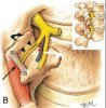
When you perform lumbar RFA, if you are the type that checks an oblique view, how do you line up the end plates? For example, your are burning the right L4 MB at the junction of the right L5 SAP and L5 TP:

1. You square up the superior endplate of L5 with the inferior endplate of L4
2. You square up the superior endplate of L5 with the inferior endplate of L5
3. You don't bother to line up endplates....

I do option 2. Bogduk's publications do option 2. I see that Dreyfuss, however, does option 1. What are your preferences and rationale?

Specifically I'm talking about oblique views here.

Members don't see this ad.
 
4. I don't go oblique.

I use AP fluoro to Dreyfuss position 2 for MBB, then walk the tip upto position 1; If there is a significant amount or arthritis, I will walk over the TP and pull it back to lie on os at the posterior aspect of the TP junction.

If it does not look right or does not stimulate right, I will get a lateral.

For AP and lateral imaging, I square the superior endplate of the level I am RF'ing.
 
Members don't see this ad :)
i do most of my RFs with a slight oblique and slight caudal tilt (meaning that my intensifier is leaning back towards feet) - i don't align endplates - not that important in my book (if you look at dissection or just a plain old spine/skeleton - and lay your curved needle in the position that you think is ideal for RF then you will see what i mean)

dorsal ramus L5 it doesn't really matter and i do that in the AP view

the only time i check lateral is when patients get reactions to stimulation that don't make sense to me - just to make sure i am not to deep - ie: patients groin is "itchy" with stimulation...
 
15 deg oblique for L1-L4 medial branches with a 15-25 degree caudad beam angulation. This approximates the curvature and places the needle tip (curved) in direct contact with the middle 4/6 of the SAP, well away from the sometimes enormous MA ligaments. The L5 dorsal ramus approach I use is 5-10 deg oblique and 10 deg caudad.
 
in direct contact with the middle 4/6 of the SAP, well away from the sometimes enormous MA ligaments.

I am having trouble visualizing this. Can you download this picture, mark the spot you are talking about, and upload it in a reply? In the picture m = mamillary process, a = accessory process, straight arrow shows the ligament, arrowheads are the posterior ramus, and the curved arrow is at the SAP. Thanks!

P.S. "4/6", not 2/3?
 

Attachments

  • mb.JPG
    mb.JPG
    22.9 KB · Views: 178
I start in declined pillar view and advance the cannula until I touch bone between the SAP and TP. Then, I flip to a lateral oblique and advance until the tip of the cannula is covering the pedicle shadow. Then, I flip to a lateral and double check my trajectory and position: Stim, burn X 2, repeat at next level.
 
Top