Pictures of the Week

This forum made possible through the generous support of SDN members, donors, and sponsors. Thank you.
IMG_0095.jpeg


Not a lot of pain, hands not working well.

Members don't see this ad.
 
Members don't see this ad :)
Thoughts on this contrast pattern? Young guy with severe left L4-5 foraminal stenosis secondary to disc. Sent by surgeon for L4 TFESI. No relief. Now feels new pain in the ball of the foot (before injection it was inner ankle and big toe).

Capture.PNG
 
Thoughts on this contrast pattern? Young guy with severe left L4-5 foraminal stenosis secondary to disc. Sent by surgeon for L4 TFESI. No relief. Now feels new pain in the ball of the foot (before injection it was inner ankle and big toe).

View attachment 241391
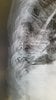
That's epidural. You can see the fat blebs. Probably retroneural. Injection was fine
 
So post a lateral. Needle tip is well outside pedicle on your AP. Well below pedicle. Endplate not squared off.

Ah, so you want to glow in the dark? Take a million shots and get everything absolutely perfect while an uncomfortable needle is in your patient? This injection won't make a textbook, but it's fine
 
Ah, so you want to glow in the dark? Take a million shots and get everything absolutely perfect while an uncomfortable needle is in your patient? This injection won't make a textbook, but it's fine
Your “fat blebs” are not aligned with the borders of the epidural space. Ap and lateral does not correlate with excess radiation, it correlates with minimum standards.
 
  • Like
Reactions: 1 user
Your “fat blebs” are not aligned with the borders of the epidural space. Ap and lateral does not correlate with excess radiation, it correlates with minimum standards.

It's epidural.
 
Thoughts on this contrast pattern? Young guy with severe left L4-5 foraminal stenosis secondary to disc. Sent by surgeon for L4 TFESI. No relief. Now feels new pain in the ball of the foot (before injection it was inner ankle and big toe).

View attachment 241391

Looks like it’s outlining the DRG. I’d say it’s epidural. Do you put local in the injectate? Did he get temporary relief?
 
Members don't see this ad :)
I typically enter oblique, hit bone and slip inferiorly and a bit medial, then check AP and inject. If contrast is off or if the injection is otherwise difficult, I'll check a lateral to get a better sense of things. My average ESI only has oblique and AP views saved.

If I don't check a lateral on everyone am I really not meeting minimum standards? Not being confrontational... I genuinely would like to know.
 
Lateral view shows no contrast. Im unconvinced it is not extraforaminal. Get tip 6 oclock under the pedicle on repeat tfesi and shoot contrast and save pics in ap/lat with contrast. Go live in AP to ensure no vascular flow.

No lateral means not minimum standards.
Take pride in your tour work and do it right. Shortcuts are not good and we have too many posers in our field.

agree on wrapping pedicle

I've seen wrap inferior, in fact yesterday, this is epidural
 
  • Like
Reactions: 1 user
Screen Shot 2018-11-05 at 3.57.24 PM.png

47 y/o runner. Menisci and ACL are ok. Just doing a moderately fast treadmill run and developed sudden onset right knee pain. Able to slow down and complete 20 more minutes of running. Now having intermittent severe pain when walking lasting 5 seconds when tweaking it just right. How long til he heals and treatment recommendations for return to sports ASAP. Doing RICE. Will not take meds.
 
View attachment 241472
47 y/o runner. Menisci and ACL are ok. Just doing a moderately fast treadmill run and developed sudden onset right knee pain. Able to slow down and complete 20 more minutes of running. Now having intermittent severe pain when walking lasting 5 seconds when tweaking it just right. How long til he heals and treatment recommendations for return to sports ASAP. Doing RICE. Will not take meds.
Don’t really look at knee MRIs, can you explain pleaseee?
 
65 yo S/P left knee arthroplasty. I performed genicular RFA in May and he had relief of all pain except superior to patella. Tried topical compounded neuromodulating cream, gabapentin, diclofenac topical w/o improvement. Pain was relieved with the injection of local above the patella, lateral to his scar. He finally reported that it was so bad he wished I would just cut the leg off.

I explained that a superficial branch of femoral is most likely the cause of his pain since the local relieved his pain. Contacted expert on this forum oreosandsake but nerve not reliably visualized on ultrasound to ablate. I offered pulsed RFA to patient with caveat this is not a proven therapy. Last week I injected about 0.5 ml local and that relieved his pain so I had target for RFA narrowed down. Below are images of needle placement. Did 2 overlapping pullback lesions for 6 minutes each lesion. He returned today reporting that he has had no pain since procedure. I placed needles at an angle to try and maximize area treated.

IMG-20181108-WA0000.jpg
IMG-20181108-WA0002.jpg
 
  • Like
Reactions: 1 users
this sounds like treating a scar neuroma, but do you typically pulse lesion those, or regular RFA, in your practice?

Most scar neuromas I see are abdominal. I don't have ultrasound so if they don't respond to local/steroid I send them for pulsed RFA under US guidance. There is concern about overlying skin with thermal in patients that are not obese. What are you doing?
 
I was not directly involved, just asked to review. Chronic neck pain. Seen by several providers over years through various insurance plans. Last year saw a colleague with same complaint but also mentioned gait issues. Exam showed poor tandem gait, brisk patella reflexes. Negative babinksi. MRI C-spine ordered with plan for injections, but image showed C3 lesion without significant canal stenosis. Patient ended up seeing neurology to rule out multiple sclerosis. MRI brain negative for lesions. Neuro felt clinical symptoms were unlikely MS. She did tell neuro gait issues came about sometime after RFA so they obtained outside procedure notes indicating RFA at C3 leading neurology to feel RFA is the most likely cause. I did not see any fluoro images retrieved.
 
Neurologist likely has never seen RFA and this is so unlikely I can not believe it. I was born after the dinosaurs and I swear I didn’t kill them off. Just as likely. Likely lesion existed before any care offered and may be reason patient was seeking care.
 
Neurologist likely has never seen RFA and this is so unlikely I can not believe it. I was born after the dinosaurs and I swear I didn’t kill them off. Just as likely. Likely lesion existed before any care offered and may be reason patient was seeking care.
Why so unlikely? We had a patient in fellowship who had cervical RFA by a CRNA out in the boonies who was playing doctor, under deep sedation with propofol. Big cord lesion. Thankfully she wasn’t totally paralyzed by it. Mainly CRPS-like pain as a consequence.
 
I labeled it "suspected" to avoid any judgement. If I was directly involved I would've asked if there was a prior MRI. I figure most docs would've obtained cervical MRI prior to injections. If this lesion was absent, I'd agree with neurology.
 
  • Like
Reactions: 1 user
I think this could be due to an RF. not 100% convinced, but not impossible. could have stuck the cord with lido before the actual RF for all we know. i would like to think that even the most idiotic of us wouldnt try an interlaminar at C3-4.

this is EXACTLY why you need a cervical spine MRI before facet procedures. i would argue the same holds true for lumbar
 
im not following your logic here.

would those findings affect your clinical decision making if you thought that the patient definitely had cervical facet arthropathy? or are you thinking medico-legal?
 
I was not directly involved, just asked to review. Chronic neck pain. Seen by several providers over years through various insurance plans. Last year saw a colleague with same complaint but also mentioned gait issues. Exam showed poor tandem gait, brisk patella reflexes. Negative babinksi. MRI C-spine ordered with plan for injections, but image showed C3 lesion without significant canal stenosis. Patient ended up seeing neurology to rule out multiple sclerosis. MRI brain negative for lesions. Neuro felt clinical symptoms were unlikely MS. She did tell neuro gait issues came about sometime after RFA so they obtained outside procedure notes indicating RFA at C3 leading neurology to feel RFA is the most likely cause. I did not see any fluoro images retrieved.

"Sometime" after or right after RFA? If from RF wouldn't the lesion be more linear and angulated than midline given the approach and target?
 
"Sometime" after or right after RFA? If from RF wouldn't the lesion be more linear and angulated than midline given the approach and target?

Not sure - notes don't indicate a specific date. The neurology evaluation was well after the procedure. I doubt somebody tried an epidural over C3. Given how skinny the patient appears from the MRI, I think something could've gone wrong.
 
Referred for revision. Not working.
 

Attachments

  • 20181127_174020.jpg
    20181127_174020.jpg
    82.5 KB · Views: 96
Referred for revision. Not working.

Who is the doc that put it in at T9-10 to start with? Not a good choice to enter there. You can stylet it and likely get it up higher, or throw it away and come in with two leads from other side entry site. Would want op note to see if doc documented why this was done with one lead and at site of entry.
 
Probably a surgeon put it in who for some reason didn’t do a laminectomy. Doesn’t look anchored either.
 
Who is the doc that put it in at T9-10 to start with? Not a good choice to enter there. You can stylet it and likely get it up higher, or throw it away and come in with two leads from other side entry site. Would want op note to see if doc documented why this was done with one lead and at site of entry.

Have any of you ever entered there? I don't see why you wouldn't just go T12-L1 (or lower). Very odd situation. He is s/p L3-L5 decompression. No reason not to go in at L1-2 even. I'm not revising him anyways; he doesn't want to mess with it. I offered to try a removal of the IPG and possibly the lead if I could safely remove it but he wants to do nothing.
 
Have any of you ever entered there? I don't see why you wouldn't just go T12-L1 (or lower). Very odd situation. He is s/p L3-L5 decompression. No reason not to go in at L1-2 even. I'm not revising him anyways; he doesn't want to mess with it. I offered to try a removal of the IPG and possibly the lead if I could safely remove it but he wants to do nothing.
Problem solves itself.
Explant would take 5 minutes except for closing. The lead is already out of the spine (mostly).
 
  • Like
Reactions: 1 user
Have any of you ever entered there? I don't see why you wouldn't just go T12-L1 (or lower). Very odd situation. He is s/p L3-L5 decompression. No reason not to go in at L1-2 even. I'm not revising him anyways; he doesn't want to mess with it. I offered to try a removal of the IPG and possibly the lead if I could safely remove it but he wants to do nothing.

Make sure the device representatives are aware of the positioning and try programming on those 4 distal electrodes prior to the revision. They likely did not see the lateral imaging. I also try to let them know so they can tell the implanter because that person needs feedback on this. The lead should just pull out though pretty readily if/when things are explanted.
 
Top