- Joined
- May 30, 2010
- Messages
- 1,651
- Reaction score
- 1,607
Coming in for a second opinion.

Did some asshat try and put the wire in at C3-4? At least it is behind the cord.
Any reason I can't take this paddle out? It's just floating out in soft tissue so it looks like it would be pretty simple to just cut it out. Am I missing something?
Shoot to kill the surgeon. (figuratively- med mal and insurance fraud). Medical board, atty.
Wet = Modic IMO
But modic changes refer to the endplate, not the disc, right? This radiologist also referred to the L2-3 modic changes as endplate changes, not a wet disc.
Moving on.
Funny bones C6-T3. No symptoms of radic or myelopathy. Seen for back pain and someone got neck MRI anyways.View attachment 288440
Does the patient have a history of vascular or pulmonary issues? The AP diameter develops as a function of pressure and blood flow I think? It looks as if there's some sort of intrathoracic pressure issue going on.Spoke with Rads. No syndrome.
View attachment 289145View attachment 289146
\
H/o back surgery
H/o post-polio syndrome
H/o ???
Name that third disease.
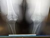
View attachment 289145View attachment 289146
\
H/o back surgery
H/o post-polio syndrome
H/o ???
Name that third disease.
Pagets
Spinal cord stimulator deficiency.View attachment 289145View attachment 289146
\
H/o back surgery
H/o post-polio syndrome
H/o ???
Name that third disease.
Spinal cord stimulator deficiency.
Meaty, beefy, big, and bouncy.She has pain.
View attachment 292129
Young guy. Axial extension-based pain with bilateral L5 pseudoarticulations that I suspect are the primary pain generator. I want to inject for diagnostic and therapeutic reasons but unsure where exactly I should target my needle -- thinking the inferolateral corner of each L5 transverse process. Any thoughts?
Perhaps not relevant to your issue, but are you certain that is L5 and not S1?
Sent from my iPhone using Tapatalk
Good points.Excellent point to discuss. Had an L3 kypho yesterday. Rads read it as L4 and 6 lumbar vertebrae.
There are several conventions on naming lumbar vertebrae.
1.Iliac crest is L4
2. Aorta splits at L4
3. Lowest vertebrae over a fully formed disc is L5.
4. First vertebrae without ribs is L1.
5. Count from C1 (when available).
All are correct as long as the treatment is done at the level intended to treat. But can be devastating when a Radiologist and a surgeon (pain doc) are using different naming conventions and the wrong level gets treated.
True, however thoracic imaging not always available.I discussed this with Tim Maus from Mayo Radiology a few years ago. He said if there is any ambiguity that you must count down from T1 as T1 is always T1. Anything else is subject to variability. Hypoplastic thoracic ribs, lumbar ribs, sacralization, lumbarization.
Sent from my iPhone using Tapatalk