
- Joined
- May 30, 2005
- Messages
- 21,250
- Reaction score
- 12,364
74 y/o female with extensive lumbar surgical history. Ongoing intractable leg and back pain. Limited to 50 feet with rolling walker, starting to fail in ADLs. Appears much more frail than typical 74 y/o.
Tried multiple opiates. On low dose short acting and Cymbalta. Failed Neurontin, Lyrica, Pamelor, Doxepin, Keppra.
Pain 8/10, worse with standing/walking, but unrelieved at rest. 50/50 back and both legs.
Plan was SCS trial if I could get through the T12-L1 or T11-12 spaces.
Reports:
2/23/17
STUDY lumbar spine radiographs.
COMPARISON: CT from November 11, 2015
TECHNIQUE: 2 views of the lumbar spine
CLINICAL INDICATION: Thoracic and/or lumbar sacral neuritis or radiculopathy
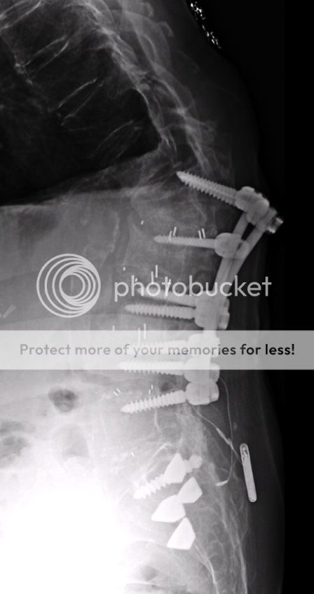
RESULT: For the purposes of this dictation caudal most lumbar like vertebral
body represents L5. Abandoned screw is seen in the right sacrum. Pedicle
screws and associated posterior fixation rods extending from the L1 level to
the L5 level. No peri-hardware lucencies or findings of hardware
complication. Interbody graft at multiple levels in the lumbar spine.
Laminectomy defects at multiple levels. Possible worsening compression
deformity of the L2 vertebral body when compared to the prior exam. Diffuse
osteopenia. Gibbus deformity of the lower thoracic/upper lumbar spine.
Stimulator device overlies the sacrum and lower lumbar spine. Fixation
hardware traverses the SI joints. Endplate osteophytes are seen multiple
levels in the lumbar spine. Imaged bowel gas pattern is nonobstructive. Lung
bases are hyperinflated and clear.
IMPRESSION:
Possible new compression deformity of the L2 vertebral body.
Osteopenia, postsurgical and degenerative change.

2/27/17
MAGNETIC RESONANCE IMAGING
Lumbar spine without contrast
CLINICAL INDICATION
Severe back pain
Compression fracture
Abnormal x-ray
COMPARISON: Correlation with radiographs of the lumbar spine performed
February 23, 2017.
CT myelogram of the lumbar spine performed
f the lumbar spine performed November 11, 2015
TECHNIQUE
Multiplanar, multi-sequence MRI of the lumbar spine was performed without
intravenous Gadolinium contrast.
FINDINGS
Posterior fusion hardware again extends from L1-L5. Post-surgical changes of
laminectomy again extend from T12-L1 through L3-L4 and from L4-L5 through
L5-S1. A portion of a screw is again seen in the right aspect of S1.
Surgical hardware extending across a bilateral sacroiliac joints is
incompletely imaged.
T10-T11
Diminutive disc bulge. Mild bilateral facet arthropathy. No significant
canal stenosis. Mild bilateral foraminal stenosis.
T11-T12
Diminutive disc bulge. Mild bilateral facet arthropathy. No significant
spinal canal or foraminal stenosis.
T12-L1
Small disc bulge. Mild bilateral facet arthropathy. No significant canal or
foraminal stenosis. There is mild loss of disc height and mild vacuum disc
phenomenon.
L1-L2
Overall, there is disc desiccation at this level, but there is also some T12
hyperintensity within the ventral aspect of the intervertebral disc.
Degenerative endplate irregularity. Relatively prominent Modic type
I/inflammatory degenerative endplate marrow change within the inferior aspect
of L1 and superior aspect of L2. There is also likely some Modic type
III/sclerotic degenerative endplate marrow change at this level. Mild
degenerative loss of height of the L1 and L2 vertebral bodies, which appears
to be new since prior exam. Small left paracentral disc protrusion, at the
site of an annular fissure mildly indenting the left ventral aspect of the
thecal sac. The thecal sac is widely patent, due to prior laminectomy. No
significant right foraminal stenosis. Within the limitations of the hardware
artifact, it is suggested that there is moderate left foraminal stenosis.
There appears to have been prior right facetectomy at this level.
L2-L3
No significant disc herniation, canal or foraminal stenosis is appreciated.
There appears to have been prior bilateral partial facetectomy.
L3-L4
There appears to have been prior partial bilateral facetectomy. No
significant disc herniation, canal or foraminal stenosis is appreciated.
L4-L5
No significant disc herniation, canal or foraminal stenosis.
L5-S1
No significant disc herniation, canal or foraminal stenosis.
The visualized distal spinal cord demonstrate normal caliber and signal. The
conus medullaris terminates in normal position. There appears to be a 4 mm T2
hyperintense lesion in the right hepatic lobe on image 18 of the sagittal
T2-weighted sequence.
A 3 mm T2 hyperintense lesion in the mid left kidney.
Mild STIR hyperintensity in the paraspinal soft tissues at L1-L2, more
prominent toward the right than the left.
IMPRESSION
1. New mild loss of height of the L1 and L2 vertebral bodies, with T2/STIR
hyperintensity in a portion of the L1-L2 intervertebral disc and relatively
extensive Modic type I/inflammatory degenerative endplate marrow change at
L1-L2, in combination with mild apparent Modic type III/inflammatory
degenerative endplate marrow change at this level. There is mild edema in the
paraspinal soft tissues at L1-L2. In light of lucencies seen surrounding the
bilateral pedicle screws at L1 on prior CT myelogram, it is thought most
likely that these changes reflect progression of degenerative change at L1-L2
related to motion at this level. Changed related to discitis/osteomyelitis
are thought unlikely, unless this is a strong clinical concern. A new left
paracentral disc protrusion at the site of an annular fissure is seen at
L1-L2, without significant associated canal stenosis. CT is recommended to
re-evaluate the surgical hardware and reassess areas of lucency around the L1
pedicle screws.
2. No definite acute compression fracture is appreciated.
3. Extensive post-surgical change and mild multilevel degenerative changes
are again seen, as described above. No significant canal stenosis. Up to
moderate foraminal stenosis appears to present. Details as above.
4. Tiny lesions in the liver and left kidney, indeterminate but perhaps
reflecting cysts.

I am not happy with my Radiology folks requesting repeat CT when myelogram showed lucency 12/15 on CT. No indication for further studies, patient declines further spine surgery.
Plan: SCS trial.
Tried multiple opiates. On low dose short acting and Cymbalta. Failed Neurontin, Lyrica, Pamelor, Doxepin, Keppra.
Pain 8/10, worse with standing/walking, but unrelieved at rest. 50/50 back and both legs.
Plan was SCS trial if I could get through the T12-L1 or T11-12 spaces.
Reports:
2/23/17
STUDY lumbar spine radiographs.
COMPARISON: CT from November 11, 2015
TECHNIQUE: 2 views of the lumbar spine
CLINICAL INDICATION: Thoracic and/or lumbar sacral neuritis or radiculopathy
RESULT: For the purposes of this dictation caudal most lumbar like vertebral
body represents L5. Abandoned screw is seen in the right sacrum. Pedicle
screws and associated posterior fixation rods extending from the L1 level to
the L5 level. No peri-hardware lucencies or findings of hardware
complication. Interbody graft at multiple levels in the lumbar spine.
Laminectomy defects at multiple levels. Possible worsening compression
deformity of the L2 vertebral body when compared to the prior exam. Diffuse
osteopenia. Gibbus deformity of the lower thoracic/upper lumbar spine.
Stimulator device overlies the sacrum and lower lumbar spine. Fixation
hardware traverses the SI joints. Endplate osteophytes are seen multiple
levels in the lumbar spine. Imaged bowel gas pattern is nonobstructive. Lung
bases are hyperinflated and clear.
IMPRESSION:
Possible new compression deformity of the L2 vertebral body.
Osteopenia, postsurgical and degenerative change.


2/27/17
MAGNETIC RESONANCE IMAGING
Lumbar spine without contrast
CLINICAL INDICATION
Severe back pain
Compression fracture
Abnormal x-ray
COMPARISON: Correlation with radiographs of the lumbar spine performed
February 23, 2017.
CT myelogram of the lumbar spine performed
f the lumbar spine performed November 11, 2015
TECHNIQUE
Multiplanar, multi-sequence MRI of the lumbar spine was performed without
intravenous Gadolinium contrast.
FINDINGS
Posterior fusion hardware again extends from L1-L5. Post-surgical changes of
laminectomy again extend from T12-L1 through L3-L4 and from L4-L5 through
L5-S1. A portion of a screw is again seen in the right aspect of S1.
Surgical hardware extending across a bilateral sacroiliac joints is
incompletely imaged.
T10-T11
Diminutive disc bulge. Mild bilateral facet arthropathy. No significant
canal stenosis. Mild bilateral foraminal stenosis.
T11-T12
Diminutive disc bulge. Mild bilateral facet arthropathy. No significant
spinal canal or foraminal stenosis.
T12-L1
Small disc bulge. Mild bilateral facet arthropathy. No significant canal or
foraminal stenosis. There is mild loss of disc height and mild vacuum disc
phenomenon.
L1-L2
Overall, there is disc desiccation at this level, but there is also some T12
hyperintensity within the ventral aspect of the intervertebral disc.
Degenerative endplate irregularity. Relatively prominent Modic type
I/inflammatory degenerative endplate marrow change within the inferior aspect
of L1 and superior aspect of L2. There is also likely some Modic type
III/sclerotic degenerative endplate marrow change at this level. Mild
degenerative loss of height of the L1 and L2 vertebral bodies, which appears
to be new since prior exam. Small left paracentral disc protrusion, at the
site of an annular fissure mildly indenting the left ventral aspect of the
thecal sac. The thecal sac is widely patent, due to prior laminectomy. No
significant right foraminal stenosis. Within the limitations of the hardware
artifact, it is suggested that there is moderate left foraminal stenosis.
There appears to have been prior right facetectomy at this level.
L2-L3
No significant disc herniation, canal or foraminal stenosis is appreciated.
There appears to have been prior bilateral partial facetectomy.
L3-L4
There appears to have been prior partial bilateral facetectomy. No
significant disc herniation, canal or foraminal stenosis is appreciated.
L4-L5
No significant disc herniation, canal or foraminal stenosis.
L5-S1
No significant disc herniation, canal or foraminal stenosis.
The visualized distal spinal cord demonstrate normal caliber and signal. The
conus medullaris terminates in normal position. There appears to be a 4 mm T2
hyperintense lesion in the right hepatic lobe on image 18 of the sagittal
T2-weighted sequence.
A 3 mm T2 hyperintense lesion in the mid left kidney.
Mild STIR hyperintensity in the paraspinal soft tissues at L1-L2, more
prominent toward the right than the left.
IMPRESSION
1. New mild loss of height of the L1 and L2 vertebral bodies, with T2/STIR
hyperintensity in a portion of the L1-L2 intervertebral disc and relatively
extensive Modic type I/inflammatory degenerative endplate marrow change at
L1-L2, in combination with mild apparent Modic type III/inflammatory
degenerative endplate marrow change at this level. There is mild edema in the
paraspinal soft tissues at L1-L2. In light of lucencies seen surrounding the
bilateral pedicle screws at L1 on prior CT myelogram, it is thought most
likely that these changes reflect progression of degenerative change at L1-L2
related to motion at this level. Changed related to discitis/osteomyelitis
are thought unlikely, unless this is a strong clinical concern. A new left
paracentral disc protrusion at the site of an annular fissure is seen at
L1-L2, without significant associated canal stenosis. CT is recommended to
re-evaluate the surgical hardware and reassess areas of lucency around the L1
pedicle screws.
2. No definite acute compression fracture is appreciated.
3. Extensive post-surgical change and mild multilevel degenerative changes
are again seen, as described above. No significant canal stenosis. Up to
moderate foraminal stenosis appears to present. Details as above.
4. Tiny lesions in the liver and left kidney, indeterminate but perhaps
reflecting cysts.

I am not happy with my Radiology folks requesting repeat CT when myelogram showed lucency 12/15 on CT. No indication for further studies, patient declines further spine surgery.
Plan: SCS trial.
Last edited: