Contrast in ESI

This forum made possible through the generous support of SDN members, donors, and sponsors. Thank you.

interjectionreflection

Full Member
2+ Year Member
Joined
Aug 20, 2021
Messages
80
Reaction score
26
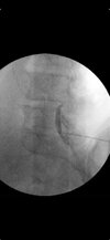
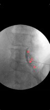
Hi all. I came across this picture during a review and the authors labeled this image as not epidural. I wanted to get folks thoughts on this. I admit I'm still a relatively new staff so If this looks this is blatantly not an epidural pattern I am happy to take pointers and learn. I feel fairly confident in identifying patterns that look classic like textbook images but sometimes for whatever reason (bad spines, volume of contrast used, could be technique too) I get patterns that don't look classic for epidural, subdural or otherwise. In fellowship for some reason they usually looked fairly obvious. I feel uncomfortable with these "in between" looking patterns and was wondering if people have shots of non-textbook looking epidural flows that are still epidural or tricky to identify patterns. All of the articles I find have pretty classic shots but none have in between. Even my colleagues who have 10 and 20+ years of experience disagree on some flows patterns when I ask them.
 

Attachments

  • 1000095245.jpg
    1000095245.jpg
    37 KB · Views: 215
I always get two views with one definitely being an AP
 
Looks epidural.

Looks like the pattern you’ll get if you’re a few degrees off plane from the lamina in CLO.
 
I have the same experience as a newer attending - now that I'm on my own, I'm finding relatively frequent non-textbook contrast patterns and second-guessing myself (especially for TFESIs).
 
1000095245 copy.jpeg


A lot of this contrast looks like it is not epidural. This looks like ipsilateral oblique and not CLO. I cannot identify a SP. The only VILL I can make out is on the other side of the spine. The area circled above looks like contrast behind the spine, running down the lamina. This is a poorly documented procedure without additional images.
 
View attachment 396058

A lot of this contrast looks like it is not epidural. This looks like ipsilateral oblique and not CLO. I cannot identify a SP. The only VILL I can make out is on the other side of the spine. The area circled above looks like contrast behind the spine, running down the lamina. This is a poorly documented procedure without additional images.
That circled area is anterior to S1 lamina. Tips of footballs marked. VILL clearly defined by posterior margin of contrast. Classic CLO spread.
 

Attachments

  • markup_1000008331.jpg
    markup_1000008331.jpg
    45 KB · Views: 126
View attachment 396058

A lot of this contrast looks like it is not epidural. This looks like ipsilateral oblique and not CLO. I cannot identify a SP. The only VILL I can make out is on the other side of the spine. The area circled above looks like contrast behind the spine, running down the lamina. This is a poorly documented procedure without additional images.
This is why I still LORTA when doing clo.

You have multiple experts here tht say it's acceptable and 1 dissected.

Versus all would.probably not argue over a lateral..
 
That circled area is anterior to S1 lamina. Tips of footballs marked. VILL clearly defined by posterior margin of contrast. Classic CLO spread.
Nope, horrible pic. Only pedicles I can see are on the left of the picture at L4 and L5. This image will not do.
This is why IPSIS states to have 5 saved images.
 
Nope, horrible pic. Only pedicles I can see are on the left of the picture at L4 and L5. This image will not do.
This is why IPSIS states to have 5 saved images.
and meanwhile we all get cancer.

i get it if you want to publish a picture, but if i am at the right depth, and i get a LOR (even with TFESI) i am not wasting my time and lifespan on a million views. (ie: laterals for a TFESI with great AP spread)
 
and meanwhile we all get cancer.

i get it if you want to publish a picture, but if i am at the right depth, and i get a LOR (even with TFESI) i am not wasting my time and lifespan on a million views. (ie: laterals for a TFESI with great AP spread)
With the above pic, should a complication occur....
PM me.
 
Clearly epidural, with contrast covering inferior L4 to S2. That pt could have been decompressed at L4, have a little scoliosis or a million reasons why the pic isn’t perfect, but it’s still epidural.

I inject that picture routinely.

I do NOT recommend you save 5 images, an insane ask of a guy/gal doing 50-60 procedures under fluoro per week for many yrs.
 
Looks epidural with the top spread (beneath lamina, not moving into subdural space)
Would you mind posting the source and what the authors discussed for context
 
Looks epidural with the top spread (beneath lamina, not moving into subdural space)
Would you mind posting the source and what the authors discussed for context
Image and Contrast Flow Pattern Interpretation for Attempted Epidural Steroid Injections


It's by a well-known Pysiatrist. He points to the CLO image as an example of a "subdural with cystic flow" I've never heard of that. It's reassuring to know that this CLO looks epidural or posterior to the epidural space to most of us. I was beginning to question my training. Lol.

If you have any resources that show epidural spreads that are atypical but still epidural, I would very much appreciate them.
 
Adding in the other panels and legend.

View attachment 396082

Shows the importance of examining ALL of the data and why one view is no view. I’m sure that the contrast gurus that I respect would label this intradural. The final test would be, can you aspirate the contrast that you just injected? If yes, it’s confirmatory.

The next questions are, in the setting of a lumbar ESI:

1. What is the risk of injecting 10 ml?
2. How will intradural spread affect the desired results?

I think this happens quite frequently (either pure intradural or mixed intradural/ epidural) and in the interest of time, many of us ignore it. But, should we?
 
Adding in the other panels and legend.

Adding in the other panels and legend.

View attachment 396082

With the benefit of the AP view, I would have my suspicions about this if I was doing the procedure because the contrast is so midline and concentrated (and not diffusing whatsoever), I would already start wondering about if this was intra or subdural. one way you can often confirm this is to aspirate and you will often get a good portion of the contrast back if you are intradural (versus epidural you will get nothing back). The contralateral oblique looked okay but the other 2 views look much less reassuring

In this particular situation, I would start walking the needle back at half millimeter at a time until I could no longer aspirate any contrast or saline (what I think of as a reverse loss-of-resistance). Alternatively you could pull back fully behind the ligament and redo your loss-of-resistance, although I often prefer the former versus the latter as at least I know exactly where on that as a starting point.

Unfortunately, when working at L5-S1 you are often going to be dealing with a very thin ligamentum flavum and often a minuscule posterior epidural space which is why it is relatively rare that I would do an interlaminar at L5-S1 unless I can clearly see a good landing spot on their MRI
 
Shows the importance of examining ALL of the data and why one view is no view. I’m sure that the contrast gurus that I respect would label this intradural. The final test would be, can you aspirate the contrast that you just injected? If yes, it’s confirmatory.

The next questions are, in the setting of a lumbar ESI:

1. What is the risk of injecting 10 ml?
2. How will intradural spread affect the desired results?

I think this happens quite frequently (either pure intradural or mixed intradural/ epidural) and in the interest of time, many of us ignore it. But, should we?
With the benefit of the AP view, I would have my suspicions about this if I was doing the procedure because the contrast is so midline and concentrated (and not diffusing whatsoever), I would already start wondering about if this was intra or subdural. one way you can often confirm this is to aspirate and you will often get a good portion of the contrast back if you are intradural (versus epidural you will get nothing back). The contralateral oblique looked okay but the other 2 views look much less reassuring

In this particular situation, I would start walking the needle back at half millimeter at a time until I could no longer aspirate any contrast or saline (what I think of as a reverse loss-of-resistance). Alternatively you could pull back fully behind the ligament and redo your loss-of-resistance, although I often prefer the former versus the latter as at least I know exactly where on that as a starting point.

Unfortunately, when working at L5-S1 you are often going to be dealing with a very thin ligamentum flavum and often a minuscule posterior epidural space which is why it is relatively rare that I would do an interlaminar at L5-S1 unless I can clearly see a good landing spot on their MRI
In the original reference the lateral is not a true lateral so not sure what that would change for whatever its worth but I do wonder if it would make it look more epidural if it were a true lateral.

I have the same experience as a newer attending - now that I'm on my own, I'm finding relatively frequent non-textbook contrast patterns and second-guessing myself (especially for TFESIs).
This is from another reference link describing a subdural patterns. This looks very different than the one I asked about originally hence why I asked my question to more seasoned staff. Glad to hear im not the only one that is finding that.

1733768873949.png
 
In the original reference the lateral is not a true lateral so not sure what that would change for whatever its worth but I do wonder if it would make it look more epidural if it were a true lateral.


This is from another reference link describing a subdural patterns. This looks very different than the one I asked about originally hence why I asked my question to more seasoned staff. Glad to hear im not the only one that is finding that.

View attachment 396093

Subdural vs INTRAdural. Intradural not commonly discussed.
 
In the original reference the lateral is not a true lateral so not sure what that would change for whatever its worth but I do wonder if it would make it look more epidural if it were a true lateral.


This is from another reference link describing a subdural patterns. This looks very different than the one I asked about originally hence why I asked my question to more seasoned staff. Glad to hear im not the only one that is finding that.

View attachment 396093
Can you post this link?
 
Having a brain fart, subdural is same thing as IT right?


The article actually equates intradural and subdural as synonymous.

Subdural is below dura but still above the arachnoid. Intrathecal is below arachnoid but above pia.

My understanding is that there are 3 layers to the dura and what layer you inject into determines the contrast pattern. Central layer —> sharply delineated central pattern. Dural boundary cell layer—> tram track pattern. Perhaps intradural and subdural are synonymous but depending upon the dural layer injected, the contrast pattern is different.
 
Just to add my 2cents, late to the game of course, is that the original picture is fishy and not clear epidural spread. It's too far obliqued and poor resolution to see the layers properly. The contrast also looks too "thick" from my experience. I usually see a very thin line. I would be concerned it's not epidural. Of course, I would look at other views to confirm. After seeing the AP posted later, I would say no to epidural spread. It's far too thick/dense midline to be epidural. I don't usually take true laterals unless I feel I'm losing the needle tip in the contrast on my approach.
 
Question for the hoard - has anyone performed a C7-T1 ILESI and seen contrast completely skip the level they were injecting at and for several levels above and below, but yet appear 100% epidural at distal levels? Got loss unquestionably and thought this was another 5 minute procedure.

Was collimated and when I went to push the dye live, there was no contrast spread at all coming from my needle tip in CLO. I had no idea where it went so I assumed the worst/vascular. Did more contrast live, again no spread anywhere. When I removed collimation to start over, there was clear as day epidural contrast spread up and down that started around C5 above and around T2 (levels not exact). Confirmed epidural spread with vaculoated pattern on AP.

I will never collimate for this procedure again having seen this. Has anyone had this happen to them before?
dont think it was the collimation, but i have seen this as well. if it looks epidural, just inject away with dex. pretty rare to get vascular spread with a interlaminar CESI and even if you do, injecting dex isnt the end of the world
 
and if you are injecting the cord, you should be getting some visceral response from the patient.


unless they under deep sedation...


always collimate unless you want to undo it for an image or two when you dont see contrast. your situation was 1 in a 100. the amount of extra radiation exposure to yourself and the patient is infinitely more significant.
 
I can’t imagine injecting the cord. It would already be too late with contrast. I use dex for cesi and I very frequently get the chest pressure and burning sensations. I always have this fear that somehow post contrast, I am intra-cord when they complain.
 
I can’t imagine injecting the cord. It would already be too late with contrast. I use dex for cesi and I very frequently get the chest pressure and burning sensations. I always have this fear that somehow post contrast, I am intra-cord when they complain.
That sensation is usually just the Omnipaque.
Burning sensation is perineal from dex.
 
That sensation is usually just the Omnipaque.
Burning sensation is perineal from dex.
Given your experience, how do you differentiate cord injection from just the contrast or injectate ifself? I would say 75% of my pts complain of some form of pain with injection so I tend to push extremely slowly - they report immense pressure or tightness in the chest or back and I am confident I’m not in the cord because they say it worked at their follow up but their response is so visceral during injection that sometimes I even question myself.
 
Hopefulgasman-

That is not my experience FWIW, performing these procedures or proctoring them over [emoji[emoji[emoji6]][emoji[emoji6][emoji6]]][emoji[emoji[emoji[emoji6][emoji6]][emoji[emoji[emoji6]][emoji[emoji6]]]][emoji[emoji[emoji[emoji6][emoji6]][emoji[emoji[emoji6]][emoji[emoji6]]]][emoji[emoji6][emoji6]][emoji[emoji[emoji6]][emoji[emoji6][emoji6]]]][emoji[emoji6][emoji6]][emoji[emoji[emoji[emoji6][emoji6]][emoji[emoji[emoji6]][emoji[emoji6]]]][emoji[emoji6][emoji6]][emoji[emoji[emoji6]][emoji[emoji6][emoji6]]]]] years

Maybe it’s how you are describing the experience?

Clearly what you’re doing is fine as people get better

Are you going C[emoji[emoji[emoji[emoji6][emoji6]][emoji[emoji[emoji6]][emoji[emoji6]]]][emoji[emoji[emoji[emoji6][emoji6]][emoji[emoji[emoji6]][emoji[emoji6]]]][emoji[emoji6][emoji6]][emoji[emoji[emoji6]][emoji[emoji6][emoji6]]]][emoji[emoji6][emoji6]][emoji[emoji[emoji[emoji6][emoji6]][emoji[emoji[emoji6]][emoji[emoji6]]]][emoji[emoji[emoji[emoji6][emoji6]][emoji[emoji[emoji6]][emoji[emoji6]]]][emoji[emoji6][emoji6]][emoji[emoji[emoji6]][emoji[emoji6][emoji6]]]][emoji[emoji6][emoji6]][emoji[emoji[emoji[emoji6][emoji6]][emoji[emoji[emoji6]][emoji[emoji6]]]][emoji[emoji6][emoji6]][emoji[emoji[emoji6]][emoji[emoji6][emoji6]]]]]]/T[emoji[emoji[emoji6]][emoji[emoji6][emoji6]]]? Midline or lateral? Tell us more
 
I have the same experience. But the reality is, if there was no response with needle advancement under fluoro, then I would think highly unlikely cord is involved. However the patient’s response with active administration of dex is striking.
 
I have the same experience. But the reality is, if there was no response with needle advancement under fluoro, then I would think highly unlikely cord is involved. However the patient’s response with active administration of dex is striking.
Correct. It’s never the contrast. It’s always the Dex.
 
if you touch the cord, they will have a zinging sensation into the arm and most likely the leg. very different than some chest pressure. and it wont be when you inject, it would be be when you advance the needle. dont ask me how i know.

this is exactly why you cannot have any sedation on board. you want to roll the dice with tetraplegia, be my guest.....
 
You guys just need to switch to depo. Happens almost every time with dex.
I feel like I live in bizarro world sometimes, bc I agree with this and cannot for the life of me understand how this isn’t known across the board.

Dexamethasone causes chest pain regularly when given in a cervical epidural, and yes, it causes vaginal burning in women as well, but not penile symptoms in men.

It never causes chest pain in a lumbar ILESI.

Depo causes none of these symptoms, and in a stenotic spinal canal is most likely superior in its efficacy than dex.

The sole benefit IMO of dexamethasone in an ILESI at any level is the cost per dose over Depo, the latter being more expensive.

I find Depo 40-80mg + 2cc saline at C7-T1 to be a wonderfully effective procedure, and I think all cervical procedures and surgeries outperform thoracolumbar. At least, that’s how I see it.

I’ve used dexamethasone and quit bc it doesn’t work as well as Depo and causes very disconcerting side effects, chest pressure being the most common, and I’ve sent 3-4 ppl to the hospital bc of aarhythmia and caused two TIA as a result.

That’s never happened with Depo.
Not that this is high volume procedure but I use dex in my cervical transforaminals
And IA C1-2

I’ve switched to depo for all ILESIs
 
Not that this is high volume procedure but I use dex in my cervical transforaminals
And IA C1-2

I’ve switched to depo for all ILESIs
I use dex at C1-2 as well. True. I do very few of them at this point in my career.
 
Top