

-
CycleTrack and SDN are teaming up to make medical school admissions more transparent and accessible! Read the announcement to learn how we’re supporting the future of CycleTrack’s free application-tracking tools .
You are using an out of date browser. It may not display this or other websites correctly.
You should upgrade or use an alternative browser.
You should upgrade or use an alternative browser.
Step II help me read this CT
Started by Dr McSteamy
Get help with your application
Use all the free resources available to you from SDN: articles, guides, expert advising, forums discussions, and school research.
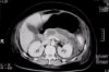
looks almost like a question i had in UW 😉
The picture obviously as you can see isn't the clearest thing in the world. What makes it harder to read the CT images on question banks is that you often don't know the level of the cut which often helps when doing a proper radiology read. Anyhows, the liver looks normal along with what appears to be the gallbladder. The kidneys also didn't seem to show anything out of the ordinary and the big black thing was probably just the stomach. The way I arrived at a pancreatic pathology was seeing that it was larger than what I'm used to seeing along with the white/gray level seen within the pancreas. Never having had radiology, that would have been my guess. Of course, the question stem helps a lot because then you would tend to ignore the small vessels which aren't so readily seen in that size image. What I wrote would obviously be ripped apart by a radiologist but since I'm not going into radiology, I think its okay
The picture obviously as you can see isn't the clearest thing in the world. What makes it harder to read the CT images on question banks is that you often don't know the level of the cut which often helps when doing a proper radiology read. Anyhows, the liver looks normal along with what appears to be the gallbladder. The kidneys also didn't seem to show anything out of the ordinary and the big black thing was probably just the stomach. The way I arrived at a pancreatic pathology was seeing that it was larger than what I'm used to seeing along with the white/gray level seen within the pancreas. Never having had radiology, that would have been my guess. Of course, the question stem helps a lot because then you would tend to ignore the small vessels which aren't so readily seen in that size image. What I wrote would obviously be ripped apart by a radiologist but since I'm not going into radiology, I think its okay
i posted this because maybe some of the seniors here already took the radio elective.
yeah. it's from UW.
the pt was on bipolar medication, which screwed with the pancreas.
but that's not my concern.
I also picked the pancreas because i thought it didn't look right. lucky guess.
But I posted this because i want to learn how to read the clues from the CT.
I'm wondering if that darker gray area in the pancreas is the abnormal?
Yeah the darker gray area in the pancreas is abnormal. It looks like there's a cystic collection at the head of the pancreas. And size wise, it seems a lot bigger than normal.
Valproic acid has been implicated with pancreatitis, so I'm guessing that's the drug used for the Bipolar disorder in your question?
Disclaimer: I'm NOT a radiologist.
Valproic acid has been implicated with pancreatitis, so I'm guessing that's the drug used for the Bipolar disorder in your question?
Disclaimer: I'm NOT a radiologist.
That larger amorphous mass extending from the left kidney anteriorly and medially is the pancreas. It appears significantly enlarged, with areas of heterogeneity (ie darker grey within the lighter white color) suggestive of fluid collections or significant inflammation.
There also appears to be free air. Hard to see with the picture, but there is a small collection medially and some that appears along the dependent portions of the abd cavity.
I am not a radiologist, but the pancreatic abnormality is quite striking.
There also appears to be free air. Hard to see with the picture, but there is a small collection medially and some that appears along the dependent portions of the abd cavity.
I am not a radiologist, but the pancreatic abnormality is quite striking.
