Capillaries and hydrostatic/osmotic pressure

This forum made possible through the generous support of SDN members, donors, and sponsors. Thank you.
D

deleted388502

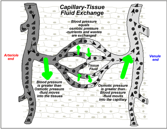
Alrighty so I'm a tad confused on this whole deal with capillaries losing some of the fluid due to osmotic pressure being greater than hydrostatic pressure near venules.

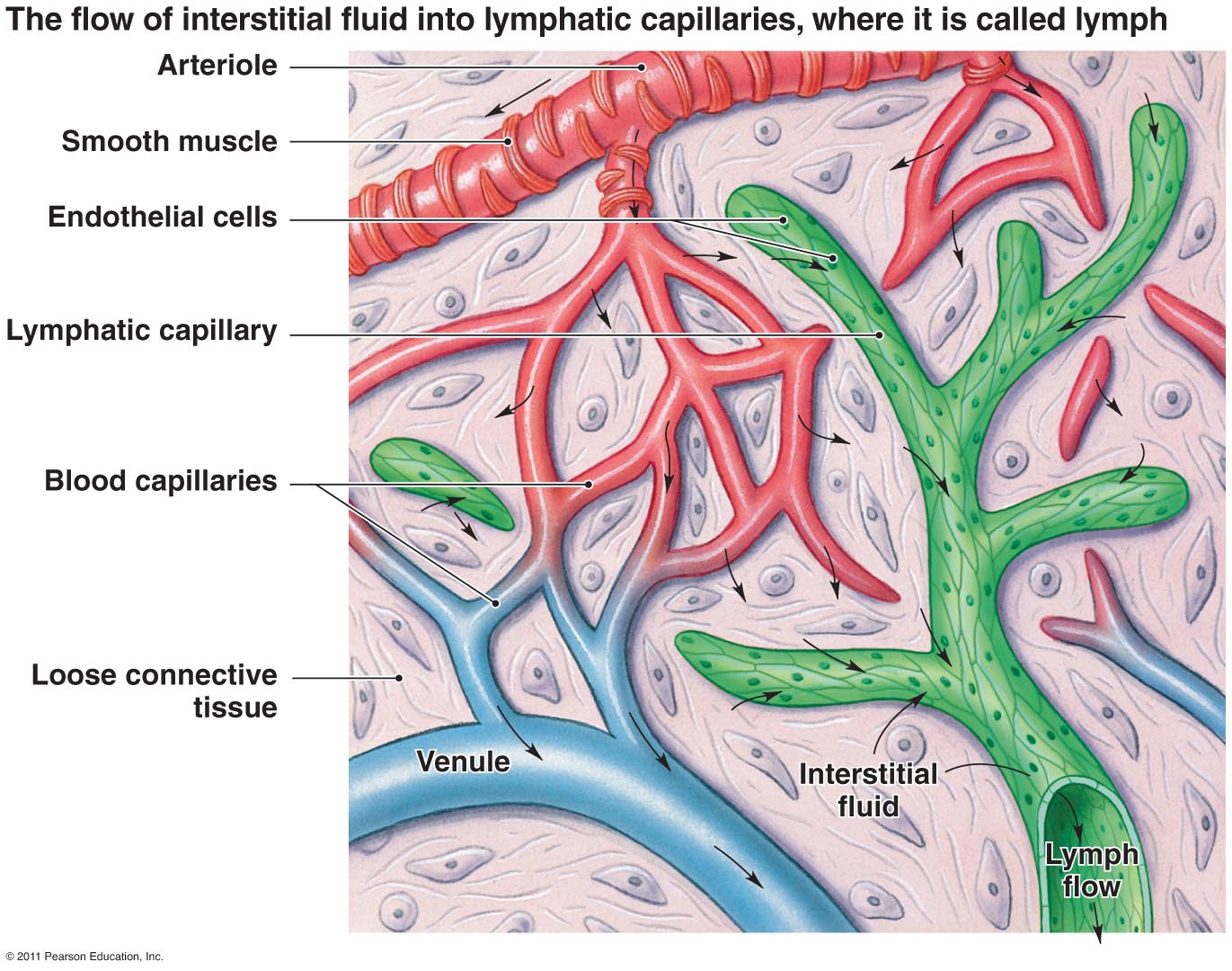
Could someone clear this up? I know the diagram in the EK textbook also shows that 10% is lost to the lymph, and I got a bit lost there - does this just mean that so called "good" nutrients that should be going through the capillaries are lost with the large proteins and other things taken up by the lymphatic system?

I understand that the blood is being pushed from artery --> arteriole --> capillary via hydrostatic pressure, but I don't understand how osmotic pressure is overcoming the hydrostatic pressure near the venule end.

Also, when we say capillaries are the site of nutrient exchange in interstitial tissue - can someone just specify what interstitial tissue is NOT included in this, and what nutrients are not included? I know fat is transported via lacteals and carbohydrates/proteins are taken via capillaries, so does it just include proteins and carbohydrates?

Thanks!

Members don't see this ad.
 
Last edited by a moderator:
  • Like
Reactions: 1 user
"The flow is from high to low" is what my physio and kinesio teachers always taught me...
 
I think you are talking about the the pressure in the cappillaries. When the blood flows into the arteriol end, it has higher pressure. Cappillaries is just a single layer of endothelium cells and it is not sealed. That is how it can exhange material with the surrounding tissues (any tissue outside the circulartory system are all called interstitial tissues. For example, muscles, lungs, livers, etc) The higher pressure at the arterial end pushes out the plasma out into the tissue. The tissue has high osmolarity compared to the plasma and also tend to draw the plasma out into the tissue. Now, the tissue is filled with water. The plasma has a lot of proteins in it, and these are large and can not pass the capillary wall. Their function is to provide osmolarity on the cappillary side. As water is continuously pushed out of the capillary end into the tissue, the plasma becomes more concentrated, and therefore high osmotic pressure. This pressure tends to draw water back into the capillaries at the venule end. Even though most water is drawn back into the cappilarries during the process, some water is still left in the tissue. Actually, some small proteins are pushed out of the capillaries and remains in the tissue. Now, here is where lymphatic system comes in. The lymphatic vessles picks up the left-over water and proteins from the tissue, and transfer it it into large lymphatic vessls and merge into and the thoracic lymthatic duct. At the end the lymphatic duct transport the water and small protein back into the circulatory system.
 
Members don't see this ad :)
Oncotic pressure is just osmotic pressure contributed by proteins with capillaries serving as the semipermeable membrane

I know. I was wondering if he was specifically talking about oncotic pressure, since that is a type of osmotic pressure which deals with the proteins of the capillaries. Or if he was mentioning about osmotic pressure in general.
 
I know. I was wondering if he was specifically talking about oncotic pressure, since that is a type of osmotic pressure which deals with the proteins of the capillaries. Or if he was mentioning about osmotic pressure in general.
Sorry, bro, no bad intention was intended. I think she should be talking about the capillary.
 
What EK is referring to is net fluid flow within a capillary. Let's first look at it from a physical standpoint and then we can apply it to the biological system.

What forces will be acting on the fluid in the capillary? Overall, there are forces pushing fluid out of the capillaries and forces pulling fluid into the capillaries. We can start with forces pushing fluid out. The first force is the pressure from the heart (and gravity if you are standing) called the hydrostatic pressure, which is a force over the entire area of the capillaries. The net force is in the direction of the flow, but fluid also has a force pushing it against the walls of the capillary. Since the capillaries are not perfectly sealed in most areas, some fluid will be pushed out. This fluid is referred to as extracellular fluid or interstitial fluid, which eventually gets picked up by blind ended tubes (lymph system) and becomes part of lymph that eventually dumps this fluid back into the circulatory system.

The next force that tends to pull fluid out of the cell is called the osmotic pressure. Osmotic pressure refers to a pressure caused by an imbalance in solutes. In this case we want to look at the osmotic pressure outside the capillary. This is the force due to all the solutes that are outside of the capillary. Their charges will pull the water out of the capillary. This is exactly like putting a cell into salt water, which is hypertonic to the cell. It causes all the water to flow out of the cell and the cell will shrivel up.

Now, there are also pressures pulling fluid back into the capillaries. The main one to focus on for the MCAT is also an osmotic pressure. This is due to the proteins/ions/solutes in the capillary. There is a special term for this called the oncotic pressure or colloidal osmotic pressure. This pressure force pulls fluid back into the capillary. This is similar to putting a cell in dehydrated water which is hypotonic to the cell so the oncotic pressure of the cell pulls water into it. BTW, a confusing convention with osmotic pressure is that it fluid flows from low osmotic (solute) --> high osmotic (solute) pressure.

OK, so the main forces pushing fluid out are the osmotic pressure of the extracellular/interstitial space (high osmotic pressure) pulling the fluid out and the hydrostatic pressure pushing the fluid out.

The main MCAT force pulling the fluid back into the capillary is the osmotic pressure caused by the solutes in the capillary (aka oncotic pressure).

Now, when fluid enters the capillary from an arteriole it initially has a net force [(hydrostatic pressure)+(osmotic pressure of the EC space) - (oncotic pressure)] pushing fluid out of the capillary. As the fluid travels through the capillary more fluid flows out. This increases the concentration of the solutes in the capillary (the opposite of dilution) as it is traveling towards the venule. Now the net pressure [(hydrostatic pressure)+(osmotic pressure of the EC space) - (oncotic pressure)] is negative so the fluid flows back into the capillary on the venus side of the capillaries.

Just to sum up. The net fluid flow at the arteriolar end of the capillary is out of the capillaries. The net fluid flow at the venule side of the capillary is into the capillary.

The diagram below is great because it is simple and really drives the point home.

Tiw8Vni.gif
 
Last edited:
Also, when we say capillaries are the site of nutrient exchange in interstitial tissue - can someone just specify what interstitial tissue is NOT included in this, and what nutrients are not included?

Intersitium = area that is outside of the cells that is bathed in extracellular fluid. The main nutrient is glucose and oxygen. The main waste is CO2 and other metabolites.

I know the diagram in the EK textbook also shows that 10% is lost to the lymph, and I got a bit lost there - does this just mean that so called "good" nutrients that should be going through the capillaries are lost with the large proteins and other things taken up by the lymphatic system?

There is no preference per say for "good" and "bad" nutrients/waste. The capillaries at the arteriolar side are coming from an oxygenated heart and high in nutrients. The gradient pushes those nutrients out with the fluid flow if the molecule is small enough (glucose etc.). In the interstitum = extracellular space, there is a gradient of metabolites that can flow into the capillary with the fluid that is pulled into the capillary at the venule side as described in the previous post.

Also, proteins are not generally flowing in and out of capillaries. Proteins in the extracellular space are made mostly by connective tissue cells. Proteins in the capillary are mostly made from the liver. Do not get confused with that.

Most of the nutrients/waste you are talking about travels in and out of the capillary via the fluid flow that is caused by the pressure gradient.
 
Last edited:
This picture should also help put everything into context. It is hard to put capillaries, interstitium, and lymph into context when looking at it in isolation:

XqYaxOd.jpg


Note the capillaries have an arterial side (red) and venous side (blue) where the net fluid flow differs. This fluid now enters the INTERSTITUM which in the body is mostly made of connective tissue which consists of a sparse amount of cells producing HUGE amounts of extracellular fluid. Just to get you thinking, what organelle do you think connective tissue/interstitial cells (purple/pinkish things with dark dots/nucleus in it) have a lot of? Rough ER because they are making a ton of extracellular proteins. Now these proteins DO NOT enter the capillary. Capillary proteins made mostly by the liver do not leave the capillaries.

The fluid that leaves the capillaries is eventually picked up by the lymph vessels (green) and then flows to be dumped back into the circulatory system thereby continuing the cycle.
 
Last edited:
Top