High Yield Neuro Clinical Vignette

This forum made possible through the generous support of SDN members, donors, and sponsors. Thank you.

MudPhud20XX

Full Member
10+ Year Member
Joined
Nov 26, 2013
Messages
1,349
Reaction score
193
I will start first, so feel free!

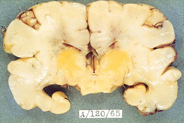
6 years old boy visits hospital with his Mom. He complains about his headache and seems to walk strangely. Upon MSK exam, you noticed the boy is having upper extremity muscle weakness. When you use a cotton wisp to assess the patient's sensation, there is no abnormal sensation. What is the most likely diagnosis?

Members don't see this ad.
 
An obese 35 y/o female comes in to the ER with complaints of difficulty walking, severe headaches, and visual disturbances. She has no history of meningitis or trauma. Fundoscopy reveals papilledema. Her medications includes treatment for acne, anxiety, and high blood pressure. After workup, a diagnosis is made and the physician prescribes acetazolamide. What mechanism is most likely behind this pathology?

a) Increased blood pressure
b) Scarring of arachnoid granulations
c) Increased activity of epyndymal cells
d) Medication interaction
e) Neoplasm with psammoma bodies on histology
 
Alright, looks like I stirred the pot a bit with this one. I got the info from FC.. The answer is a uncal transtentorial herniation, which causes compression of the midbrain leading to the following signs:
  • An ipsilateral fixed, dilated pupil with an eye "down and out" from compression CN III
  • Contralateral hemonymous hemianopsia from compression ipsilateral PCA
  • ipsilateral hemiparesis due to compression of the contralateral crus cerebri -> a FALSE localizing sign

They state that this is the common "triad" of symptoms for a uncal transtentorial herniation

@MudPhud20XX , @dfib slim, @Slade009
 
  • Like
Reactions: 1 users
An obese 35 y/o female comes in to the ER with complaints of difficulty walking, severe headaches, and visual disturbances. She has no history of meningitis or trauma. Fundoscopy reveals papilledema. Her medications includes treatment for acne, anxiety, and high blood pressure. After workup, a diagnosis is made and the physician prescribes acetazolamide. What mechanism is most likely behind this pathology?

a) Increased blood pressure
b) Scarring of arachnoid granulations
c) Increased activity of epyndymal cells
d) Medication interaction
e) Neoplasm with psammoma bodies on histology

Well, if I knew exact meds it might be easier.. but my best guess is B.

Scarring of arachnoid granulations decreasing CSF outflow leads to increased ICP and hydrocephalus (IIH, aka Pseudotumor Cerebri). This explains the papilledema, headaches, and walking difficulties. Vitamin A is known to damage arachnoid granulations, and is used as a component of acne treatment so I suspect that may be the cause.
 
Members don't see this ad :)
A mother brings in her 5 y/o son because she noticed that he started growing pubic hair and enlarged genitals. On examination the boy has signs of precocious puberty and you notice he has to move his head up in order to visualize your hands while testing the upper visual field. What is the underlying cause? How is the lesion affecting his vision? Why precocious puberty?
Germinoma of pineal gland - compression of sup. colliculi & pretectal area causing Parinaud synd. (impaired upward conjugate gaze), secretion of hCG causes precocious puberty, +/- hydrocephalus due to compression of cerebral aqueduct.

I'm not that smart, just had this exact UW Q yesterday lol
 
An obese 35 y/o female comes in to the ER with complaints of difficulty walking, severe headaches, and visual disturbances. She has no history of meningitis or trauma. Fundoscopy reveals papilledema. Her medications includes treatment for acne, anxiety, and high blood pressure. After workup, a diagnosis is made and the physician prescribes acetazolamide. What mechanism is most likely behind this pathology?

a) Increased blood pressure
b) Scarring of arachnoid granulations
c) Increased activity of epyndymal cells
d) Medication interaction
e) Neoplasm with psammoma bodies on histology

Ok so it can't be B because they say she has no history of meningitis, there isn't any fever nor is there any neck stiffness.

Based on the fact that she's a female with papilledema and (the give away) she was only prescribed Acetazolamide leads me to think she has a Pseudotumor Cerebri.
Answer is D: Women of childbearing age (a risk factor) with acne taking Vitamin A excess (another risk factor) have increased chances of developing a Pseudotumor Cerebri.

how this occur ??

Germinomas are histologically similar to the sex-organ tumors dygerminoma (female) and seminoma (male). They are histologically and functionally on the border of Trophoblasts and scattered Beta-HCG-secreting Syncytiotrophoblasts may be present causing the high levels.
 
  • Like
Reactions: 1 user
Well, if I knew exact meds it might be easier.. but my best guess is B.

Scarring of arachnoid granulations decreasing CSF outflow leads to increased ICP and hydrocephalus (IIH, aka Pseudotumor Cerebri). This explains the papilledema, headaches, and walking difficulties. Vitamin A is known to damage arachnoid granulations, and is used as a component of acne treatment so I suspect that may be the cause.

I wrote this myself so I was trying to drop hints in the stem. The Answer is C. Vitamin A causes increased production of CSF via increased activity of epyndymal cells. You had the right idea with Vitamin A. The disease present is pseudotumor cerebri which is seen in excess Vitamin A intake. Can be seen with acne medications. Scarring of the arachnoid granulations can be a consequence of previous meningitis but the patient does not have a hx of this. D is wrong because although it is the medication causing it, it isn't an interaction between her medications for the other conditions.

I tried to drop hints/distractors in the question stem cause I know from taking the USMLE a few days ago that the questions are similar to this style.
 
  • Like
Reactions: 1 user
Germinoma of pineal gland - compression of sup. colliculi & pretectal area causing Parinaud synd. (impaired upward conjugate gaze), secretion of hCG causes precocious puberty, +/- hydrocephalus due to compression of cerebral aqueduct.

I'm not that smart, just had this exact UW Q yesterday lol

Nice. You gots it.
 
  • Like
Reactions: 1 user
A 78 yr old woman with a hx of atrial fibrillation is brought to the emergency department after failing down a flight of stairs. Current medication include warfarin. She appears grossly lethargic and is responsive only to painful stimuli. Her blood pressure is 187/99 mmHg, and pulse is 48/min. A contrast CT scan is shown.

1. What is the most likely diagnosis?

2. Explain the hemodynamic abnormalities in this pt.

upload_2015-6-1_9-54-56.png
 
A 2 yr old boy is returned to the physician b/c of vomiting and ataxia. Physical examination shows papilledema and disproportionate growth of his head. An MRI of the head shows a large tumor involving the anterior horn of the Rt. lateral ventricle. A diagnosis of choroid plexus carcinoma is made. What particular brain structure is most likely to be affected by direct extension of this tumor?
A. Cerebellum
B. Caudate nucleus
 
wait, can anyone explain why hCG is being secreted? Is it the pineal gland tumor making hCG, thus neoplastic syndrome???

Because it's a germinoma, it can be associated with trophoblastic cells that secret hCG, so yeah, it's a neoplastic syndrome. It's extremely rare to have a primary CNS germinoma & even more rare to have one that secretes hCG, but this is the kind of b.s. that they love because they can relate it physiology.
 
  • Like
Reactions: 1 user
Members don't see this ad :)
A 2 yr old boy is returned to the physician b/c of vomiting and ataxia. Physical examination shows papilledema and disproportionate growth of his head. An MRI of the head shows a large tumor involving the anterior horn of the Rt. lateral ventricle. A diagnosis of choroid plexus carcinoma is made. What particular brain structure is most likely to be affected by direct extension of this tumor?
A. Cerebellum
B. Caudate nucleus

Head of caudate is right there, no?
 
  • Like
Reactions: 1 user
Head of caudate is right there, no?
yes it is. I chose cerebellum and got it wrong. Keep in mind that the lateral ventricle is adjacent to cauduate nucleate, which is the reason it's giving the boy ataxia right? Since caudate nucelus is part of the basal ganglion involved in the movement right?
 
  • Like
Reactions: 1 user
A 78 yr old woman with a hx of atrial fibrillation is brought to the emergency department after failing down a flight of stairs. Current medication include warfarin. She appears grossly lethargic and is responsive only to painful stimuli. Her blood pressure is 187/99 mmHg, and pulse is 48/min. A contrast CT scan is shown.

1. What is the most likely diagnosis?

2. Explain the hemodynamic abnormalities in this pt.

View attachment 192590

1. Given she's an elderly woman and the image I would say that it looks like a Subdural Hematoma. My differentials were a stroke which was on my mind because of her Hx and CN4 Palsy because it presents with trouble looking down (e.g at stairs, reading a newspaper) but it also presents with vertical diplopia which was absent.

2. Increased ICP, right? Oh wait, that isn't hemodynamics.

What I want to know is why her BP was high?

yes it is. I chose cerebellum and got it wrong. Keep in mind that the lateral ventricle is adjacent to cauduate nucleate, which is the reason it's giving the boy ataxia right? Since caudate nucelus is part of the basal ganglion involved in the movement right?

Yes, that's correct.
 
I was thinking about it but the way I thought it out got me lost. This is how I thought it all wrong:
High ICP -> Respiratory depression -> Low O2 -> Hypoxia -> Vasodilation (except for in the lungs, they vasoconstrict) -> Low BP (+ reflex tachycardia)

When in actuality this equation clears it all up (written in my notes):
Cerebral Perfusion Pressure= MAP - ICP
 
  • Like
Reactions: 1 user
tumblr_n3yyedTls91rq470wo1_1280.jpg

A newborn boy dies at the age of 2 weeks. The 26 year old mother was unaware of the pregnancy and received no prenatal care. A section of the newborn brain as seen at autopsy is shown above. Which of the following conditions was the mother most likely being treated for while pregnant?
A. Seizures
B. Cystic Acne
C. Atrial fibrillation
D. UTI
E. Hypercholesterolemia
F. Bipolar disorder
Dang, I can't really tell where the lesion is.... I think I am seeing some yellow discoloration below the lateral ventricles... Are those basal ganglion lesions?? Not sure. I would eliminate B, C, D, E. It's probably either seizure, acne or bipolar med.
 
tumblr_n3yyedTls91rq470wo1_1280.jpg

A newborn boy dies at the age of 2 weeks. The 26 year old mother was unaware of the pregnancy and received no prenatal care. A section of the newborn brain as seen at autopsy is shown above. Which of the following conditions was the mother most likely being treated for while pregnant?
A. Seizures
B. Cystic Acne
C. Atrial fibrillation
D. UTI
E. Hypercholesterolemia
F. Bipolar disorder

Is it B (Vitamin A) for spontaneous abortions. Or is it C Warfarin (Fetal Hemorrhage)? My final answer is B. By the way nice clear pic.
 
  • Like
Reactions: 1 user
Is it B (Vitamin A) for spontaneous abortions. Or is it C Warfarin (Fetal Hemorrhage)? My final answer is B. By the way nice clear pic.
wait but this newborn died after birth, so is this still considered as spontaneous abortion?
 
  • Like
Reactions: 1 user
tumblr_n3yyedTls91rq470wo1_1280.jpg

A newborn boy dies at the age of 2 weeks. The 26 year old mother was unaware of the pregnancy and received no prenatal care. A section of the newborn brain as seen at autopsy is shown above. Which of the following conditions was the mother most likely being treated for while pregnant?
A. Seizures
B. Cystic Acne
C. Atrial fibrillation
D. UTI
E. Hypercholesterolemia
F. Bipolar disorder

Ok I think I got it. It can't be ischemia because it looks too light and odd to be that. I think it looked like fat but realized it's Kernicterus. Now that it's narrowed down the answer should be D.

EDIT:
I just want clarify for readers that may not understand or still in the process of gaining knowledge.

UTI's (very common in females, because of the closeness of the opening of the Vagina and Anus to the opening of the Urethra, and also because of their shorter Urethra) -> TMP-SMX (most commonly prescribed for UTI's) -> Drug-induced neonatal bilirubin toxicity -> Buildup of Bilirubin in the brain -> Kernicterus.
 
Last edited:
  • Like
Reactions: 1 user
Top