Official Step 1 High Yield Concepts Thread

This forum made possible through the generous support of SDN members, donors, and sponsors. Thank you.

Transposony

Do or do not, There is no try
10+ Year Member
Joined
Nov 10, 2011
Messages
1,811
Reaction score
999
Let's discuss our doubts/offer clarifications about mechanisms/concepts for Step 1

ASK ANY QUESTIONS here.

To kick start the thread here is something I didn't know:

1. Penicillin-binding proteins (PBPs) are actually enzymes (transpeptidases & carboxypeptidases) which cross-link peptidoglycan. Penicillins binds to these enzymes and inactivating them thereby preventing cross-linkiing of peptidoglycan.

2. Periplasmic space (Gram -ve) contain proteins which functions in cellular processes (transport, degradation, and motility). One of the enzyme is β-lactamase which degrades penicillins before they get into the cell cytoplasm.
It is also the place where toxins harmful to bacteria e.g. antibiotics are processed, before being pumped out of cells by efflux transporters (mechanism of resistance).

There are three excellent threads which you may find useful:

List of Stereotypes

Complicated Concepts Thread

USMLE images

Members don't see this ad.
 
Last edited:
  • Like
Reactions: 5 users
VHL disease with pheochromocytoma, hemangioblastomas of the medulla and spinal cord, and renal and hepatic cysts, and d retinal angiomas.
Tuberous sclerosis, with lymphangioleiomyomatosis of lungs, angiomyolipomas and cystic disease in kidneys, cerebral cortical tubers, retroperitoneal lymphangiomyoma, and cardiac RHABDOMYOMA ,Epiloia , mulberry tumor (retinal astrocytoma), Subependymal nodule, Poliosis, subungual nodule in fingers.

good Q about them

•A 20-year-old, mentally ******ed woman saw the physician because she had flank pain for 1 week. Physical
examination showed right costovertebral angle tenderness. Patches of leathery-appearing (shagreen patches) and
hypopigmented (ash-leaf patches) skin were scattered over her body. There was a subungual nodule on her right index
finger. Abdominal CT scan showed bilateral renal cysts and tumor masses. MRI of the brain showed subependymal
nodules and 1- to 4-cm cortical foci with loss of the gray-white distinction. CT scan of the chest showed a 3-cm mass
involving the interventricular septum. Two years later, she has sudden, severe headache. MRI now shows a nodule
obstructing the cerebral aqueduct. Neurosurgery is performed, and a subependymal giant-cell astrocytoma is removed.
What is the most likely diagnosis?


(A) Down syndrome
(B) Krabbe disease
(C) Neurofibromatosis type 1
(D) Neurofibromatosis type 2
(E) Tuberous sclerosis
(F) Von Hippel–Lindau disease

answer is Tuberous sclerosis. it is one of the phakomatoses—rare inherited disorders in which hamartomas and neoplasms
develop throughout the body, along with cutaneous abnormalities. Patients with tuberous sclerosis have cortical tubers,
which are hamartomas of neuronal and glial tissue; other characteristic findings include renal angiomyolipomas, renal
cysts, subungual fibromas, and cardiac rhabdomyomas. In Down syndrome (trisomy 21), patients may develop acute
leukemia, but not brain neoplasms, and individuals who survive to middle age develop Alzheimer disease. Krabbe disease
is a leukodystrophy that results in deficiency of galactocerebroside β-galactosidase and an onset of neurologic
deterioration in infancy. Neurofibromatosis type 1 is characterized by deforming cutaneous and visceral neurofibromas,
cutaneous café-au-lait spots, and neurofibrosarcomas. In neurofibromatosis type 2, acoustic schwannomas, meningiomas,
gliomas, and ependymomas are present. Von Hippel–Lindau disease is characterized by hemangioblastomas in the
cerebellum, retina, and spinal cord, and by pheochromocytomas.
 
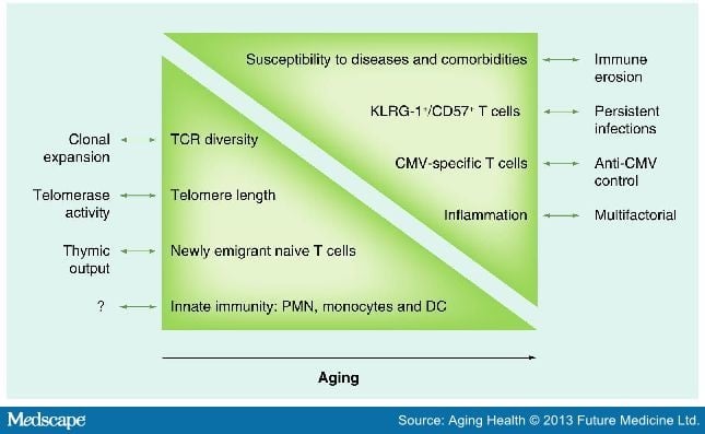
Immunosenescence : Old age changes associated with immune system.
T cells (particularly naïve T cells) are less numerous because of age-related atrophy of the thymus, whereas B cells overproduce autoantibodies, leading to the age-related increase in autoimmune diseases and gammopathies. Thus older people are generally considered to be immunocompromised and have reduced responses to infection (fever, leukocytosis) with increased mortality.

JCI64096.f1.jpg


780507-fig1.jpg


Inflammaging :
Old age is also associated with increased background levels of inflammation including blood measurements of C-reactive protein (CRP), erythrocyte sedimentation rate (ESR), and cytokines such as interleukin 6 (IL-6) and tumor necrosis factor α (TNF-α).
 
Last edited:
  • Like
Reactions: 2 users
This is pretty good.

The way I was taught and usually think about this is:

The body has various ways of regulating total body sodium in a way that total body sodium usually doesn't change much. Low serum concentration of sodium usually indicates a solvent (water) problem more than a solute problem. Hyponatremia usually means you're holding on to too much water (SIADH, etc.) Too much water leads to cerebral edema and its consequences-lethargy, confusion, coma, seizures, etc. Too high a sodium concentration is also usually a water problem (elderly bedridden person can't drink water, DI, etc.) and can disrupt electrolyte balance and lead to some similar issues. Correcting too fast high to low can give you cerebral edema. Correcting low to high can give you central pontine myelinolysis.

Potassium is part of the cocktail they give in lethal injection. High K+ stops the heart because it interferes in the outward K+ channel flow by decreasing its membrane potential. You will get problems with repolarization (prolonged-->QT prolongation; issues with repolarization-->hyperacute T waves)

Hypokalemia usually results in problems with repolarizing after muscle contraction. You get muscle cramping and stuff.

Insulin can cause potassium uptake by cells.

Calcium is important for a few main things: muscle contraction (tetany in hypocalcemia), neurotransmitter release (psychic moans), bone formation (bones). Calcium deposits with oxalate in kidneys to form stones (calcium citrate is more soluble than calcium oxalate, so citrate is also involved). I'm not exactly sure about the reason for abdominal pain (groans) but here's my hypothesis: high calcium interferes with serotonin release from synaptic vesicles. In the brain this could lead to depression (psychic moans). The enteric nervous system is mostly serotonergic. Disturbances in this could cause dysfunctions in motility and dysfunctions in the pathways that normally perceive pain and insults to the gut. Remember how we use odansetron for hyperemesis of pregnancy, chemo nausea, etc. and how we use SSRIs for IBS? I'm not sure if this is the reason, but it's the way I imagined it.

Obviously don't get mad if everything here is not totally 100% accurate, but it's just how I think about it.

Hope this helped.
 
Members don't see this ad :)
Arnold Chiari:
A congenital anomaly resulting from a normal-sized cerebellum developing in an abnormally small posterior fossa with a low tentorial attachment. There is a narrow craniospinal junction and malformation of the posterior fossa leading to downward displacement of the medulla, fourth ventricle, and cerebellum into the cervical spinal canal, as well as elongation of the pons and fourth ventricle. This results in impaction of the foramen magnum, compression of the cervicomedullary junction by the ectopic tonsils, and interruption of normal flow of cerebrospinal fluid (CSF). Two types, type I is asymptomatic (till adulthood) and type II is symptomatic and commonly associated with syringomyelia and meningomyelocele respectively. Symptoms arise from dysfunction of brainstem and lower cranial nerves.

Dandy Walker:
Also congenital but this time there is agenesis or hypoplasia of the cerebellar vermis, cystic dilatation of the fourth ventricle, and enlargement of the posterior fossa with aqueductal obstruction leading to hydrocephalus or it may be associated with atresia of the foramen of Magendie and foramen of Luschka.
The enlarged fourth ventricle balloons out backward as a membrane-wrapped cyst and lifts and displaces the posterior portion of the brain, as well as cause an internal obstruction of normal CSF flow, with resultant supratentorial hydrocephalus.
Unlike Arnold Chiari, the posterior fossa is large ( there's a lot of space in the post fossa for the 4th ventricle to WALK around and expand).

Arnold Chiari I or II both are associated with syringomyelia.

Only type-II has the myelomeningocoele, but they both have syringomyelia.

That's actually high-yield.
The only thing you've gotta know is that the meningomyelocele is type-II.

If it's type-II, they'll practically always tell/show you there's the myelomeningocoele, and the patient will likely be an infant.

With type-I, they'll likely give you a teenager / young adult with the typical bilateral loss of temperature/pain sensation in a cape-like distribution. That indicates the syringomyelia at the anterior white commissure.
 
beta blockers contraindicated in cocaine use

Cocaine blocks reuptake of adrenergics (NE/E),
If you block beta-receptors then your catecholamines have to bind to alpha receptors instead (alpha 1 effects are much more salient and faster than alpha 2). You'll cause vasoconstriction and increased BP by giving a beta-blocker when catecholamines are floating around.

In cocaine use associated with sympathetic symptoms (e.g., tachycardia, mydriasis), the first step is an ECG. If normal, you want to give a benzo (lorazepam) first-line; if that doesn't work add an alpha-blocker (phentolamine).

Cocaine increases the risk of coronary vasospasm and coronary artery thrombus. If ECG shows signs of ischaemia (e.g., ST-T-wave changes), the next best step is coronary angiography to rule out thrombus. In either event, if ischaemia is present, in addition to the lorazepam, give nitrates or dihydropyridine CCB, PLUS aspirin.
 
Renal cysts, hemangiomas, and benign adenomas are usually asymptomatic in von Hippel- Lindau disease.
- Von Hippel-Lindau disease is autosomal dominant.The gene that causes von Hippel-Lindau disease (VHL) is localized to chromosome 3p25-26.
The normal gene has a key role in cellular response to hypoxia and acts as a tumor suppressor. Presymptomatic and prenatal diagnosis by direct mutation analysis is possible for most patients. may be associated with pheochromocytoma due to MEN-IIA
 
@Transposony from uptodate : ''The phosphorus concentration in malignant cells is up to four times higher than in normal cells. Thus, rapid tumor breakdown often leads to hyperphosphatemia, which can cause secondary hypocalcemia. When the calcium concentration times phosphate concentration (the calcium phosphate product) exceeds 60 mg2/dL2, there is an increased risk of calcium phosphate precipitation in the renal tubules, which can lead to acute kidney injury. In addition, precipitation in the heart may lead to cardiac arrhythmias. Renal replacement therapy may be needed ''
 
@Transposony from uptodate : ''The phosphorus concentration in malignant cells is up to four times higher than in normal cells. Thus, rapid tumor breakdown often leads to hyperphosphatemia, which can cause secondary hypocalcemia. When the calcium concentration times phosphate concentration (the calcium phosphate product) exceeds 60 mg2/dL2, there is an increased risk of calcium phosphate precipitation in the renal tubules, which can lead to acute kidney injury. In addition, precipitation in the heart may lead to cardiac arrhythmias. Renal replacement therapy may be needed ''
But where does phosphate comes from?
 
What do you mean , Phosphorus = Phosphate , the cancer cells prolly take up more from diet or somethin... @Chilladosis yeah but that's mostly in chronic states , in the acute setting you get the nephropathy and cardiopathy described , which is actually kinda the same thing , precipitation - deposition of calcium-phosphate ''stuff''
 
  • Like
Reactions: 1 user
Great.
To summarize the differentiating feature are:
Atropine poisoning --> dry skin (Muscarinic block as sweating is sympathetic muscarinic mediated)
TCA overdose --> dry skin with arrhythmia and hypotension (Muscarinic and alpha 1 block)
Amphetamine toxicity --> warm and sweaty skin (as sweating is sympathetic muscarinic mediated)
They all have s/s of sympathetic over-activity.
B blockers will not prevent diaphoresis.
 
Members don't see this ad :)
difference b\w Candida esophagitis and Herpes simplex esophagitis ??
Really? They don't look the same. Like anywhere else, herpes is going to look like vesicles that can ulcerate. Candida looks like white on an erythematous base. Also, like any fungal infection, candidiasis anywhere is going to give you more of a left shift and advanced herpes disease is going to give more of a lymphocytosis picture,
 
  • Like
Reactions: 1 user
Really? They don't look the same. Like anywhere else, herpes is going to look like vesicles that can ulcerate. Candida looks like white on an erythematous base. Also, like any fungal infection, candidiasis anywhere is going to give you more of a left shift and advanced herpes disease is going to give more of a lymphocytosis picture,

Tell em @sloop
 
What do you mean , Phosphorus = Phosphate , the cancer cells prolly take up more from diet or somethin... @Chilladosis yeah but that's mostly in chronic states , in the acute setting you get the nephropathy and cardiopathy described , which is actually kinda the same thing , precipitation - deposition of calcium-phosphate ''stuff''
It comes from ATPs.
Although cancer cells produce far less ATP per molecule of glucose by aerobic glycolysis (Warburg effect), they produce it much faster.
 
Really? They don't look the same. Like anywhere else, herpes is going to look like vesicles that can ulcerate. Candida looks like white on an erythematous base. Also, like any fungal infection, candidiasis anywhere is going to give you more of a left shift and advanced herpes disease is going to give more of a lymphocytosis picture,
Candida esophagitis causes Tan yellow plaques in lower of esophagus. while Herpes simplex esophagitis causes sharply demarcated ulcerations that have a brown-red base in the lower of esophagus.
 
Really? They don't look the same. Like anywhere else, herpes is going to look like vesicles that can ulcerate. Candida looks like white on an erythematous base. Also, like any fungal infection, candidiasis anywhere is going to give you more of a left shift and advanced herpes disease is going to give more of a lymphocytosis picture,

Perhaps a better question is asking to differentiate CMV vs HSV esophagitis
 
HSV is punched out lesions and CMV is linear lesions right?


Sent from my iPhone using SDN mobile app
 
  • Like
Reactions: 1 users
why is Asthma associated with High DLCO? UW says its due to increased capillary blood volumes? is that bc of the increased effect of IPP on Venous return?
 
why is Asthma associated with High DLCO? UW says its due to increased capillary blood volumes? is that bc of the increased effect of IPP on Venous return?
AFAIK DLCO is normal in Asthma.
Increased DLCO is seen in conditions that increase effective blood flow to the functional lung such as heart failure, diffuse alveolar hemorrhage, pulmonary infarction and idiopathic pulmonary hemosiderosis (IPH).
 
  • Like
Reactions: 1 users
why is Asthma associated with High DLCO? UW says its due to increased capillary blood volumes? is that bc of the increased effect of IPP on Venous return?

I believe asthma is typically high but can also have a normal DLCO value. The way one of the clinicians explained it to us was that it was based on the severity of the particular asthma.

In asthma air gets trapped in the alveoli and over time you get hyperinflation of the lung. Increased gas volume leads to increased DLCO.

If the patient has mild asthma the DLCO would likely appear closer to normal values.
 
Breast cancer risk factors - what are they in order of most to least important in terms of increasing risk
 
Pathoma and FA discrepancy: pathoma mentioned that colon Ca via the mismatch repair pathway doesnt arise from prexisting adenoma's but FA mentions that sporadic colorectal ca from the mismatch pathway arise from serrated polyps.
 
UW mentions hypovolumia causes hyperuricemia by increasing uric acid reabsorption in the PCT, which can precipitate a gouty attack. for ****k's sake how?
 
UW mentions hypovolumia causes hyperuricemia by increasing uric acid reabsorption in the PCT, which can precipitate a gouty attack. for ****k's sake how?
Isn't this similar to how BUN increases in pre-renal azotemia? Decrease in RPF leads to increase in Na reabsorption which causes water to and urea to follow.
 
also i made a thread earlier on SDN about gallstones and cholesterol, is cholesterol soley solubilized in bile by converting cholesterol into bile acids via 7alphaOhlase? or is that part of it and the other part is that cholesterol is by itself in the bile, and is made soluble purely bc of phospholipids and bile acids/salts somehow solubilizing them chemically? little confused here


edit: also why are pigment stones seen specifically in TPN and alcholic cirrhosis?
 
Last edited:
I also would imagine it might have been asking how to treat drug induced Parkinsonism...benztropine is the tx of choice for that. Haloperidol should def be the answer for what causes the drug induced Parkinsonism.


Sent from my iPhone using SDN mobile app
 
Can someone explain like im 5 why high precision means more statistical power?

I pretty much just combined two concepts. Random error decreases precision. Increased precision causes increased statistical power. So- decreased random error --> increased precision --> increased statistical power (I think of it like you would have more statistical power aka be more sure you're right if there is less random error). Idk if it's technically true but it helped me memorize those main concepts lol


Sent from my iPhone using SDN mobile app
 
Fluoroquinolones.
Ertapenem
Some spelling errors here, but for what I have read and understood:

Also cefipime (4th generation cephalosporin) treats pseudomonas. Amikacin (only tetracycline hepatically excreted MOA: inhibits aminoacyl tRNA binding) is processed by the liver and used for pseudomonas. There is another set of drugs, the abbreviation is "TCP - takes care of pseudomonas", I think it stands for ticracillin, carbacillin, penicillin. It must be covered by two drugs, that's why there are so many drugs listed for it.

For pseudomonas as a bug, known that along with staph aureus, it is the #2 cause for problems with burn patients (#1 in 2nd week), cystic fibrosis, diabetics, and neutropenics. (normal count is 60% - "never(60%) let (30%) mom (<10%) eat (<10%) beans (<10%)". From the little flow chart in FA (one of the few pages I actually like in it), pseudomonas is gram negative, rod, lactase negative, oxidase positive, has a biofilm, green pigment, fruity smell when plated. Clinically, it will present in malignant otitis externa, whirlpool folliculitis (World Q), catheterized patients are the most common cause of it.

To summarize the differentiating feature are:
Atropine poisoning --> dry skin (Muscarinic block as sweating is sympathetic muscarinic mediated)
TCA overdose --> dry skin with arrhythmia and hypotension (Muscarinic and alpha 1 block)
Amphetamine toxicity --> warm and sweaty skin (as sweating is sympathetic muscarinic mediated)
They all have s/s of sympathetic over-activity.
Atropine poisoning - it's anticholinergic/sympathetic, so dry hot, dilated pupils are a classic clue ("eyes wide with fright"). Jimson weed (the dude who is cutting down logs type question) is identical to it. Give a cholinergic drug for reversal, my go to is physostigmine.

TCA drugs - remember they are anticholinergic as well present with the symptoms above, there is a World question on this.

Amphetamines - MOA: inhibit reuptake of dopamine(?), I believe. Systolic BP will almost touch 200, it's usually a teenager.

(as sweating is sympathetic muscarinic mediated)
I would think sweating, since it's "wet", would be parasympathetic, and parasymapathetic receptors are dominated by muscarnic receptors?

It's a continuous process and fun since it one of those rare Step 1 subjects which you are actually going to use in clinical practice.
wish I had kept up with them =\
I knew nothing regarding Pharm when I first started my prep, I just did 20 minutes a day first thing when I start studying, 5 new drugs a day and then review old drugs that I studied previously. It is very tedious at first, but you get the hang of it and will get easier as the days progress. It's a really important subject that you have to do piecemeal, don't neglect it. Even if your 1 month out from your exam, you can finish 150 drugs if you do 5 per day.
 
Last edited:
Top