Section Bank Bio #47

This forum made possible through the generous support of SDN members, donors, and sponsors. Thank you.

wheatthinners

Full Member
7+ Year Member
Joined
Jul 25, 2014
Messages
68
Reaction score
10
.

Members don't see this ad.
 
Last edited:
B does not appear to be correct. The reason I use "appear" is because you can't exactly tell the absorbance in Fig. 1. You can only conclude that it's "about" 0.1 or so, which is equal to the absorbance in Fig. 2. So it's got the same affinity in either case. Now, you don't need to know the numbers to know that B is wrong. You must know that B is wrong because IgG is used as a control. If it changed from Fig. 1 to Fig. 2, it would be a terrible experiment and there would be no way to compare the results.
 
@aldol16

question about the control: it controls for the specificity of the enzyme-linked antibody to FSHpep in (A) and FSH in (B). That is, we would want to ensure that the absorbance was from FSH-Ab binding. So the low to none absorbance from IgG is desired. Am I thinking about this correctly?
 
question about the control: it controls for the specificity of the enzyme-linked antibody to FSHpep in (A) and FSH in (B). That is, we would want to ensure that the absorbance was from FSH-Ab binding. So the low to none absorbance from IgG is desired. Am I thinking about this correctly?

From the data given in the question, this is a valid interpretation. So what they're doing here is using an anti-body to tag the protein they want and then using an anti-body for the first anti-body to tag the first anti-body with a fluorescent tag. So the confound you want to avoid here is what if your second anti-body just happens to tag antibodies non-specifically? Then you're kind of screwed, in biological parlance. So you use a negative control, in which you don't expect anti-body binding to IgG. And lo and behold, there's no antibody binding.

There's also the possibility of the second anti-body binding other things, of course - I'm not sure why they don't account for that in this paper. You would have to read the paper to find out - the MCAT gives you very little extraneous data here.
 
Members don't see this ad :)
@theonlytycrane

From the data given in the question, this is a valid interpretation. So what they're doing here is using an anti-body to tag the protein they want and then using an anti-body for the first anti-body to tag the first anti-body with a fluorescent tag. So the confound you want to avoid here is what if your second anti-body just happens to tag antibodies non-specifically?

There's also the possibility of the second anti-body binding other things, of course
The way this is typically done to avoid nonspecific interactions is you'll have for example a mouse derived anti-ProteinX antibody which binds your protein of interest. Then the secondary antibody could be a goat derived anti-mouse-Fc HRP linked antibody that will provide the colormetric change.
 
PEhpb9n.png


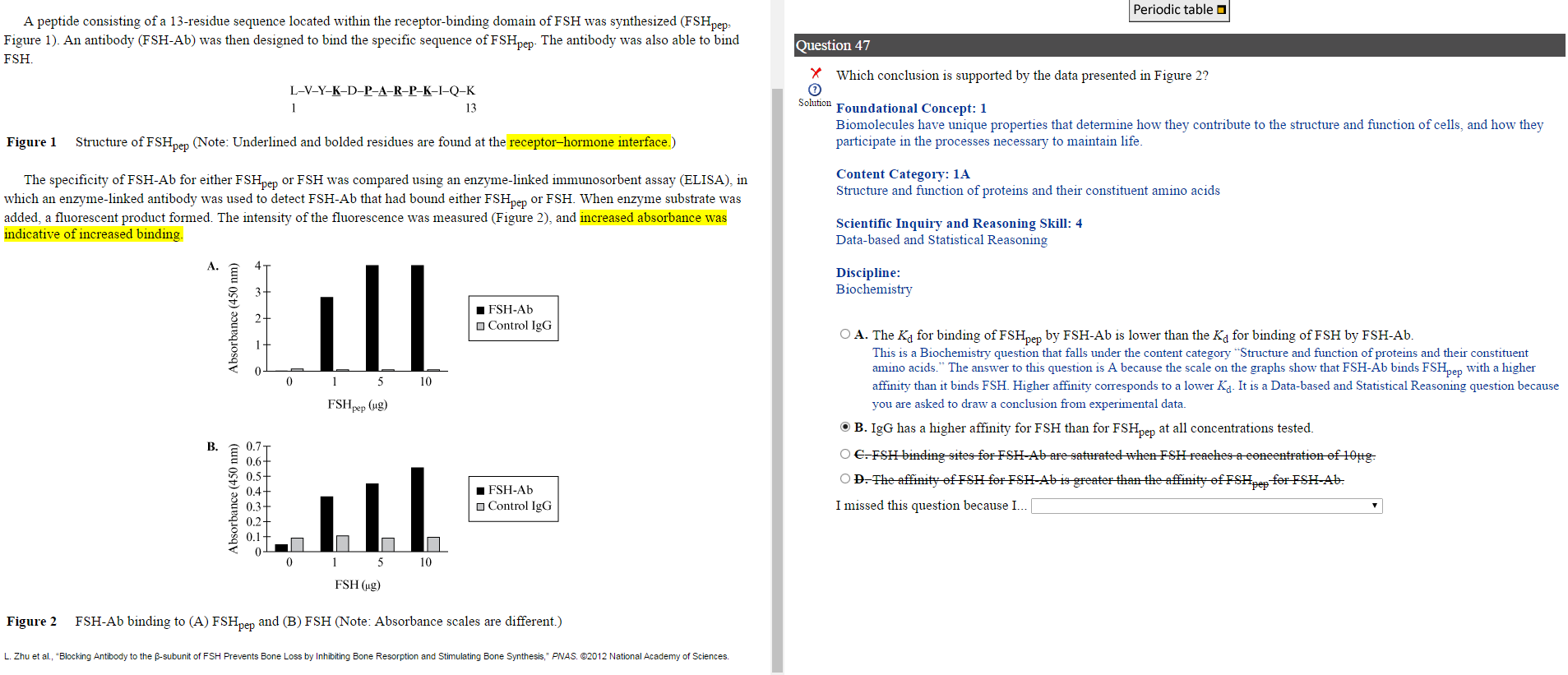
I understand why A is correct. But why would B be incorrect? I was debating between these 2, because they both seemed correct to me. Thank you.

I did the exact same thing: narrowing it down to A or B and then I went with A because I knew that answer was right.

As far as B is concerned, if you look at Figure 2, IgG is showing absorbance (and thus binding) even when there is 0 (no) FSH present and then that number stays the same even when [FSH] is increased. Thus, the IgG isn't showing any real affinity for FSH at all, regardless of the concentration tested.
 
I thought it had to do with the y axis values. As you can see the absorbances are much higher for FSH pep than for FSH.
 
Top