- Joined
- Dec 29, 2012
- Messages
- 247
- Reaction score
- 4
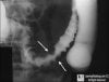
68 yo male patient presents to your clinic with postprandial pain. X-ray below, whats the dx?

Decreased cross sectional area due to arterioles being non-compliant?
I'm horrible with vasculitis/heart so this is a complete shot in the dark
right but what is it about struge weber that makes the patient have an increase in preload and a decrease in afterload?
AVM associated with Sturge weber -> increased preload. Not sure what exactly causes reduced after load though.
AVM associated with Sturge weber -> increased preload. Not sure what exactly causes reduced after load though.

AVM associated with Sturge weber -> increased preload. Not sure what exactly causes reduced after load though.
which type of drug long term can cause this.
Which type of drug long term can cause this.
Don't know what I'm looking at here... the speckles in the mediastinal area, or the fact that the lung field is really black.
Higher yield lung pathology that's drug related is typically pul fibrosis... in which case, it could be Bleomycin, Bulsulfan, Amiodarone.... or long term methotrexate.
Probably wrong.
GJ. mechanism please?
")
I was thinking the same thing but it doesn't look like fibrosis. maybe gas? there's something wrong with the diaphragm
perforated gastric ulcer is all i can tell you unfortunately lol. we had a question just like this on our exam
by the way, i just found this thread today. thank you so much for this, it is soo helpful.
i will be on the lookout for images I can add
I was thinking the same thing but it doesn't look like fibrosis. maybe gas? there's something wrong with the diaphragm
Yup, the diaphragm is a good place to focus.
My xray/CT/MRI interpreting skills are not that good (and neither is my anatomy haha) but if you perforate the diaphragm in a surgical procedure or maybe a stab wound, wouldn't u get a similar presentation? but you're looking for a drug right?
Sharklasers already got NSAID though... and the mechanism he provided seemed to make sense to me.
Oh? sorry i didn't see that. how does it do that?
There's air in the stomach (bloaty feeling we often get after crazy nachos), generated from gastric bacteria... and I was thinking since NSAID caused the perforation, the only place the air would go after reching the intra-peritoneal space is up..right up underneath the diaghram.
There's air in the stomach (bloaty feeling we often get after crazy nachos), generated from gastric bacteria... and I was thinking since NSAID caused the perforation, the only place the air would go after reching the intra-peritoneal space is up..right up underneath the diaghram.

this dude's stomach is rock hard...what pathology, and what would you see on histology to confirm said pathology?
signet ring cells?
There's air in the stomach (bloaty feeling we often get after crazy nachos), generated from gastric bacteria... and I was thinking since NSAID caused the perforation, the only place the air would go after reching the intra-peritoneal space is up..right up underneath the diaghram.
Yessirrr... what's the pathology or name of the disease?
intracerebral avm ipsi to the red face
Just missed this one on a qbank, haven't seen it in awhile.

are these ST depressions caused by angina?
Hint:
Patients are often asymptomatic or may have nonspecific symptoms.
I see some ST depression and T wave inversion - that points to ischemia though. Stable angina, or less than 75% occlusion of the coronary artery?
Digoxin toxicity? That's basically always my guess when I have no idea what the ECG is showing LOL
Heart block? 1st degree?
You see the slow drop after the S wave, like a steady gradual drop, and a step rise? That's supposed to be a sign of LVH (HTN for ex.). But idk if that's what this is. It's not seen in every complex. That's all I can see.
What about a BBB?
WPW? although I don't see a clear delta wave...
Hopefully I don't **** this question up, going to try to have a bit of a stem attached
40 y/o caucasian female with no significant PMHx presents to PCP w/exertional dyspnea. She returned a month ago from a mission trip to south asia. She mentions to you that the trip was uneventful and that she enjoyed building huts in the dirt and eating seafood with the locals, though she had a few days of stomach cramping and loss of appetite. CBC reveals
HBG - 9.8
HCT - 30
MCV - 87
PLT - 200k
WBC - 7k
The PBS is attached.
Over the next month, if you decide to watch her condition and see what happens, the most likely development will be
A) Peripheral neuropathy
B) Ringed sideroblasts
C) Elevated free erythrocyte protoporphyrin
D) Dilated cardiomyopathy
I think I made there be one most likely answer... But if I didn't I apologize :x
Correct, I had a hard time with the delta waves as well yet it claims that this is classic.
The explanation:
The EKG is classic for WPW syndrome. WPW syndrome is caused by pre-excitation of the ventricles due to an accessory pathway (the bundle of Kent). Patients are often asymptomatic or may have nonspecific symptoms. On EKG, look for the delta wave (slurred upstroke in the QRS complex), short PR interval, and widened QRS as seen in the exhibit.

that's crazy... it doesn't show a classic pic of WPW... on second take I can slightly see it on the last QRS spike
To be fair I think the 3rd depolarization looks a lot nicer than your V2,V3.
But I'd agree your I,V5, V6 is what I'd call classic