


Advertisement - Members don't see this ad
A woman in her early 30s, previously healthy, presented with progressive right-sided hearing decrease and persistent tinnitus over several months.
No facial numbness, no facial palsy, no imbalance, and no other cranial nerve deficits were reported. Performance status normal.
Audiogram (08/06/2025):
Right ear: Sensorineural hearing loss, PTA ≈ 32 dB HL, AI ≈ 59%.
Speech discrimination 100% at 60 dB, suggesting serviceable hearing (AAO-HNS Class B).
Left ear: Essentially normal thresholds, PTA ≈ 15 dB HL.
She retains usable hearing in the affected ear, an important prognostic factor when selecting RT vs surgery.
MRI Brain with contrast (T1Gd, T2, CISS sequences) dated 11/11/2025.
Right vestibular schwannoma, Koos grade III
o Enhancing CPA mass extending into the IAC.
o Volume: ~4 cc (well within SRS/FSRT suitability range).
o Displaces but does not severely compress the brainstem.
o No cystic degeneration.
o Clear visualization of 7th and 8th nerve entry zone (last two images).
o No hydrocephalus.

we discussed with her observation (since she does not have previous serial MRIs). she insisted on doing RT.
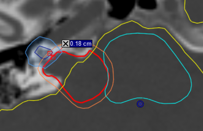
what would be the best dose/fractionation regimen, knowing that the distance cochlea-tumor is <2mm ?
- 25 Gy in 5 fr at 80%
- 18 Gy in 3 fr at 80%
- 54 Gy in 28 fr
- other
No facial numbness, no facial palsy, no imbalance, and no other cranial nerve deficits were reported. Performance status normal.
Audiogram (08/06/2025):
Right ear: Sensorineural hearing loss, PTA ≈ 32 dB HL, AI ≈ 59%.
Speech discrimination 100% at 60 dB, suggesting serviceable hearing (AAO-HNS Class B).
Left ear: Essentially normal thresholds, PTA ≈ 15 dB HL.
She retains usable hearing in the affected ear, an important prognostic factor when selecting RT vs surgery.
MRI Brain with contrast (T1Gd, T2, CISS sequences) dated 11/11/2025.
Right vestibular schwannoma, Koos grade III
o Enhancing CPA mass extending into the IAC.
o Volume: ~4 cc (well within SRS/FSRT suitability range).
o Displaces but does not severely compress the brainstem.
o No cystic degeneration.
o Clear visualization of 7th and 8th nerve entry zone (last two images).
o No hydrocephalus.
we discussed with her observation (since she does not have previous serial MRIs). she insisted on doing RT.
what would be the best dose/fractionation regimen, knowing that the distance cochlea-tumor is <2mm ?
- 25 Gy in 5 fr at 80%
- 18 Gy in 3 fr at 80%
- 54 Gy in 28 fr
- other


