- Joined
- Apr 29, 2011
- Messages
- 2,171
- Reaction score
- 863
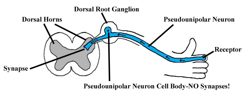
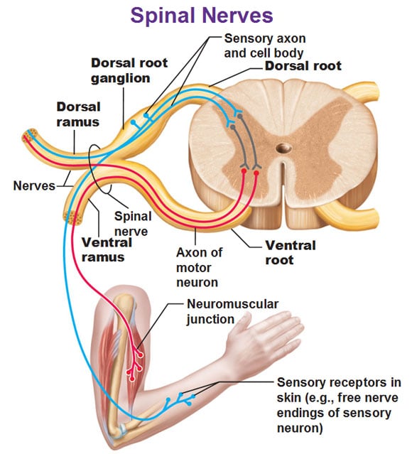
Does the DRG serve the function of the yellow line in the picture below?

Is there anything else notable about the DRG that I should know?
Is there anything else notable about the DRG that I should know?