EKG help

This forum made possible through the generous support of SDN members, donors, and sponsors. Thank you.

chocomorsel

Senior Member
Removed
15+ Year Member
Joined
May 24, 2006
Messages
6,750
Reaction score
6,259
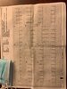
What do y'all think of this EKG? Healthy, 40 year old male with Covid + in May, recovered, since then has had intermittent fluttering and has Hx or chronic Migraine.

Paroxysmal Atrial Tachycardia versus 2:1 Atrial Fluttler?

Members don't see this ad.
 

Attachments

  • IMG-2034.jpeg
    IMG-2034.jpeg
    224.1 KB · Views: 237
Last edited:
Not sure, could just be sinus tachycardia with a biphasic t wave in lead II making it look funny.
 
  • Like
Reactions: 2 users
Members don't see this ad :)
Have heard of several patients developing heart block in acute Covid infections.
 
  • Like
Reactions: 1 user
flutter in II, III, and aVF
 
  • Like
Reactions: 1 users
Looks like sinus tachycardia to me, but I'd be interested if he's a steady 120 or has variability. Also if you're really unsure you can just give some adenosine and try to slow down the AV node to unveil flutter waves.

V1 P wave morphology is important

"
Typical Atrial Flutter (Common, or Type I Atrial Flutter)
Involves the IVC & tricuspid isthmus in the reentry circuit. Can be further classified based on the direction of the reentry circuit (anticlockwise or clockwise):

Anticlockwise Reentry: Commonest form of atrial flutter (90% of cases). Retrograde atrial conduction produces:

  • Inverted flutter waves in leads II,III, aVF
  • Positive flutter waves in V1 – may resemble upright P waves
Clockwise Reentry. This uncommon variant produces the opposite pattern:

  • Positive flutter waves in leads II, III, aVF
  • Broad, inverted flutter waves in V1"
 
Last edited:
  • Like
Reactions: 1 user
Looks like sinus tach with biphasic T waves in the inferior leads


1592519983885.png
 
  • Like
Reactions: 2 users
Members don't see this ad :)
It is paroxysmal and no one tried any vagal maneuvers to slow it down. It popped back to NSR on its own.

You have a NSR ekg for comparison?


Man, if that's 2:1 flutter then I'm just gonna resign myself to missing it 100% of the time unless the ventricular rate is 150 bpm
 
  • Like
Reactions: 2 users
You have a NSR ekg for comparison?


Man, if that's 2:1 flutter then I'm just gonna resign myself to missing it 100% of the time unless the ventricular rate is 150 bpm
My cards buddy thinks its Flutter with poor R wave progression. Says I need a "high quality ECG" to tell PAT from Aflutter.
 
Im leaning towards sinus tach and the problem is not in the T waves. P waves seem P wave pulmonale in some leads. I have seen this a bunch of times in CoViD patients.
Eg. Hypoxic pulm vasoconstriction, increased PA pressures etc etc
OR
Needs of diuretics
#my2cents
 
  • Like
Reactions: 1 user
Looks like sinus tach to me. Could you ask your cards buddy what about this makes it PAT vs AFlutter?
 
I’ve changed my mind. Definitely look like flutter waves or some sort of SVT.
 
  • Like
Reactions: 1 user
Top