- Joined
- Dec 8, 2005
- Messages
- 574
- Reaction score
- 2
Hello all!
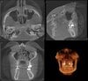
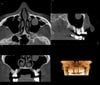
Coming over from oral radiology in peace. Just wanted to get some clarification from the people mostly working in this region on a fairly innocuous topic. In dentistry on CBCTs we see mucous retention pseudocysts all the time, we pretty much ignore them unless they are obstructing the OMC. However there seems to be confusion regarding mucous retention pseudocysts vs polyps when the entity arises from the superior aspect of the sinus and hangs down. Some radiologists call it a polyp some call it a pseudocyst. We dont have a way with CBCT to accurately measure the Hounsfield units to help determine internal contents. I've attached a couple examples of what we often call polyps. Wanted to get your opinion on this topic and also your thoughts on you treat these any more aggressively or differently than a pseudocyst? Generally seems to be little more than semantics but just wanted to get some clarification. Thanks!
Coming over from oral radiology in peace. Just wanted to get some clarification from the people mostly working in this region on a fairly innocuous topic. In dentistry on CBCTs we see mucous retention pseudocysts all the time, we pretty much ignore them unless they are obstructing the OMC. However there seems to be confusion regarding mucous retention pseudocysts vs polyps when the entity arises from the superior aspect of the sinus and hangs down. Some radiologists call it a polyp some call it a pseudocyst. We dont have a way with CBCT to accurately measure the Hounsfield units to help determine internal contents. I've attached a couple examples of what we often call polyps. Wanted to get your opinion on this topic and also your thoughts on you treat these any more aggressively or differently than a pseudocyst? Generally seems to be little more than semantics but just wanted to get some clarification. Thanks!