
Step I NBME 17 question

Started by mj413121

This forum made possible through the generous support of SDN members, donors, and sponsors. Thank you.
Get help with your application

Use all the free resources available to you from SDN: articles, guides, expert advising, forums discussions, and school research.

mj413121

Full Member
7+ Year Member
Advertisement - Members don't see this ad
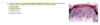
Could anyone explain the picture in this question for me? Thank you
 

Attachments

  • Screen Shot 2018-05-27 at 2.44.42 PM.png
    Screen Shot 2018-05-27 at 2.44.42 PM.png
    203.2 KB · Views: 207
What you can see is

1.Hyperkeratosis (thickened stratum corneum)
2.Parakeratosis (you can see the nuclei very clearly in the stratum corneum)
3.Dysplasia (notice keratinocytes hyperchromatic and large nuclei go up almost all the way to the top. This isnt so in normal skin)

all this fits actinic keratosis
 
What you can see is

1.Hyperkeratosis (thickened stratum corneum)
2.Parakeratosis (you can see the nuclei very clearly in the stratum corneum)
3.Dysplasia (notice keratinocytes hyperchromatic and large nuclei go up almost all the way to the top. This isnt so in normal skin)

all this fits actinic keratosis
Thank you!