Need help to understand these radiology images for class

This forum made possible through the generous support of SDN members, donors, and sponsors. Thank you.

rjcp

New Member
Joined
Nov 1, 2019
Messages
2
Reaction score
0
Could someone please help me to understand these images and alterations for my imagiology class presentation?
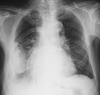
We just have these two images and these information: Male, 89y, Atrial fibrilation and previous history of myocardial infarction.
It's the first time that i have "imagiologia" class and I am a little bit lost, thank you very much.
 

Attachments

  • Sem Título.png
    Sem Título.png
    638.1 KB · Views: 144
  • Sem Título1.png
    Sem Título1.png
    422.9 KB · Views: 145
Last edited:
What language is imagiiology or imagiology?

The images are a frontal chest radiograph and a parasagittal image of a chest CT through the right hemithorax in lung windows. The main abnormality is in the major fissure.
 
Cognovi it was poorly written, it should be "imagiologia" and that is a portuguese word for the name of the class. It is the first basic class about imaging that I have in medical school, where we start learning how to look, interpret and propose differential diagnosis based on medical images. It's like a basic pre-radiology class that we have before having radiology in the clinical years.

The chest radiograph gives me the idea that the alterations visible could be compatible with pneumonia but the ct gives the idea of some kind of mass like some blood leakage. coul it be some pleural effusion due to heart failure?

Maybe I am asking dumb questions but my knowleadge about this area is very low because we are start learning it now.
Thank you.
 
I think you have it. The chest radiograph shows an opacity that could be compatible with pneumonia but the CT shows the opacity localizes to the pleural space, in the major fissure, rather than lung parenchyma. This is probably a loculated pleural effusion, sometimes called pleural pseudotumor.

Aren‘t pseudotumors associated with HFrEF, which would be expected with someone post-MI plus afib?
 
Top