USMLE OFFICIAL NBME 20 - Questions & Answers - Explanations & Discussions ?

This forum made possible through the generous support of SDN members, donors, and sponsors. Thank you.

MegaKleos

Full Member
Joined
Sep 19, 2018
Messages
12
Reaction score
1
So let's continue the trend of this forum and discuss the doubts of the test. I intend on taking this as soon as it is out. Please post below if you wish to discuss and let's get together and solve our doubts.

DISCLAIMER: As has been said in the forum previously, posting COPYRIGHTED content of the NBME is a CRIME. Please do not do so. The intention of this thread is NOT to promote that but rather to discuss in a way that does not break the law. According to copyright act of united states, under DMCA, there is a special provision for "FAIR USE". Which means that people are able to discuss, critic and have an academic conversation as long as it is within reason and of course verbatim questions are unacceptable. To the admins of the forum, please dont delete this, i dont intend on posting anything against the law and hence please dont delete this thread.

Members don't see this ad.
 
Finished the test and my review today. A little bummed to see questions repeated from the retired NBME exams. There were a couple I missed that I would really appreciate an explanation for as I haven't been able to find much in my resources or a cursory lit review.

************SPOILERS**********

Why would decreasing respiratory rate cause an increase in intracranial pressure?

How do thiazides create a thin milky discharge from both breasts?

How do you narrow down between psychogenic polydipsea and nephrogenic diabetes insipidus for a patient that may or may not be on lithium and no vasopressin test?

************END SPOILERS**********

If anyone is trying to work out the curve and compare this to other tests I scored 7 points worse on this than UWSA1, but it was my highest NBME so far (among 13,15,16,17,18). An 83% translated to a 244.
 
  • Like
Reactions: 1 user
Thanks so much for the reply! I was trying to be pretty vague about the questions but the 2nd one was in a woman who had a HTN and T2DM and was only listed to be on HCTZ. She came in with a 3 month history of a thin milky discharge and the question asked for the responsible mechanism. The correct answer was drug effect.

Bonus add on question: Why is the chance for HLA match between siblings in organ donation 25%?
 
A 25 yr old woman has flu like illness that is followed by decreased exercise tolerance and shortness of breath , particularly lying down . Findings on examination of tissue obtained on myocardial biopsy suggestive of viral infection. Which of the following is the most likely cause of her illness?

Adenovirus
Herpesvirus
Myxovirus
Orthomyxoviridae
Picornavirus

Are is picorna ? But why not adenovirus
 
Members don't see this ad :)
The most important cause (at least on Step 1) of viral myocarditis is coxsackie, which is a picornavirus
 
For the toxoplasmosis in HIV patient question, why is it non-hodgkins and not cerebral toxoplasmosis? I get that they started her on treatment but couldn't it also be advanced toxoplasmosis?
 
What do the sxs of 1 month joint pain, fatigue, lower and upper extremity edema and frequent headaches point to? Why would the spleen be enlarged? I'm confused what disease pattern this is. This is the question where they also give you a CT
 
  • Like
Reactions: 1 user
For the toxoplasmosis in HIV patient question, why is it non-hodgkins and not cerebral toxoplasmosis? I get that they started her on treatment but couldn't it also be advanced toxoplasmosis?
From what I remember, the CD count was below 50 or something and it was a single enhancing lesion which points more towards lymphoma. Toxo is usually multi ring enhancing lesions. Correct me if im wrong
 
  • Like
Reactions: 1 users
6 months after a klebsiella pneumonia infection (with cavitary lesion and fluid level), what happens to lung parenchyma? Why is it not normal (b/c doesn't the lung heal)? Why is the answer pulmonary fibrosis?
 
During a procedure, the thoracic duct is damaged in the post. mediastinum. This is most likely to impair normal lymphatic return of which structure?

Heart, left breast, left upper extremity, right kidney, right lung.

Answer is right kidney but wouldn't left upper extremity and breast also be correct answers?
 
Last edited:
6 months after a klebsiella pneumonia infection (with cavitary lesion and fluid level), what happens to lung parenchyma? Why is it not normal (b/c doesn't the lung heal)? Why is the answer pulmonary fibrosis?

I put liquefactive necrosis but to my understanding abscesses destroy the tissue resulting in liquefactive necrosis, so I guess after the infection resolved the area just becomes fibrosed?
 
6 wk old girl w/ few day history of vomiting small amounts of milk 2-3x per day. Appears well, 50th percentile for length and weight. Physical exam = no abnormalities. Which is causing patient's findings?

Esophageal spasm
Gut Malrotation
Immature lower esophageal sphincter
Neuromuscular abnormality of esophagus
Pyloric stenosis
Tracheoesophageal fistula

Could anyone help explain how to narrow down to this answer? Physical exam being normal helps rule out a few but not sure how to distinguish between a few like esophageal spasm, neuromuscular abnormalities and immature esophageal sphincter. Thank you.
 
70 yr old man with cerebral infarction 8 yrs ago, The question is neurologic deficits most likely present? But my question is about the photograph, is that a cerebral peduncle infarction at the right side?
 
During a procedure, the thoracic duct is damaged in the post. mediastinum. This is most likely to impair normal lymphatic return of which structure?

Heart, left breast, left upper extremity, right kidney, right lung.

Answer is right kidney but wouldn't left upper extremity and breast also be correct answers?
I was looking for that too, and I think it is because is posterior mediastinum and the other options: heart is in middle mediastinum, upper extremity and breast drains in brachiocephalic veins which are in superior mediastinum: https://www.researchgate.net/figure...diastinum-is-the-space-occupied_fig1_31326044 . I accept other suggestions :S
 
  • Like
Reactions: 1 user
Members don't see this ad :)
During a procedure, the thoracic duct is damaged in the post. mediastinum. This is most likely to impair normal lymphatic return of which structure?

Heart, left breast, left upper extremity, right kidney, right lung.

Answer is right kidney but wouldn't left upper extremity and breast also be correct answers?

Thanks for your help on my question! The left upper extremity and breast are drained by the axillary lymph node. The kidney is drained by the thoracic duct. The heart has it's own lymph system going on surrounding the heart
 
  • Like
Reactions: 1 user
70 yr old man with cerebral infarction 8 yrs ago, The question is neurologic deficits most likely present? But my question is about the photograph, is that a cerebral peduncle infarction at the right side? Yes, it's a infarct at the basis pedunculi in the midbrain (above the pyramidal decussation of the lateral corticospinal tract).

During a procedure, the thoracic duct is damaged in the post. mediastinum. This is most likely to impair normal lymphatic return of which structure? Heart, left breast, left upper extremity, right kidney, right lung. Answer is right kidney but wouldn't left upper extremity and breast also be correct answers?
I was looking at this too. I think it's because the apical lymph nodes even on the left side may not all drain into the thoracic duct. From radiopedia: " Apical (terminal) lymph nodes drains into: subclavian trunk (may drain into jugulosubclavian venous trunk, subclavian vein, jugular lymphatic trunk, right lymphatic duct (left into thoracic duct), inferior deep cervical nodes)". Also the parts of the left breast can drain parasternally to the right breast. No option for the kidney to go anywhere but to the para-aortic --> cisterna chyli --> thoracic duct.

6 wk old girl w/ few day history of vomiting small amounts of milk 2-3x per day. Appears well, 50th percentile for length and weight. Physical exam = no abnormalities. Which is causing patient's findings?
No abnormalities, only some vomiting, looks well w/ no failure to thrive. Combine all that w/ a high rate of GERD/regurg in neonates due to immature LES gives you the answer.

6 months after a klebsiella pneumonia infection (with cavitary lesion and fluid level), what happens to lung parenchyma? Why is it not normal (b/c doesn't the lung heal)? Why is the answer pulmonary fibrosis?
Klebsiella and TB both form cavitary lesions w/ fibrosis. This is what makes ppl w/ prior Klebsiella and TB infections prone to aspergillomas.

How do thiazides create a thin milky discharge from both breasts?
I looked everywhere to see if there's any association between HCTZ and galactorrhoea and found nothing. Closest thing I could find was that some HTN medications such as methyldopa and verapamil are associated w/ galactorrhoea and she was mentioned to have HTN (though none of these drugs were mentioned in the stem....).

What do the sxs of 1 month joint pain, fatigue, lower and upper extremity edema and frequent headaches point to? Why would the spleen be enlarged? I'm confused what disease pattern this is. This is the question where they also give you a CT

I believe the disease pattern is SLE (joint pain, headache, upper and lower edema, and slight fever fits) or at least something autoimmune. States of chronic inflammation, like SLE, can lead to splenomegaly. My confusion here is the CT clearly shows an enlarged spleen, but the question is asking which of the following additional findings was most likely seen in this pt. I don't really understand why splenomegaly is the best choice when since the question told you through the CT that the pt has splenomegaly. Is it common that NBME/USLME asks q like this, or am I missing something w/ it?
 
Last edited:
  • Like
Reactions: 2 users
1) Can someone explain why a left axis deviation would be associated w/ coarctation of the aorta? Is it because the LV overload will lead to hypertrophy of the LV and thus left axis deviation?
- Yep

2) In someone diagnosed w/ Fanconi syndrome, is there a reason why hypophosphatemia would be more likely to show up in a serum study than hypokalemia? I would assume that due to the massive luminal concentration of Na+ from lack of PCT reabsorption (and associated water) that Na+ will be reabsorbed in the more distal nephron w/ further loss of K+.

3) If you guys remember the vignette featuring a pregnant woman w/ fever and kidney pain and a histology picture of obstructive uropathy, what is the picture showing? Normal tubules?
- Intact tubules w/ inflammatory infiltrate

4) This is just a math problem, but if you have a pt who has an autosomal dominant disease and a family Hx for that disease on her father's side, what's the likelihood of her potential kids getting a cancer that affects those w/ that autosomal dominant disease ~100% of the time.

I'm very confused here since you don't get any info on the potential father. The answer was 50%, but wouldn't that assume the father was homozygous for the recessive trait? If the future father also had the disease (what if they meet at a support group for ppl w/ the condition), then the likelihood of her kids getting the cancer is much higher. I must be missing something here?
- Just assume the most likely scenario of a homozygous recessive father.

Edit: Some kind ppl answered some of these q's for me on another site, so I'll add their answers here in case others were also wondering. I'm still somewhat unsure about the Fanconi syndrome one since RTA type II is also associated w/ hypokalemia and it at least makes sense to be that more K+ would be dumped later to reabsorb Na+ and H2O, but clearly that's not what's happening.
 
Last edited:
  • Like
  • Wow
Reactions: 1 users
Someone can explain this question: the 56 years old man that is brought to the emergency department after 30 minutes of chest pain...What is the dx? Pulmonary hypertension?, and I can't understand why the interstitial oncotic pressure is decrease?in the correct answer.
 
70 yr old man with cerebral infarction 8 yrs ago, The question is neurologic deficits most likely present? But my question is about the photograph, is that a cerebral peduncle infarction at the right side?
70 yr old man with cerebral infarction 8 yrs ago, The question is neurologic deficits most likely present? But my question is about the photograph, is that a cerebral peduncle infarction at the right side? Yes, it's a infarct at the basis pedunculi in the midbrain (above the pyramidal decussation of the lateral corticospinal tract).
70 yr old man with cerebral infarction 8 yrs ago, The question is neurologic deficits most likely present? But my question is about the photograph, is that a cerebral peduncle infarction at the right side?


this is a little bit confusing to me , if the infarction in the right cerebral peduncle and above decussation , shouldnt that be a CONTRALATERAL UMNL (hemiparesis ) on the LEFT? in the answers the patient Spastic hemiparesis is on the RIght side ? i think that would only make sense if the area of infarction on the LEFT . 8 years ago the area of infarction would be smaller compared to the healthy one !? would appreciate if anyone has explanation . thank you
 

Attachments

  • Screen Shot 2019-04-10 at 2.55.16 PM.png
    Screen Shot 2019-04-10 at 2.55.16 PM.png
    110.3 KB · Views: 112
Last edited:
My bad! I didn't go back to look at the picture so mixed up L and R. On the left side, you see where he had the infarction 7 years ago and the tissue is now dead and gone. Right side is normal. As this slice is above the decussation, an issue on the left side will produce right sided hemiparesis, like you said. Here's a pic of a normal stained around the same level from the internet.

257674
 
Someone can explain this question: the 56 years old man that is brought to the emergency department after 30 minutes of chest pain...What is the dx? Pulmonary hypertension?, and I can't understand why the interstitial oncotic pressure is decrease?in the correct answer.
If I remember correctly, he had cardiogenic shock. LV stopped working, pressure backed up into pulm circuit. Pulm circuit roughly is made of 3 "parts" - the capillaries, interstitial space, and the alveoli. In cardiogenic shock, the extra blood increases capillary hydrostatic pressure, driving fluid into the interstitial space. Compared to the alveoli, the interstitial space now has more fluid (thus more interstitial hydrostatic pressure and less oncotic pressure due to ratio of fluid to protein), and as a result of this unbalancing of forces, fluid moves into the alveoli --> pulmonary oedema.
 
  • Like
Reactions: 1 users
Someone can explain this question: the 56 years old man that is brought to the emergency department after 30 minutes of chest pain...What is the dx? Pulmonary hypertension?, and I can't understand why the interstitial oncotic pressure is decrease?in the correct answer.


yes , bigger picture of this question , is that he basically wants to know that you know, the difference between cardiogenic pulmonary edema --> its a protein poor fluid (transudate) and the non cardiogenic pulmonary edema as (ARDS) or inhalational injury where you have increased interstitial oncotic pressure due to leakage of proteins from capillaries and normal PCWP. think the increased hydrostatic pressure pushing against capillaries with normal (good) membrane permeability drove only fluid not proteins out and just diluted the interstitial oncotic pressure making it low as in this patient
 
  • Like
Reactions: 1 user
My bad! I didn't go back to look at the picture so mixed up L and R. On the left side, you see where he had the infarction 7 years ago and the tissue is now dead and gone. Right side is normal. As this slice is above the decussation, an issue on the left side will produce right sided hemiparesis, like you said. Here's a pic of a normal stained around the same level from the internet.

View attachment 257674
Thank you that's why i was confused too
 
Hello everyone, i just took and reviewed the answers to NBME 20. I have a few questions that I am still not sure about. Any help will be appreciated, thank you in advance.

section 3, question 25: the question stem describes a pt with chest pain for the past hour. The pt has a history of HTN and type 2 diabetes. The pts vitals are pulse:120, RR:24, BP:98/60. The pt has JVD and crackles are heard in the lungs. The question asks what ECG changes will you see in this pt?
  • The correct answer is ST segment elevation.
  • Another answer choice is QT interval shortening. Can someone explain to me why this pts QT interval is not shortened. My understanding of the QT interval is that it is inversely related to HR. this pt is tachycardic thus will have a short QT interval.

Section 3 question 6: the question stem describes a pt which a hemoglobin concentration of 18.5 and then shows a peripheral blood smear.
  • What is in that blood smear which points to COPD?

Section 4 question 15: a 17YO girl with right sides weakness, slurred speech, elevated pyruvate and lactate and has a NADH dehydrogenase deficiency.
  • The question shows a pedigree and asks what is the mode of inheritance? The answer is mitochondrial.
  • My question is, why are mothers not passing on the disease to all their children. One mother passed on the disease to 2 of her 3 children while another mother passed it on to 1 of her 2 children.

Section 4 question 20: a 36YO pt with decreased urinary output for 3 days was diagnosed with bilateral hydronephrosis. The pts BP is 150/96, pulse is 112, Na: 138, K: 5.9, HCO3: 22, creatinine: 5
  • Bilateral nephrostomy tubes were placed
  • The question asks which will happen to the pts urine output and urine K?
  • I understand that his urine output will increase. What I don’t understand is why is urine K will increase also. If the nephron is secreting K then it must be reabsorbing Na which will worsen the HTN
 
  • Like
Reactions: 1 user
Alright got a few:

Biostats question: Woman with brother and mother with colon cancer wants to be screened using fecal blood testing. Physician is concerned about which of the following regarding this test:

They say that it's low sensitivity, but that doesn't make sense. Sensitivity doesn't change based on pre-test probability. If it's low sensitivity here, it should be low sensitivity everywhere, and we should never use it as a screening test. I chose "Uncertain negative predictive value," because the increased prevalence of cancer in people with this woman's risk factors would decrease negative predictive value. Why would this not be the answer? Boy

Proximal tubule reabsorption: Boy presenting with defect affecting his ability to reabsorb substances in the proximal tubule. Serum studies are most likely to show which of the following findings at this time?

I said hypokalemia, the answer is hypophosphatemia. Are they saying he has X-linked hypophosphatemia? I assumed this was Fanconi syndrome.

To answer your question about Fanconi syndrome. According to first aid, the effects of fanconi syndrome are hypophophatemia, osteopenia, and metabolic acidoosis. Since phosphate is only absorbed in the PCT, it makes since that its levels would decrease. I am assuming that since the other ions get reabsorbed at other parts of the nephron, the nephron compensates and increases re-absorption in those other parts.
 
  • Like
Reactions: 1 user
Hello everyone, i just took and reviewed the answers to NBME 20. I have a few questions that I am still not sure about. Any help will be appreciated, thank you in advance.

section 3, question 25: the question stem describes a pt with chest pain for the past hour. The pt has a history of HTN and type 2 diabetes. The pts vitals are pulse:120, RR:24, BP:98/60. The pt has JVD and crackles are heard in the lungs. The question asks what ECG changes will you see in this pt?
  • The correct answer is ST segment elevation.
  • Another answer choice is QT interval shortening. Can someone explain to me why this pts QT interval is not shortened. My understanding of the QT interval is that it is inversely related to HR. this pt is tachycardic thus will have a short QT interval.
Section 3 question 6: the question stem describes a pt which a hemoglobin concentration of 18.5 and then shows a peripheral blood smear.
  • What is in that blood smear which points to COPD?
Section 4 question 15: a 17YO girl with right sides weakness, slurred speech, elevated pyruvate and lactate and has a NADH dehydrogenase deficiency.
  • The question shows a pedigree and asks what is the mode of inheritance? The answer is mitochondrial.
  • My question is, why are mothers not passing on the disease to all their children. One mother passed on the disease to 2 of her 3 children while another mother passed it on to 1 of her 2 children.
Section 4 question 20: a 36YO pt with decreased urinary output for 3 days was diagnosed with bilateral hydronephrosis. The pts BP is 150/96, pulse is 112, Na: 138, K: 5.9, HCO3: 22, creatinine: 5
  • Bilateral nephrostomy tubes were placed
  • The question asks which will happen to the pts urine output and urine K?
  • I understand that his urine output will increase. What I don’t understand is why is urine K will increase also. If the nephron is secreting K then it must be reabsorbing Na which will worsen the HTN
Section 3 question 25: ST segment elevation is the best answer since that is what you are most likely to see. If there is a shortened QT, it would most likely be insignificant and unlikely to cause problems

Section 3 question 6: It's a normal blood smear. In COPD you have increased EPO and thus Hgb. I don't think you should see any abnormalities on blood smear.

Section 4 question 15: Heteroplasmy explains why there is variable expression of mitochondrial diseases within a family or population; thus some of the children may be asymptomatic/barely effected

Section 4 question 20: Hyperkalemia will stimulate aldosterone secretion even if renin is suppressed due to his hypertension . Although Na+ will be reabsorbed, this will be transient (should resolve once the potassium levels normalized) and since his urine output will most likely return to normal, his blood pressure should also normalize
 
Hi everyone! I have (quite a few) questions from NBME 20 that I can't seem to figure out. If anyone is able to help, I'd really appreciate it!

  1. 32 yo woman who fell from a ladder. Why is arterial laceration is the right answer?

  2. 55 yo man diagnosed with hypertension. Given an ACE inhibitor, plasma renin activity increases more. Why is the answer renal artery stenosis?

  3. 47 yo woman with increasing cough, shortness of breath, exposure to parakeets. CXR shows reticulogranular changes and biopsy has lymphocytes and granulomas. Does this person have Chlamydia psittaci pneumonia or something else?

  4. 50 yo woman who overdosed. Question is about pharmacokinetics. Can someone explain the relationship between protein binding in plasma and volume of distribution at steady state?

  5. 4 yo boy with fever, abdominal pain, bloody stools. How do you know that this is Shigella and not E. coli? I’ve only ever seen methylene blue stains mentioned in the context of E. coli.

  6. 19 yo college student brought into ED due to chest pain and difficulty breathing. Why is subcutaneous crepitus the right answer?

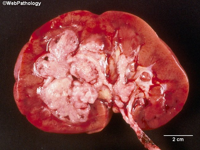
  7. 66 yo man brought to ED after MVA. Picture of kidney and histo slide. What are the picture and histo slide showing?

  8. Female newborn with respiratory distress. X-ray is given. What condition is this, and what are you supposed to see on the x-ray?

  9. 36 yo woman with 1 month history of joint pain. CT is given. What are you supposed to see on the CT, and why is the answer splenomegaly?

  10. 12 yo girl with with CBFA1 gene mutation. What condition is this?

  11. Newborn male with decreased T4. Why is the answer thyroid hormone-binding globulin deficiency?

  12. 70 yo woman with right femur fracture. Is this indirectly asking about peak bone density?

  13. 6-week-old female newborn with bilious vomit. Why is the answer annular pancreas?

  14. 23 yo woman with DIC. History of splenectomy. How do you know that Strep pneumo is the right answer? Is it just because she has a history of splenectomy?

  15. 56 yo woman with shortness of breath. Iatrogenic chylothorax. Why is the answer central line via the left internal jugular vein?

  16. This question gives you a sequence from beta globin gene and asks about a G->A mutation. What are they asking and how are you supposed to solve this problem?

  17. 16 yo student with uncontrollable sleepiness. Why is the answer direct transition from wakefulness to REM? Is it because they have narcolepsy?

  18. 53 yo woman with 1 year history of abdominal pain. Picture of stool sample is given. What in the history suggests that this is Schistosoma mansoni?
 
Last edited:
Hi everyone! I have (quite a few) questions from NBME 20 that I can't seem to figure out. If anyone is able to help, I'd really appreciate it!

  1. 32 yo woman who fell from a ladder. Why is arterial laceration is the right answer?

  2. 55 yo man diagnosed with hypertension. Given an ACE inhibitor, plasma renin activity increases more. Why is the answer renal artery stenosis?

  3. 47 yo woman with increasing cough, shortness of breath, exposure to parakeets. CXR shows reticulogranular changes and biopsy has lymphocytes and granulomas. Does this person have Chlamydia psittaci pneumonia or something else?

  4. 50 yo woman who overdosed. Question is about pharmacokinetics. Can someone explain the relationship between protein binding in plasma and volume of distribution at steady state?

  5. 4 yo boy with fever, abdominal pain, bloody stools. How do you know that this is Shigella and not E. coli? I’ve only ever seen methylene blue stains mentioned in the context of E. coli.

  6. 19 yo college student brought into ED due to chest pain and difficulty breathing. Why is subcutaneous crepitus the right answer?

  7. 66 yo man brought to ED after MVA. Picture of kidney and histo slide. What are the picture and histo slide showing?

  8. Female newborn with respiratory distress. X-ray is given. What condition is this, and what are you supposed to see on the x-ray?

  9. 36 yo woman with 1 month history of joint pain. CT is given. What are you supposed to see on the CT, and why is the answer splenomegaly?

  10. 12 yo girl with with CBFA1 gene mutation. What condition is this?

  11. Newborn male with decreased T4. Why is the answer thyroid hormone-binding globulin deficiency?

  12. 70 yo woman with right femur fracture. Is this indirectly asking about peak bone density?

  13. 6-week-old female newborn with bilious vomit. Why is the answer annular pancreas?

  14. 23 yo woman with DIC. History of splenectomy. How do you know that Strep pneumo is the right answer? Is it just because she has a history of splenectomy?

  15. 56 yo woman with shortness of breath. Iatrogenic chylothorax. Why is the answer central line via the left internal jugular vein?

  16. This question gives you a sequence from beta globin gene and asks about a G->A mutation. What are they asking and how are you supposed to solve this problem?

  17. 16 yo student with uncontrollable sleepiness. Why is the answer direct transition from wakefulness to REM? Is it because they have narcolepsy?

  18. 53 yo woman with 1 year history of abdominal pain. Picture of stool sample is given. What in the history suggests that this is Schistosoma mansoni?
#15. It'ss in First aid in the immunosection where the drainage picture is that thoracic duct drains at the junction of left subclavian and internal jugular. Rupture of thoracic duct --> chylothorax pg 98 FA
 
  • Like
Reactions: 1 user
Did you end up getting a response to this? Was the white stuff in the picture cancer? Or some form of inflammation? I couldn't figure it out.
7. 66 yo man brought to ED after MVA. Picture of kidney and histo slide. What are the picture and histo slide showing?
did you end up getting an explanation for
 

Attachments

  • kidney.png
    kidney.png
    426.6 KB · Views: 149
Did you end up getting a response to this? Was the white stuff in the picture cancer? Or some form of inflammation? I couldn't figure it out.
7. 66 yo man brought to ED after MVA. Picture of kidney and histo slide. What are the picture and histo slide showing?
did you end up getting an explanation for
Without seeing the question or answers I would guess the point is that he had a catastrophic aneurysmal bleed causing the MVA and they are getting at the link between PKD and cerebral aneurysms.
 
  • Like
Reactions: 1 users
Did you end up getting a response to this? Was the white stuff in the picture cancer? Or some form of inflammation? I couldn't figure it out.
7. 66 yo man brought to ED after MVA. Picture of kidney and histo slide. What are the picture and histo slide showing?
did you end up getting an explanation for

I haven't yet. :(

Without seeing the question or answers I would guess the point is that he had a catastrophic aneurysmal bleed causing the MVA and they are getting at the link between PKD and cerebral aneurysms.
The question asks what the most likely predisposing factor in developing the ureteral lesion is. The answer is cigarette smoking. The attached histo slide also goes with the kidney picture if that helps.

Is there a link between smoking and PKD? I can't seem to remember. If so, I think you're right!
 

Attachments

  • kidney histo.PNG
    kidney histo.PNG
    366.8 KB · Views: 105
Sorry still a bit confused. That picture does not resemble PKD to me based on other gross kidney pictures of PKD i've looked up. Are you sure?
Im at a loss at what the white material is supposed to be. Is that necrotic?
 
I haven't yet. :(


The question asks what the most likely predisposing factor in developing the ureteral lesion is. The answer is cigarette smoking. The attached histo slide also goes with the kidney picture if that helps.

Is there a link between smoking and PKD? I can't seem to remember. If so, I think you're right!

Its transitional cell carcinoma, which smoking is a risk factor for; it can involve the renal pelvis/calyces. The histo image shows the papillary nature of the tumor (however it can also be flat or nodular according to pathoma).

Related image
 
  • Like
Reactions: 1 user
HELLO
I HAVE SOME QUESTIONS
1- the question on tryptohan synthase ? why is it missense mutation . why not deletion ?
2- the boy with ankyrin gene mutation ( hereditary spherocytoces) why is he heterozygous but not homo ?
3-the question on vitamin D deficeny ? why is the answer cholecalciefrol but not 7 hydroxycholesterol ?
 
55 yo man diagnosed with hypertension. Given an ACE inhibitor, plasma renin activity increases more. Why is the answer renal artery stenosis?

Note that they auscultated a renal bruit in the stem. The ace would lower BP causing less perfusion of the stenotic kidney causing increased plasma renin.
 
  • Like
Reactions: 1 user
Exam 2, item 19:

Monoclonality of endometrial carcinoma assessed with x-chromosome-linked isoenzymes.

What are they talking about here? I got the question right by elimination but don't understand.
 
Exam 2, item 19:

Monoclonality of endometrial carcinoma assessed with x-chromosome-linked isoenzymes.

What are they talking about here? I got the question right by elimination but don't understand.

I think they were talking about G6PD (which is on the X-chromosome). It's in Pathoma.
 
  • Like
Reactions: 1 user
Exam 2, item 19:

Monoclonality of endometrial carcinoma assessed with x-chromosome-linked isoenzymes.

What are they talking about here? I got the question right by elimination but don't understand.

It's about random X inactivation. Chapter 3, basic principles of neoplasia in pathoma discusses it
 
  • Like
Reactions: 1 user
This may be a silly thing that I am over-thinking / not thinking about accurately, but I am a little confused about the 13 y/o F who was immunosuppressed for her kidney xplant.

The stem volunteers that she has lymphadenopathy with hepatosplenomegaly and that she has a monomorphous population of B lymphs on histo. The question is what is likely to be found within the cells biopsied. The answer is EBV. My question is why isn't CMV a good answer here? I was under the impression from Sketchy Micro that CMV hangs out in mononuclear cells (B, T, macros) and activates in immunocompromised people, leading to hepatosplenomegaly and (presumably, though my memory may be faulty here) lymphadenopathy. I also associate splenomegaly more than hepatomegaly with EBV since reactive T cells go to hang out in the spleen. Any pointers here to help a brother out? Clearly I'm missing something.
My thinking was monomorphous B cells = lymphoma which is associated with EBV in the immunocompromised.
 
The following responses, or lack thereof, are a collection of thoughts from both myself and my roommate (another M2):

Hi everyone! I have (quite a few) questions from NBME 20 that I can't seem to figure out. If anyone is able to help, I'd really appreciate it!

  1. 32 yo woman who fell from a ladder. Why is arterial laceration is the right answer?
    Anytime you have a person who bumps their head, gets back up, and then has severe issues or dies like 6 hours later -- you have youreslf an epidural hematoma from laceration to the middle MENINGEAL artery (Goljan really emphasizes that you don't screw up and select middle cerebral). You know it has to be an arterial laceration since the dura is tightly adhered to the skull's inner surface. Goljan referred to his experience with it as needing pliers to remove the dura from the skull; graphic, but it drives the point home. Tenting seen on CT is because the epidural hematoma gets stuck between the suture lines. When it manages to break past one of the suture lines, it is my understanding that then is when you get severe sequelae, like death or whatever.
  2. 55 yo man diagnosed with hypertension. Given an ACE inhibitor, plasma renin activity increases more. Why is the answer renal artery stenosis?
    As a rule of thumb, if you give someone an ACE-I and they get a problem, they had renal artery stenosis (usually bilaterally, or so we were taught at our med school). Probably has to do with dec GFR thanks to dec AngII selective vasoconstriction of the efferent arteriole => dec sodium delivery to macula densa => inc renin release
  3. 47 yo woman with increasing cough, shortness of breath, exposure to parakeets. CXR shows reticulogranular changes and biopsy has lymphocytes and granulomas. Does this person have Chlamydia psittaci pneumonia or something else?
    My roommate (who is much more intelligent than me) claims to have discovered that this patient is experiencing hypersensitivity pneumonitis from the parakeets. I was thinking M. Avium when I selected parakeets -- I think my logic was flawed given the specifics of the patient's story.
  4. 50 yo woman who overdosed. Question is about pharmacokinetics. Can someone explain the relationship between protein binding in plasma and volume of distribution at steady state?
    If you want to clear a drug, it is probably best that it not be bound to proteins (so that it gets filtered) and it has a low volume of distribution (so it isn't in the deep, hard to reach tissues).
  5. 4 yo boy with fever, abdominal pain, bloody stools. How do you know that this is Shigella and not E. coli? I’ve only ever seen methylene blue stains mentioned in the context of E. coli.
    We also did not really like this particular question, but after reviewing Sketchy Micro, I think Shigella is the most appropriate, as it is actually regarded as highly inflammatory. Yes, E coli can be of the EHEC/STEC variety, but E coli could also be of the ETEC variety or whatever other strains it has. Ergo, E coli may be plausible, but it is not the 'most likely.' Bleh to these kinds of questions.
  6. 19 yo college student brought into ED due to chest pain and difficulty breathing. Why is subcutaneous crepitus the right answer?
    From my extensive ortho bromancho experiences, I have always regarded crepitus as the rubbing of bone on bone. We think this is a purely definitional question. Yes, crepitus could also be trapped air. Context I guess.
  7. 66 yo man brought to ED after MVA. Picture of kidney and histo slide. What are the picture and histo slide showing?
    Is this the one with the poor kidney that was cut in half against its will and has a dilated distal ureter? If so, probably showing us transitional carcinoma with mild invasion into that distal ureter. Pathoma does a pretty awesome job of talking about GU cancers (and most cancers) ((and most medicine)) IMO.
  8. Female newborn with respiratory distress. X-ray is given. What condition is this, and what are you supposed to see on the x-ray?
    Is this the one with the NG tube in what would appear to be the space supposed to be her left lung? This is a hypoplasia of the pleuroperitoneal membrane or whatever it is (words are hard idk what it is and I am too lazy to look it up right now). The guts herniate into the thorax, usually on the left side, and result in hypoplasia of the lungs (because they're horribly compressed).
  9. 36 yo woman with 1 month history of joint pain. CT is given. What are you supposed to see on the CT, and why is the answer splenomegaly?
    We can't help you nor the patient here (we don't know what's going on here). We just know her spleen is gigantanormous on the CT. Sorry.
  10. 12 yo girl with with CBFA1 gene mutation. What condition is this?
    This is a conditional called craniocleidodysplasia (or something like that). The kid on Stranger Things with the lisp has the disorder. No collar bones, too many teeth, frontal bossing => craniocleidodysplasia. CBFA1 is a gene highly implicated in osteoblast function.
  11. Newborn male with decreased T4. Why is the answer thyroid hormone-binding globulin deficiency?
    Decreased total, normal free (unbound).
  12. 70 yo woman with right femur fracture. Is this indirectly asking about peak bone density?
    Sorta, yeah. That whole thing about weight-bearing exercises, eating right, yada yada before and during that down-slope phase of life for bone density. All about reducing that 1% per year thing as best as we can. Level of activity is kinda like weight-bearing exercise. (Consider: no activity, bed-ridden -- GG to your bones; highly active, runs every other day -- good amount of weight-bearing / stress to induce remodeling and maintain integrity of the bones)
  13. 6-week-old female newborn with bilious vomit. Why is the answer annular pancreas?
    Because it was the only answer that provided a small intestinal obstruction that could be plausibly distal to where bile enters the GI tract. (I don't recall any of the other answers as plausible -- maybe my memory is failing me? I apologize if that's the case.)
  14. 23 yo woman with DIC. History of splenectomy. How do you know that Strep pneumo is the right answer? Is it just because she has a history of splenectomy?
    Encapsulated organisms run rampantly on patients who have no spleen, whether physically or functionally. (Recall the wide-array of sequalae Sickle Cell patients experience thanks to their functional autosplenectemy.)
  15. 56 yo woman with shortness of breath. Iatrogenic chylothorax. Why is the answer central line via the left internal jugular vein?
    This was the only answer that comes close to nicking the thoracic duct, specifically at its inlet, the left subclavian.
  16. This question gives you a sequence from beta globin gene and asks about a G->A mutation. What are they asking and how are you supposed to solve this problem?
    If I am thinking of the right question with you here, this had to do with intron splicing. Remember GTAG. This mutation induced an AG closer where it was supposed to be, so some of that intron just became an exon.
  17. 16 yo student with uncontrollable sleepiness. Why is the answer direct transition from wakefulness to REM? Is it because they have narcolepsy?
    Yes, the patient has narcolepsy, and this is the mechanism.
  18. 53 yo woman with 1 year history of abdominal pain. Picture of stool sample is given. What in the history suggests that this is Schistosoma mansoni?
Sorry, but my computer and me aren't getting along right now, and this answer is gonna have to be oddly out of place. This response is for item (18). When you have a traveler who has intermittent abdominal symptoms and diarrhea who has traveled to the likes of northern Africa and such, Schistosomiasis needs to be on your radar. At least, that's how I've incorporated this nugget into my mental space.

Disclaimer: I may be a dingus on some of my responses. I apologize in advance if someone else has a better answer to offer you on some of these things. I hope you forgive my occasional satire (I gotta have fun somehow :/ )

Edit: Answers are imbedded in the "quote" from your post. Sorry, I am technologically inept at times.
 
  • Like
Reactions: 2 users
HELLO
I HAVE SOME QUESTIONS
1- the question on tryptohan synthase ? why is it missense mutation . why not deletion ?
2- the boy with ankyrin gene mutation ( hereditary spherocytoces) why is he heterozygous but not homo ?
3-the question on vitamin D deficeny ? why is the answer cholecalciefrol but not 7 hydroxycholesterol ?

1. missense could induce issues at varying temperatures because of amino acid interactions or whatever. All the other answers would leave you with a non-functional protein (you can't just go around hacking off chunks of amino acids or adding in chunks of amino acids and expecting that sucker to work!).
2. AD diseases are generally regarded as heterozygous to suffice for phenotype. This is a detestable 'most likely' sort of question.
3. 7-OH cholesterol is the starting mat'l always present. Cholecalciferol requires UV rad to be generated from the 7-OH cholesterol. At least, that is my understanding. I have not reviewed the concept yet in first aid. Apologies if it is incorrect.
 
  • Like
Reactions: 2 users
1. missense could induce issues at varying temperatures because of amino acid interactions or whatever. All the other answers would leave you with a non-functional protein (you can't just go around hacking off chunks of amino acids or adding in chunks of amino acids and expecting that sucker to work!).
2. AD diseases are generally regarded as heterozygous to suffice for phenotype. This is a detestable 'most likely' sort of question.
3. 7-OH cholesterol is the starting mat'l always present. Cholecalciferol requires UV rad to be generated from the 7-OH cholesterol. At least, that is my understanding. I have not reviewed the concept yet in first aid. Apologies if it is incorrect.
thank you so much .. this was helpful
 
  • Like
Reactions: 1 user
The following responses, or lack thereof, are a collection of thoughts from both myself and my roommate (another M2):


Disclaimer: I may be a dingus on some of my responses. I apologize in advance if someone else has a better answer to offer you on some of these things. I hope you forgive my occasional satire (I gotta have fun somehow :/ )

Edit: Answers are imbedded in the "quote" from your post. Sorry, I am technologically inept at times.

Thank you (and your roommate!) for taking the time to answer everything! I really appreciate it. I also really appreciate the humor splashed in there haha.
 
  • Like
Reactions: 1 user
please can someone explain this;
23 year old woman with fever DIC hypotension.
Hx: splenectomy after trauma. Which bacteria is cause of sepsis?

I know that strep pneuomoniae is common with splenectomized patient and they need vaccination against strep to prevent infection but I think sepsis and DIC common with gram negative septicemia. So It confused me and I choosed E. coli.

Is DIC also common with gr + bacterial sepsis?
I thought this was a terrible question but I’ll give you my thought process on it:

Most common infection of asplenic pts are encapsulated bacteria, specifically strep pneumo and H flu. DIC is typically brought on by endotoxins, which are found in gram neg bacteria. However, lipotechoic acid acts as an endotoxin in gram positive bacteria, and I presume caused the DIC in this patient.

FWIW- I tend to pick strep pneumo on every asplenic pt question, and I have yet to see someone give it as an answer choice and it not be the answer. Probably will bite me in the butt on test day, but I’ll take my chances.
 
  • Like
Reactions: 2 users
I thought this was a terrible question but I’ll give you my thought process on it:

Most common infection of asplenic pts are encapsulated bacteria, specifically strep pneumo and H flu. DIC is typically brought on by endotoxins, which are found in gram neg bacteria. However, lipotechoic acid acts as an endotoxin in gram positive bacteria, and I presume caused the DIC in this patient.

FWIW- I tend to pick strep pneumo on every asplenic pt question, and I have yet to see someone give it as an answer choice and it not be the answer. Probably will bite me in the butt on test day, but I’ll take my chances.
Thanks. Great explanation
 
  • Like
Reactions: 1 user
Top