
- Joined
- May 30, 2010
- Messages
- 2,418
- Reaction score
- 2,284
- Points
- 5,626
- Attending Physician
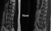
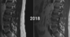
Asking for a colleague. Young patient, in his 20s, focal left-sided low back pain approx L3. His 2018 only demonstrated an L3 subcentimeter T2 hyperintense lobulated lesion, likely benign. Continues to have pain 2 years later and current MRI looks as below. I appreciate it less on recent imaging but can still appreciate subtle T2 hyperintensity on the pedicle. Different radiologist didn't call anything this time(I probably wouldn't have noticed anything if I didn't know the history).
Little doubt it's benign given unchanged appearance over 2 years. Question is -- any chance its an actual pain generator? My thought is probably not, but his pain location does apparently correspond to the location. If so, what to do about it?
Little doubt it's benign given unchanged appearance over 2 years. Question is -- any chance its an actual pain generator? My thought is probably not, but his pain location does apparently correspond to the location. If so, what to do about it?
Attachments
Last edited: