Popliteal block

This forum made possible through the generous support of SDN members, donors, and sponsors. Thank you.

Sonny Crocket

Full Member
10+ Year Member
Joined
Nov 14, 2008
Messages
208
Reaction score
75
How far up the back of the thigh are you guys doing your popliteal blocks? For ankle surgery, I really want to see the peroneal nerve join the tibial and block it there. A lot of times, however, I do not see this joining, do the block anyway but am worried about lateral ankle sparing. I guess if enough volume is I should get both.

Members don't see this ad.
 
If I can't see the femoral joining the tibial I go as high as I can while still getting a good view of the nerve
 
Members don't see this ad :)
If I can't see the femoral joining the tibial I go as high as I can while still getting a good view of the nerve

I believe you meant if you can't see the peroneal joining the tibial? The femoral nerve is definitely not in the neighborhood of the popliteal fossa.

In patients with a normal -ish body habitus, you should always be able to identify the bifurcation of the popliteal sciatic into the tibial and common peroneal nerves. Typically this occurs just proximal to the popliteal crease. The sciatic goes from deep to superficial as it goes distally towards the popliteal fossa, this often requires you to tilt the surface of the probe slightly towards the knee cap in order to direct the sound waves perpendicular to the nerve, which should make it more visible in your image. As you scan up and down the leg, you definitely need to continually adjust your tilt to optimize visualization of the siatic nerve.

If I'm doing a single shot block, I typically deposit the local right at the point of bifurcation. This will result in a faster block onset.
 
Most of the time, the bifurcation is really close to the popliteal crease. Even if the pt has a high takeoff of the peroneal nerve, you can get proximal spread if you are in the nerve sheath (epineurium). More ultrasound experience with scanning will reveal the bifurcation to you almost all the time. The bigger issue is the epineurium. If you don't get into the epineurium you'll get a slow onset partial block. A lot of NS-only blocks are like this.
 
  • Like
Reactions: 1 user
I start with the probe in the popliteal fossa and scan proximally looking for the location where the nerve bifurcates and then I block approximately 1-2 cm proximal to the bifurcation. Despite anatomical literature suggesting a wide range of places the bifurcation can occur you nearly always find it within 5-10 cm of the popliteal fossa.
 
Peripheral_Nerve_Anatomy.png
 
Most of the time, the bifurcation is really close to the popliteal crease. Even if the pt has a high takeoff of the peroneal nerve, you can get proximal spread if you are in the nerve sheath (epineurium). More ultrasound experience with scanning will reveal the bifurcation to you almost all the time. The bigger issue is the epineurium. If you don't get into the epineurium you'll get a slow onset partial block. A lot of NS-only blocks are like this.

You are not completely correct here. Please read my next post and I highly recommend obtaining a copy of my reference (only 2 pages) for a full explanation.
 
Most authors consider from the inside outward: the endoneurium, perineurium, epineurium, and paraneurium (Fig. 1A).9–11 Individual nerve fibers and their Schwann cells are surrounded by the endoneurium. Groups of endoneurium-sheathed fibers, called fascicles, are surrounded by a thin, dense, multilayered connective tissue sheath, known as the perineurium. Connective tissue surrounding each fascicle is termed interfascicular or inner epineurium, surrounded by the epifascicular or outer epineurium (Fig. 1B). This outermost layer defines the nerve trunk and protects the peripheral nerve against mechanical stress. A loose connective tissue fills the space between the nerve and the surrounding tissue in connection with the epineurium. This tissue is called the paraneurium, mesoneurium, adventitia, or gliding apparatus.9–11 It suspends the nerve trunk within the soft tissue, fuses with similar loose connective tissue around vessels in neurovascular bundles, and brings the segmental blood supply of the nerve. The paraneurium is usually not described in textbooks.11 It may be overlooked in cadaver dissection. The paraneurium allows longitudinal nerve motion and caliber changes if adjacent joints are extended or flexed. It protects the nerve and allows it to glide against the surrounding tissue. Any attempt to touch the paraneurium leads to fibrosis, covering the nerve in a longitudinal direction. The next level is fusion with the fascicular epineurium and the 2 sheaths cannot be differentiated. This explains why the paraneurium has escaped the attention of surgeons and is thought to be the epineurium by anesthesiologists. This common nerve sheath was described after careful dissection of the sciatic nerve12 and the brachial plexus.13 Sala-Blanch et al.4 have to be commended for reporting that the most important parameter for rapid-onset PSNB was the spread of LA around both nerve components rather than the intraepineural injection itself. That result highlights the virtual space between the paraneurium and the epifascicular epineurium. We are convinced that the paraneurium can confine the LA solution around and along the epineurium and give a doughnut sign on US without nerve swelling (Fig. 2). We believe that in the case of swelling, when nerve fascicles are separated from each other, the needle should be withdrawn. We do not claim that the paraneural injection is the highway that leads to 100% success and no complications. We assume that the paraneural space is a target for the needle that can lead us to an adequate risk/benefit ratio.


http://www.anesthesia-analgesia.org/content/114/5/929.full
 
  • Like
Reactions: 1 user
I see no need to hit the epinuronium. This applies to all my blocks.
 
Members don't see this ad :)
For ankle surgery I like to block it right after it splits. In my mind I feel like that gives you more surface area to see the local anesthetic. If I block it after it joins up, I feel like I am missing the medial portion of both nerves. If I block after I can get circumferential spread around both nerves. Probably a moot point, but it makes me feel better and I feel like it sets up faster that way.
 
For ankle surgery I like to block it right after it splits. In my mind I feel like that gives you more surface area to see the local anesthetic. If I block it after it joins up, I feel like I am missing the medial portion of both nerves. If I block after I can get circumferential spread around both nerves. Probably a moot point, but it makes me feel better and I feel like it sets up faster that way.

Corrrect and Agree. Several studies say the same thing as above.
 
You may not need it but if you've had 30+h blocks then you've hit it.

Touching the nerve is one thing but planning on penetrating the epineurium is another.
As my diagram shows you should come in contact with the Popliteal/Sciatic nerve but there is no need to actually try and push the needle into the nerve.
 
Semantics, Misnomer, or Uncertainty: Where Is the Epineurium on Ultrasound?
Endersby, Ryan MD, FRCPC; Albrecht, Eric MD, DEAA; Perlas, Anahi MD, FRCPC; Chan, Vincent MD, FRCPC

Regional Anesthesia and Pain Medicine . 37(3):360–361, May/June 2012.
doi: 10.1097/AAP.0b013e318253b48f

Author Information

Department of Anesthesia and Pain Medicine Toronto Western Hospital University Health Network University of Toronto Toronto, Ontario, Canada

Accepted for Publication: 29 February 2012

To the Editor:

We read with interest the excellent study by Tran et al,1 showing that a single "subepineural" injection at the bifurcation of the sciatic nerve in the popliteal fossa is associated with a higher success rate and a shorter performance time than separate injections around the tibial and peroneal nerve divisions. They described the injection below a common sheath as subepineural. However, we would like to suggest that whether this layer represents a true epineurium or a separate adventitial layer or common sheath external to the epineurium is a matter of debate.2 The study by Vloka et al, which was quoted to support the conclusion of Tran et al that the injections were subepineural, was strictly an anatomic study with no correlation to ultrasound images.3 Vloka et al studied the anatomy of the sciatic nerve bifurcation in cadavers, and they in fact described the presence of multiple "epineural" layers. An inner epineural layer was identified, which they called the "interfascicular epineurium," extending between nerve fascicles, while a second tissue layer that surrounds the entire nerve was called the "epifascicular epineurium." In addition, a third loose connective tissue layer was identified external to the epineurium and was referred to as the "adventitia of the nerve."

Judging by the images presented in the study by Tran et al, it is unclear to us whether the "subepineural" injection refers to the interfascicular epineurium, the epifascicular epineurium, or possibly an injection deep into the adventitial layer but outside the epineurium of the 2 individual nerves themselves. Furthermore, there is a substantial amount of animal and human evidence that suggests subepineural (ie, intraneural) injections are usually accompanied by some degree of nerve swelling.4–6 In contrast, Tran et al stated that neural swelling was specifically avoided in both study groups. We believe that their images and the absence of neural swelling are both consistent with an injection outside the epineurium (possibly deep to the adventitial layer of the nerve) rather than a true subepineural injection.

In our opinion, this is an important matter and not just a matter of semantics. There is currently a heated debate in our specialty about the relative safety (or lack thereof) of intraneural injections. We should collectively strive to use common and clear terminology as to what constitutes intraneural (subepineurial) injection and what does not. We believe that Tran et al described a highly effective approach to block the sciatic nerve at its bifurcation that does not involve intraneural injection, as the title of the article would suggest.

Future studies that correlate sonographic images with anatomic findings at the level of the sciatic nerve bifurcation may shed more light on this issue.

Ryan Endersby, MD, FRCPC

Eric Albrecht, MD, DEAA

Anahi Perlas, MD, FRCPC

Vincent Chan, MD, FRCPC

Department of Anesthesia and Pain Medicine

Toronto Western Hospital

University Health Network

University of Toronto

Toronto, Ontario, Canada

Editor's Note: Thank you, Drs. Endersby et al and Tran et al, for commenting on this important issue. Readers should be aware that a study directly related to this issue will be published in the near future.
 
My mistake -- I guess it's called a Paraneural sheath now, not epineurium. The point is that you have to get a pop, and the local has to indent the nerve.
 
Injection Inside the Paraneural Sheath of the Sciatic Nerve: Direct Comparison Among Ultrasound Imaging, Macroscopic Anatomy, and Histologic Analysis
Andersen, Henning Lykke MD; Andersen, Sofie L. MD; Tranum-Jensen, Jørgen MD

Regional Anesthesia and Pain Medicine . 37(4):410–414, July/August 2012.
doi: 10.1097/AAP.0b013e31825145f3

.......
Results: Grossly, we identified a thin, transparent, and fragile tissue layer surrounding the epineurium. Sonographically, this layer was identified with injectate as a hyperechoic layer detaching from the surface of the sciatic nerve. Histologically, the sheath was seen as a multilayered circular fascia as part of the paraneural tissue. An injection of 10 mL inside the sheath spread 10 to 15 cm closely along the nerve, however, not completely circumferential, compared with 5 to 6 cm if the injection was outside the sheath. Characteristics of the ultrasound-guided injection technique are described.

Conclusions: There is a distinct tissue layer surrounding the popliteal sciatic nerve as a paraneural sheath that has distinct gross anatomic, histologic, and sonographic features. This sheath may have implications for regional anesthesia involving the popliteal sciatic nerve. We suggest that the ultrasound-guided injection technique described here could be used in future clinical studies investigating the importance of the paraneural sheath.
 
You may not need it but if you've had 30+h blocks then you've hit it.

i don't aim for 30+ hr blocks... for 2 reasons:

1) I don't like to worry 'bout them.

2) Motor block is much higher with an epinuronium block. Patients hate motor blocks lasting more than a couple of hours-- main reason why I find catheters so nice. You can dial in a good concentration (.1% ropivicaine) and have no pain and while attaining full motor function.
 
A response at 0.4mA means you've gone through the epineurium 100% of the time so it has been done millions of times unknowingly.

I have done it Hundreds and Hundreds of times as well. But, with U/S I no longer need to penetrate the nerve; I do try and push the needle through the very ede of the fascial layer or paraneurium but only when my needle angle allows it to be performed safely.

I no longer use or recommend the nerve stimulator for a popliteal block. Instead, inject the local around the nerve but the needle should make contact with each component, tibial and common peroneal, at least once (I prefer at least 2 direct contacts with the nerve) during the block.
 
My mistake -- I guess it's called a Paraneural sheath now, not epineurium. The point is that you have to get a pop, and the local has to indent the nerve.

If you are careful with the needle and do this block frequently then you know the needle tip should be visible prior to any "pop" or the needle ends up intraneural.

When and if you have a good view of the needle tip it is easy to glide the tip of the needle along/into the very edge of the fascial layer surrounding the individual component. You will quickly see the nerve move away from your needle during injection. Repeat this process around each individual nerve component so that all aspects of the nerve is surrounded by local anesthetic and your success will approach 100% with duration exceeding 24 hrs (Bup plus decadron).
 
  • Like
Reactions: 1 users
A response at 0.4mA means you've gone through the epineurium 100% of the time so it has been done millions of times unknowingly.

no it doesn't. We like to think that the mA required to get a stimulus is a marker for needle to nerve distance, but it doesn't always hold up. This is especially easy to see if you combine an U/S with nerve stimulator. While it's likely that you've gone through the epineurium if you get a twitch at less than 0.40 mA, it is not guaranteed.
 
no it doesn't. We like to think that the mA required to get a stimulus is a marker for needle to nerve distance, but it doesn't always hold up.

That's not what i said: the sciatic nerve is a different beast to get a stim you have to penetrate the epineuriome because of the high quantity of interstial tissue

http://www.ncbi.nlm.nih.gov/pubmed/21862889
 
Curr Opin Anaesthesiol. 2012 Oct;25(5):596-602. doi: 10.1097/ACO.0b013e328356bb40.
Where should the tip of the needle be located in ultrasound-guided peripheral nerve blocks?
Choquet O, Morau D, Biboulet P, Capdevila X.
Source
Department of Anesthesiology and Critical Care Medicine, Lapeyronie University Hospital, INSERM 1046, Montpellier I University, Montpellier, France. [email protected]
Abstract
PURPOSE OF REVIEW:
Data now exist describing the appropriate positioning of the needle tip and pattern of local anaesthetic spread after injection. The recent literature has been analysed in search of studies on the optimal procedure for common approaches centred on block efficacy, performance time, and safety.
RECENT FINDINGS:
Large peripheral nerves are surrounded by a gliding layer, the adventitia or paraneurium. Ultrasonically, a circumneural spread corresponds to adventitial extraneural injection. Nerve expansion with fascicular separation matches intraneural injection. Deliberate intraneural injection remains controversial, and is not advisable at the present time. For popliteal sciatic nerve blocks, positioning the needle in the common nerve sheath between the tibial and peroneal components and obtaining a circumneural spread surrounding both divisions predict rapid surgical anaesthesia. Using axillary and infraclavicular approaches, ultrasound-guided perivascular injection aiming at circumferential spread around the artery appears a valuable alternative to individual targeted nerve injections. For single injection interscalene block, an injection into the fascial sheath but far from the plexus proved to be as effective as an injection adjacent to the nerve structures. Fascial plane approaches are appealing alternatives for thin nerves that run between muscles and cannot be regularly visualized with the current resolution of ultrasound systems.
SUMMARY:
The ultrasound appearance of nerves and target injections are better understood. The specific distributions of local anaesthetic spread that predict success are significantly different from one anatomical site to another. It seems advisable to avoid intraneural injection.
 
Any ultrasound pics or videos

How do you increase the chance of success of this block
 
Any ultrasound pics or videos

How do you increase the chance of success of this block


The key is making sure you adequately block around both nerves, and dont miss one or part of one. Use 40ml 0f 0.375% to have enough volume to spread.

Start in the pop-fossa, visualize the artery and the nerve tissue sitting on top.

Scan north until you see a smaller round nerve joining that other nerve

When they are together or at the bifurcation inject around both of them (and sometimes inside as other posters have mentioned)

Going inside the first layer of connective tissue surrounding the nerve is more of a guaranteed success, but not absolutely necessary

If for whatever reason you cannot get them to come together in a nice location that you can block, block them individually where you can see them best (rarely this happens)
 
  • Like
Reactions: 1 user
The key is making sure you adequately block around both nerves, and dont miss one or part of one. Use 40ml 0f 0.375% to have enough volume to spread.

Start in the pop-fossa, visualize the artery and the nerve tissue sitting on top.

Scan north until you see a smaller round nerve joining that other nerve

When they are together or at the bifurcation inject around both of them (and sometimes inside as other posters have mentioned)

Going inside the first layer of connective tissue surrounding the nerve is more of a guaranteed success, but not absolutely necessary

If for whatever reason you cannot get them to come together in a nice location that you can block, block them individually where you can see them best (rarely this happens)

I rarely, if ever, use more than 20 mls of local for a popliteal block. My success rate is close to 100% with this volume. The "connective tissue" that you need to penetrate is the mesoneurium. If you "penetrate" that tissue plane and block the nerve (either as one large nerve or each component separately) the block never fails.
 
The Popliteal/sciatic nerve is very "forgiving" in terms of being able to tolerate an intraneural injection. I never intentionally inject intraneurally or the epineurium but if it does happen there is typically no issues postop in terms of complications (for this particluar nerve).

25FF1.png
 
  • Like
Reactions: 1 users
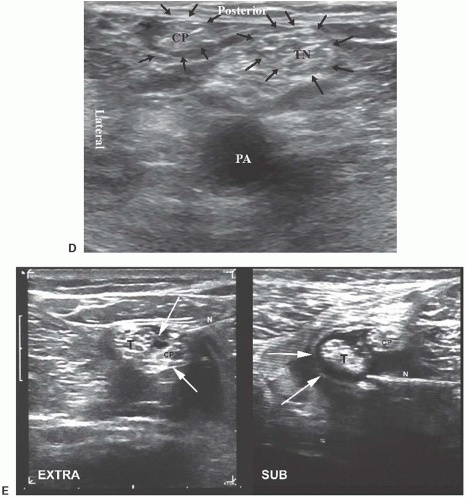
At this level, the sciatic nerve is encased by a thick mesoneurial sheath. Within the nerve, both the peroneal and tibial nerves are encased by their proper epineurium. Within the epineurium of each nerve lie the fascicles, each of which is bounded by perineurium. Currently, the term used to describe the fascial sheath of the sciatic nerve, which surrounds both components (tibial and common peroneal nerve), is a topic of contention. Authors have referred to this anatomic structure as a paraneural sheath1 or complex fascial layer,2,3 whereas others termed it a common epineural sheath4 (Fig. 32.1E). The authors’ preference is to call this the mesoneurium. Deposition of local anesthetic within the paraneural sheath at the level of the nerve bifurcation, but outside the epineurium of the tibial and peroneal nerves, may afford some advantages in the block.5

Ultrasound-Guided Popliteal and Lateral Sciatic Block
 
  • Like
Reactions: 1 user
A response at 0.4mA means you've gone through the epineurium 100% of the time so it has been done millions of times unknowingly.

Correct. And we know from the studies that the sciatic nerve is very "tolerant" of either an epineural or even, an intraneural injection. Again, it isn't necessary to do an epineural injection to obtain a very successful block but one must penetrate the "sheath" surround the individual components of the sciatic nerve or penetrate the mesoneurium of the sciatic nerve itself (when both components are combined to form one large nerve).
 
Correct. And we know from the studies that the sciatic nerve is very "tolerant" of either an epineural or even, an intraneural injection. Again, it isn't necessary to do an epineural injection to obtain a very successful block but one must penetrate the "sheath" surround the individual components of the sciatic nerve or penetrate the mesoneurium of the sciatic nerve itself (when both components are combined to form one large nerve).

Can u please give me any reference article to the above quote. Thanks
 
Would recommend you inject at the site of bifurcation where they still share a common sheath: Ultrasound-guided popliteal block through a common paraneural sheath versus conventional injection: a prospective, randomized, double-blind study. - PubMed - NCBI

It's relatively straightforward (esp out of plane), lowers your intraneural risk because you're in the common sheath, not in the sheath of each nerve, and it works. I would recommend injecting an ml or two and then moving the probe a few inches up the thigh as you keep injecting. If you're seeing spread, you're in the sheath. If you don't, you're not. I can't recall ever having a failed pop block.
 
  • Like
Reactions: 1 users
This is crazy. 20 to 30 ml is all that is needed.

Not sure I would call 40ml "crazy" but I do agree its alot and use 30 myself. This advice was given to someone who isnt exactly precise with the local it seems and therefore could use a little margin of error.
 
Not sure I would call 40ml "crazy" but I do agree its alot and use 30 myself. This advice was given to someone who isnt exactly precise with the local it seems and therefore could use a little margin of error.

40 ml is crazy;)

If you need to use that much volume then your block technique needs refinement.
 
  • Like
Reactions: 1 users


I do it right at the bifurcation between both nerves and see both nerves peel away.
If you can't find the bifurcation, keep scanning, you're not in the right place until you can see it.
20-25 mL 0.25% is all you should need
 
  • Like
Reactions: 1 user
The volumes for blocks vary wildly. I typically use 15ml for an interscalene block but one of my colleagues uses 50ml. BTW thanks for the excellent ultrasound images posted here.
 
The volumes for blocks vary wildly. I typically use 15ml for an interscalene block but one of my colleagues uses 50ml. BTW thanks for the excellent ultrasound images posted here.

Part of the goal of the block to make it as long lasting as possible. The more mgs of bupi you put in the block the longer its going to last. Dont you think Im getting more duration out of my 30ml of 0.5% bupi plus decadron compared to the 15ml of 0.5%? What is the downside to more (assuming you leave enough room to not cause toxicity)?
 
Correct. And we know from the studies that the sciatic nerve is very "tolerant" of either an epineural or even, an intraneural injection. Again, it isn't necessary to do an epineural injection to obtain a very successful block but one must penetrate the "sheath" surround the individual components of the sciatic nerve or penetrate the mesoneurium of the sciatic nerve itself (when both components are combined to form one large nerve).

Which other nerves are tolerant to intraneural injections and which blocks do I have to be extra careful
 
Part of the goal of the block to make it as long lasting as possible. The more mgs of bupi you put in the block the longer its going to last. Dont you think Im getting more duration out of my 30ml of 0.5% bupi plus decadron compared to the 15ml of 0.5%? What is the downside to more (assuming you leave enough room to not cause toxicity)?

50cc for an interscalene though? Seems excessive... Maybe the goal is to put so much volume in as to compress the carotid, cause the patient to lose O2 to their brain and pass out. Supposedly patients don't feel ANYTHING with that block. ;)
 
Top