Remember when we used to share "good cases!" on here? Well, here's one.

This forum made possible through the generous support of SDN members, donors, and sponsors. Thank you.
I don’t think I’m quite ready to call OB to admit yet.

Sorry for the snarkiness. I was feeling a little RustedFox ;)

I accept all snark :) shed be going icu regardless

Members don't see this ad.
 
C’mon son, you gotta give us a real BP. Nobody is getting a 150/130 BP and saying “okay, that sounds legit. No need to repeat.”

I repeated it and it was like 152/126.
Repeated it again and diastolic was > 120. But given the rest of the exam I didn’t feel like her true one was 150/90. Especially when she was just treated for pre-epttampisidibia.
 
Last edited:
  • Like
Reactions: 2 users
Not trained in EM, can I still play?

Can we get some labs? Cbc, cmp, ua, utox, spot urine protein/cr, coags.

Agree with labetalol and mag; can we add postpartum eclampsia to the ddx?
 
Members don't see this ad :)
Would favor this, and it’s basically my ddx. Only thing I’d add that probably isn’t relevant is that eclampsia is also a post partum Dx (up to 6-12weeks depending on source) should be considered with end organ damage and that bp. Still needs labs and Icu, call to her ob, abx, would probably get cta ctv and discuss with neuro if time course and hard findings were convincing.

So I thought initially pre-eclampsia w/ odd neurologic features, but not eclampsia as she wasn't having a seizure. Not a generalized seizure at least. Maybe an SAH or IPH. Ischemic stroke possible but I couldn't find the lesion based on her exam. It was kind of confusing.

immediate CT Head neg
I loaded her with 6g Mag over 20 mins. It took some talkin' with the pharmacist because they didn't have a Mg level back, and I said "that's ok....she needs it now...."
I ordered all the regular labs, including TSH (LOL I don't know why), serum Tox stuff, UTox, and that's about it

Over the next 30 minutes, her vitals got better
HR now 105
BP now 125/85

her labs come back
BMP normal
Mg 2.1
LFTs normal except AlkP 280?
WBC 19 w/ normal diff
Hg 7.2
Coags normal
TSH OK
UA 3+ protein, 1+ glucose, otherwise neg
Serum Tox neg
UTox + meth


About two hours in she is getting worse. I can barely arouse her to noxious stimuli. Easily shows me two fingers with right hand. cannot with the left hand. She is becoming plegic on the left, upper > lower. No convulsive movements. She is per-intubation. The rest of her non-neuro exam has not changed.

At this point I "activate stroke" even though I had all the testing I needed. This is how we get a neurologist on the robot quickly.
Teleneuro says "agree no tPA, get stat CTA"
So I get one.

Clean cerebral arteries.

I tube her.

I start transfusing her because I don't know what else to do, and the teleneuro says admit her to the ICU.
 
  • Like
Reactions: 1 user
Would depend on diagnosis at my hospital. ICU if CVA treated with tPA, venous sinus thrombosis, TTP, septic or hemorrhagic shock. L&D under OB if routine postpartum preeclampsia, endometritis, sepsis without shock, or mild blood loss anemia with normalized vitals and hemorrhage control.

Let’s see more of the case and see where it takes us.

Do you see any of those conditions leading to this presentation? She reeks icu-bound to me
 
I believe alkaline phosphate naturally goes up during pregnancy and specifically third trimester. May just not have gone down yet. Also up in HELLP I believe, but don’t suspect in this case with normal LFTs.

Sounds like methamphetamine intoxication with subsequent withdrawal. Probably contributed to preeclampsia. Doubt eclampsia, but I suppose partial seizure possible.

PRES on the differential, but seems unlikely given degree of hypertension.

Initially had me thinking cerebellar stroke vs. venous sinus thrombosis high on the differential. Saw one similar case once. Meth could also precipitate. Too drowsy initially to stand up and check Romberg/gait? What was the tPA discussion? CVA seemed unlikely given exam? Unclear timeframe? Recent delivery? Might consider a rapid MRI given unclear and within the window for tPA.

tPA was in the discussion, and given she still was having vag bleeding/spotting it was a no-go.

Meth can cause people to act crazy. Her symptomology initially was dysarthria with odd eye movements and a non-focal motor extremity exam, which progressed to worsening obtundation, --> tube, and unable to move the left side with normal CT Head and CTA Brain.

I really thought she would have had a LVO given how her exam changed.

Did not try to walk her...she got worse in the ED.
I spoke to Mom on the phone after she had been there for about 1 hour, and it wasn't too much help. Mom admitted she did meth "a few times recently" but didn't think she did it recently. Earlier in the day she was complaining of diarrhea, then had dizziness.

I was a little frustrated with this case because she was getting worse and I really didn't know what was going on
and when I talked to the ICU they were all about "is this subclinical status epilepticus" and "we have to talk to Neurology now" and "I don't now how to treat pre-eclampsia" and I told them about the Mg infusion and he basically ignored me and said "just call OB". It was an ICU doc that nobody here really likes.

More shortly.
 
  • Like
Reactions: 1 users
tPA was in the discussion, and given she still was having vag bleeding/spotting it was a no-go.

Meth can cause people to act crazy. Her symptomology initially was dysarthria with odd eye movements and a non-focal motor extremity exam, which progressed to worsening obtundation, --> tube, and unable to move the left side with normal CT Head and CTA Brain.

I really thought she would have had a LVO given how her exam changed.

Did not try to walk her...she got worse in the ED.
I spoke to Mom on the phone after she had been there for about 1 hour, and it wasn't too much help. Mom admitted she did meth "a few times recently" but didn't think she did it recently. Earlier in the day she was complaining of diarrhea, then had dizziness.

I was a little frustrated with this case because she was getting worse and I really didn't know what was going on
and when I talked to the ICU they were all about "is this subclinical status epilepticus" and "we have to talk to Neurology now" and "I don't now how to treat pre-eclampsia" and I told them about the Mg infusion and he basically ignored me and said "just call OB". It was an ICU doc that nobody here really likes.

More shortly.

All seems pretty reasonable on your end. Agree that I didn’t have supporting evidence for eclampsia and should probably favor preeclampsia, but was wondering about either nonconvulsive status or other odd seizure pattern given the eye movements and gaze preference. I think of em on a spectrum where this lady is on the crappy end.

I don’t like your icu doc either. I’d love it if “I don’t know how to treat that” was part of emtala, though it would put me out of the job when they hired the cheapest shill.
 
Need some HELLP interns?
(nice job of hiding the platelets, just like my lab always does with ANY abnormality)
 

If she didn’t get better with the mag & BP control that makes PRES/eclampsia stuff less likely in my mind. I’d worry about central venous sinus thrombosis with the reported headache/stroke like symptoms. So some combo of MRI + CTV/MRV is needed. Since she got the CTA maybe MRI/MRV is the move.

if she had an epidural she may have had a spinal leak which increases chances for CVST.
 
  • Like
Reactions: 3 users
It could all just be meth/intox, although given the severity of symptoms, this is a diagnosis of exclusion. I think considering an LVO/CVA was pretty reasonable given the sudden focality of symptoms with hemiparesis. CTA and/or CTP with stroke activation was a good move. That being said, CVA causing those symptoms should have flagrantly abnormal CTA, so kind of ruled out in my opinion. Sounds like preE/PRES is already being treated with mg and BP control.

Other considerations would be cerebral venous sinus thrombosis vs. perhaps status epilepticus. Patient already intubated and on heavy anticonvulsant/sedation, so pretty well covered. I agree with empiric broad spectrum Abx for sepsis without a source (would consider a pelvic to assess for EMM +/- US/CTAP depending on findings), ICU admission, stat neuro consultation and plan for rapid MRI/MRV and EEG inpatient. In any world I want this patient managed by an intensevist in an ICU, an OB primary on a L&D floor would kill this patient.
 
Last edited:
  • Like
Reactions: 1 user
Dysarthria, gaze palsy, hemiparesis, unconsciousness points to a medial brain stem syndrome. Would get MRI as first priority. Infective encephalitis also possible so would treat that.

Venous thrombosis unlikely with that degree of coma but no CT findings. It’s not hypertensive PRES if the BP is now normal but getting worse. I would actually consider driving the blood pressure back up at this point and seeing if she improves.

Could also be postpartum cerebral vasoconstriction made worse by meth. MRI may find vasoconstricted vessels but not necessarily
 
Last edited:
Dysarthria, gaze palsy, hemiparesis, unconsciousness points to a medial brain stem syndrome. Would get MRI as first priority. Infective encephalitis also possible so would treat that.

Venous thrombosis unlikely with that degree of coma but no CT findings. It’s not hypertensive PRES if the BP is now normal but getting worse. I would actually consider driving the blood pressure back up at this point and seeing if she improves.

Could also be postpartum cerebral vasoconstriction made worse by meth. MRI may find vasoconstricted vessels but not necessarily

This is outside of my area of expertise, but the little bit of PRES I've seen improved over a course of days. She seemed like she was already teetering on the edge, I'm not sure I'd rule out PRES based on continued deterioration over the course of an hour or so after BP normalization. I'm open to education though. Infectious or autoimmune encephalitis is a good thought and another that came to mind. I'm assuming she got AED loaded and shipped to MRI/V after tubed. Interested to see what it showed
 
Members don't see this ad :)
Yeah I think if the initial inpatient workup is unrevealing MRI/MRV and EEG and no source of sepsis is localized, than an LP would be indicated at some point. Some really odd ball stuff enters the differential at that point like NMDA encephalitis. I probably wouldn't do the LP in the ER; however, no fever, neuro exam seems more focal, which is suggestive of a specific brain territory or vascular lesion; whereas I think of infectious encephalitis/meningitis as presenting with more generalized encephalopathy (which I guess was the patient's initial presentation before the hemiparesis).
 
This is outside of my area of expertise, but the little bit of PRES I've seen improved over a course of days. She seemed like she was already teetering on the edge, I'm not sure I'd rule out PRES based on continued deterioration over the course of an hour or so after BP normalization. I'm open to education though. Infectious or autoimmune encephalitis is a good thought and another that came to mind. I'm assuming she got AED loaded and shipped to MRI/V after tubed. Interested to see what it showed

Maybe my wording wasn’t great. What I was saying was that this is not a hypertensive encephalopathy. PRES is highly likely but hypertension in eclamptic PRES is not causative of the neurological symptoms.
 
After being told that CTA Brain was OK, and I start transfusing and calling people like the ICU and OB.

I get a call 20 minutes later from the radiologist.

CTA Neck shows a narrowing of the R vertebral artery around C4. Could be thrombus / dissection.

I load her with aspirin 325 PR while transfusing pRBCs. I order a bunch of MRIs

I make more calls....and at the end of my shift the signout is "we are getting a STAT EEG in the ED, if negative pt is admitted here, if + then she needs to be transferred." That's the way it works at my shop. It sucks.

The next day I log into the EMR to see what happened.

MRI 1.jpg
MRI 2.jpg
MRI 3.jpg

MRA.jpg


MRV negative.

she self-extubated in the night and left AMA several hours later. Unable to walk well, she asked to be wheelchaired out to the car where her family member was waiting.

Not the most difficult case, as most Neurology emergencies have few proven ER interventions to make a difference. But nonetheless interesting and upsetting as she was in her 20s and now has a baby to take care of.
 
  • Like
Reactions: 1 users
After being told that CTA Brain was OK, and I start transfusing and calling people like the ICU and OB.

I get a call 20 minutes later from the radiologist.

CTA Neck shows a narrowing of the R vertebral artery around C4. Could be thrombus / dissection.

I load her with aspirin 325 PR while transfusing pRBCs. I order a bunch of MRIs

I make more calls....and at the end of my shift the signout is "we are getting a STAT EEG in the ED, if negative pt is admitted here, if + then she needs to be transferred." That's the way it works at my shop. It sucks.

The next day I log into the EMR to see what happened.

View attachment 312338 View attachment 312339 View attachment 312340
View attachment 312341

MRV negative.

she self-extubated in the night and left AMA several hours later. Unable to walk well, she asked to be wheelchaired out to the car where her family member was waiting.

Not the most difficult case, as most Neurology emergencies have few proven ER interventions to make a difference. But nonetheless interesting and upsetting as she was in her 20s and now has a baby to take care of.

What's the break point in admission VS transfer based on? Lack of neurotele?
 
I make more calls....and at the end of my shift the signout is "we are getting a STAT EEG in the ED, if negative pt is admitted here, if + then she needs to be transferred." That's the way it works at my shop. It sucks.

This is true everywhere I have worked, continuous EEG usually can only be done in a true neuro-ICU, which is only at comprehensive stroke centers.

she self-extubated in the night and left AMA several hours later. Unable to walk well, she asked to be wheelchaired out to the car where her family member was waiting.

:rage::rage::rage::rage:

I guess I should expect no less from an ER patient at this point in my career, but still..this is literally madness.
 
  • Like
Reactions: 1 user
What's the break point in admission VS transfer based on? Lack of neurotele?

Management of status epilepticus. Our ICU docs believe that one must be on continuous EEG. Our Neurologists say that isn't necessarily true. Of course I believe our Neuro docs. Sometimes our Neurologist say no continuous EEG is needed and even the ICU docs wins out because they will refuse to consult on the case. Idiotic.
 
  • Like
Reactions: 1 user
This is true everywhere I have worked, continuous EEG usually can only be done in a true neuro-ICU, which is only at comprehensive stroke centers.

While I believe that...I think the indications for continuous EEG are narrower than our ICU docs suggest. It's all for legal reasons. They openly admit that.

For instance, people with cardiac arrest, ROSC and need for TTM have something like a 25% chance of going into subclinical status epilepticus if I recall. We never transfer these patients. Why? $$$$
 
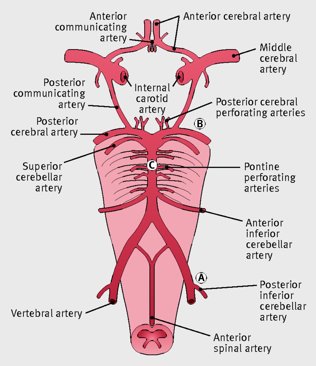
Yes...R vertebral artery dissection that was showering thrombus through the R PCA and even into the circle of willis. Hit parts of her brainstem as well. She had big bilateral stroke R > L.

Odd that thromboemboli from the vert could reach anterior brain (maybe my neurovascular anatomy isn't great), but that explains why she had the left extremity weakness in addition to the nystagmus and ataxia.

I agree, I'm not sure what kind of intervention patient could have had. Did neuro re-consider tPA after the MRIs? Did they feel there was an unclear onset or the vaginal bleeding related to delivery felt to be a contraindication?

Seems the areas of ischemia are in areas of small vessel vasculature too small to go after with a catheter (again my neurovascular anatomy knowledge is not great), at least anticoagulation to try and stabilize any further thromboemboli from the dissection would be helpful. It seems this patient is probably not gonna be real consistent with the antiplatelet therapy I'm sure they discharged her on.

I'm guessing as far as ultimate etiology, pregnancy-delivery induced softening of connective tissue in a person with unknown susceptibility (undiagnosed perhaps untypable connective tissue disorder) precipitating dissection (pregnancy/delivery known risk factor for large and medium vessel dissection).
 
While I believe that...I think the indications for continuous EEG are narrower than our ICU docs suggest. It's all for legal reasons. They openly admit that.

For instance, people with cardiac arrest, ROSC and need for TTM have something like a 25% chance of going into subclinical status epilepticus if I recall. We never transfer these patients. Why? $$$$

That's a good point, I have heard cited that NCSE is VERY under-diagnosed in ICU patients.

But I think the indication to monitor EEG in a patient with known uncontrolled seizure activity is a little stronger than in a patient who may hypothetically go into NCSE.

I've always kind of gone with the quote from the neuro-intensevist Thomas Bleck (and I'm parapharsing here) "Managing status epilepticus in a sedated patient without continuous EEG is like trying to run ACLS on a coding patient with your finger on the pulse but no continuous cardiac monitor."

That being said, I've had a few cases where I tubed a patient for status epilepticus, but it's during business hours (say 11am on a Wednesday) and the neurologist and EEG tech are actually available, stat evaluate the patient, do the EEG and determine the seizure is aborted. Usually at that point they feel comfortable recommending ICU admission rather than transfer and the intensevists are ok with that.
 
Odd that thromboemboli from the vert could reach anterior brain (maybe my neurovascular anatomy isn't great), but that explains why she had the left extremity weakness in addition to the nystagmus and ataxia.

I agree, I'm not sure what kind of intervention patient could have had. Did neuro re-consider tPA after the MRIs? Did they feel there was an unclear onset or the vaginal bleeding related to delivery felt to be a contraindication?

Seems the areas of ischemia are in areas of small vessel vasculature too small to go after with a catheter (again my neurovascular anatomy knowledge is not great), at least anticoagulation to try and stabilize any further thromboemboli from the dissection would be helpful. It seems this patient is probably not gonna be real consistent with the antiplatelet therapy I'm sure they discharged her on.

I'm guessing as far as ultimate etiology, pregnancy-delivery induced softening of connective tissue in a person with unknown susceptibility (undiagnosed perhaps untypable connective tissue disorder) precipitating dissection (pregnancy/delivery known risk factor for large and medium vessel dissection).

Imagine little clots in the R Vert. They can go just about anywhere in the brain:

F1.large.jpg


By the time the MRI was done, which was around 10 hrs after coming to the ED, tPA will no longer be effective.

My understanding of vert artery dissections is that medical mgmt is the same, if not superior to stent placement. Usually these things clot off entirely, you have one stroke and you are done. She might have been showering emboli for a few hours. Who knows.

Yea I think etiology is being pregnant (dissections are higher risk in pregnancy / post-partum states) possibly exacerbated by meth. Who knows. She was vomiting at home and maybe wretched a dissection in her neck.
 
  • Like
Reactions: 1 user
That's a good point, I have heard cited that NCSE is VERY under-diagnosed in ICU patients.

But I think the indication to monitor EEG in a patient with known uncontrolled seizure activity is a little stronger than in a patient who may hypothetically go into NCSE.

I've always kind of gone with the quote from the neuro-intensevist Thomas Bleck (and I'm parapharsing here) "Managing status epilepticus in a sedated patient without continuous EEG is like trying to run ACLS on a coding patient with your finger on the pulse but no continuous cardiac monitor."

That being said, I've had a few cases where I tubed a patient for status epilepticus, but it's during business hours (say 11am on a Wednesday) and the neurologist and EEG tech are actually available, stat evaluate the patient, do the EEG and determine the seizure is aborted. Usually at that point they feel comfortable recommending ICU admission rather than transfer and the intensevists are ok with that.

Yea this basically is what happens at my hospital.

My main complaint with the way things are done here is we have someone with less experience and expertise (ICU, Internal Medicine) over-ruling people who are the experts (Neurology) in the mgmt of SE. And thankfully I get these kinds of cases about 2-3 times a year. So it's annoying but not terrible.
 
Management of status epilepticus. Our ICU docs believe that one must be on continuous EEG. Our Neurologists say that isn't necessarily true. Of course I believe our Neuro docs. Sometimes our Neurologist say no continuous EEG is needed and even the ICU docs wins out because they will refuse to consult on the case. Idiotic.

The general practice I've seen is cEEG x24 hr and if no evidence of NCSE can d/c EEG and treat for clinical seizure activity. I've seen that duration shortened for allocation of cEEG to patients felt to be in higher need, but generally at least several hours. The hospitals where I treated SE though were all major academic / comprehensive stroke centers with ready access to cEEG, so there admittedly is an ivory tower and resource availability bias there. I'm not sure offhand of the prevalence of subclinical seizures / NCSE developing in what's initially clinical status. I agree with your general sentiments regardless.

Good case, thanks for sharing
 
Last edited:
  • Like
Reactions: 1 user
Had an interesting one recently that I took over care for about 8 hours into their stay after they had already been accepted for transfer. I won’t keep people hanging, I’ll just break up the case to allow breaks to decide what you would do with the information you have read. DON'T LOOK AT THE FILE UNTIL PROMPTED IF YOU ARE PLAYING ALONG.

Setting is a rural ER 1 hr away from nearest hospital. 60 something yr old obese lady w/ a hx of DM and HTN presents for diarrhea, nausea, and generalized weakness. Symptoms started 2-3 days prior. She is having 4-5 loose BMs a day, non bloody. She denies any recent travels or sick contacts. She denies any fevers, cough, SOB, abd pain, vomiting, chest pain, or any other symptoms.

On exam, she appears like she is not feeling well (think man-flu appearance), but non-toxic, and no significant distress. She is morbidly obese. Exam is otherwise entirely normal.

VS: 101, 94/53, 18, 98%, 98.2F Glu: 140


............What are you ordering? What is your differential............


First, we don’t have the ability to do stool studies in the department, they are send out tests, in case anyone wanted some. It would take about 10 hrs to get performed even if the stool was ready. Similar story for COVID swabs.

I’ll tell you what my colleague ordered:
CBC
CMP
Lactic Acid
Blood cultures
CXR

NSx2L
Cipro/Flagyl

remarkable labs were:
WBC 14.5
Lactic Acid 1.9
Cr 1.8
BUN 68

The rest of the labs and CXR are unremarkable.

After 2L, pt’s BP 101/63.

Unchanged after a third a final unit. Pt still looks like she is feeling unwell.


............What are y'all doing now? What do you think is going on?...........


Colleague decided to work on admitting her at this point, about 6 hours into her ER stay. He was hoping to discharge her; however, given the borderline BP, the elevated WBC, and the patient not feeling well, he erred on admission. He did not believe the patient needed a CT A/P given the lack of reported abd pain or tenderness.

He finds a hospitalist to accept transfer about 7.5 hours after presentation to the hospital 1 hr away. I show up for my shift while he is wrapping up the case. Pt is already accepted and awaiting transport to show up to take her (which will be another hour) so technically never actually handed off to me, but he does give me a one liner about the pt before he takes off. About 10 minutes after he leaves, I get called into her room because her HR has jumped up to 190. She is clearly in monomorphic Vtach on the monitor, but we get an ECG to confirm while placing pads on her and getting the crash cart. BP still 102/60. Pt looks to be in distress, tachypneic, alert and talking although a bit confused. I quickly ask the nurse to review the labs to make sure there are no significant abnormalities, electrolytes are all normal. There is no previous ECG to evaluate.


............Now what are you doing?...........


I opted for synchronized cardioversion, as I find it significantly more satisfying for the patient and myself. I gave the patient 10mg of etomidate. I chose etomidate due to it having minimal hemodynamic effect unlike propofol, has no adrenergic effect like seen with ketamine, and is very well tolerated by patients unlike ketamine. The patient converted immediately with 100J.

I waited a few minutes to allow the myocardium to relax a bit and then shot an ECG. The ECG can be viewed below. Keep in mind this is a rural facility.

ecgcase5.png


............What's your ECG interpretation? What are you going to do?............


Well the pt is clearly having a STEMI from an LAD lesion. STE in V1-V4 along w/ I and aVL and some subtle depressions in at least II and V6. There is also prominent qwaves in the anterior leads which indicates that much of the myocardium of the anterior wall has likely already infarcted. Some might think that given the q-waves, its possible this is just an aneurysmal pattern from an old anterior MI; however, the way you can differentiate this is with T/QRS ratio in any lead V1-V4 that is >0.36 (formula derived by Dr. Smith of ECG fame), or use a combined T/QRS ratio of V1-V4 >0.22.....or you could just use common sense and recognize that there are multiple leads with no q-waves that also have STE, and some subtle depressions elsewhere indicating a obvious STEMI.

So I debated giving thrombolytics and then transfer vs just transfer. ACC/AHA recommend giving if transport time expected to be >60 min, which this would likely be. Also, it is unclear when time of onset was. Do we base time of onset on when diarrhea started? Q-waves indicate this has probably been going on a few hours, at least. Given risk/benefit ratio of thrombolytics gets murkier in the 12-24 hr time period, and significantly more risky after 24 hrs, I erred on the side of no thrombolytics and transfer for PCI. Cardiology of course couldn't believe it given pt history, and asked that I repeat the ECG two more times before they finally decided to take the pt to the cath lab. Pt ended up having 100% LAD occlusion (TIMI 0 flow) and had an impella placed for cardiogenic shock. She eventually had the impella pulled, had an uncomplicated hospital stay, and was discharged on hospital day 7.

Later on in that same shift, I had a 70 something year old show up after developing a pain in her upper back after going for a swim. Of course she had a Type A dissection. She survived the OR and was eventually discharged from the hospital in great condition, as well.
 
  • Like
Reactions: 7 users
Now that's a case. Reinforces my usual practice of ignoring 'diarrhea' on ROS. I find that so many people endorse this symptom w/o really having it.
 
  • Like
Reactions: 1 user
Had an interesting one recently...

Good case(s) and strong work. Also cringed at empiric FQs for undifferentiated diarrhea.

Now that's a case. Reinforces my usual practice of ignoring 'diarrhea' on ROS. I find that so many people endorse this symptom w/o really having it.

My practice pattern also. If clinically relevant, follow-up question is "so you mean you're having completely watery stools? How many episodes in the last [pertinent timeframe]?" Suspect many of us would not have gotten an EKG at first on that lady... unless the "diarrhea" complaint was nonsense on clarification, and we're left with nausea and generalized weakness, in which case, easy EKG all day every day.
 
  • Like
Reactions: 1 user
Now that's a case. Reinforces my usual practice of ignoring 'diarrhea' on ROS. I find that so many people endorse this symptom w/o really having it.

The case also reinforces my practice of troponin and EKG for all nausea/pukey/malaise older people.
 
  • Like
Reactions: 3 users
Sorry for the delayed reply, but a few points I keep thinking about.

Good for you in localizing the lesion. I spent a lot of time during medical school memorizing all of the various stroke patterns and even some time in residency revisiting as I felt you needed to know where the clot was at in order to decide if you should give/offer tPA. Since then I’ve seen a lot of thromboembolic strokes with unusual patterns that didn’t really fit a defined distribution on exam and ended up with evidence of shower phenomena on MRI.

During “stroke activations” I no longer spend mental power really trying to accurately localize the lesion. I instead spend my time on several factors:
1) Accurately trying to find out time of symptom onset. EMS, patients and family are often wrong. I try to get the most definitive answer from the patient, but also involve all sources to help elucidate.
2) Determine in general if pattern of symptoms/deficits most likely indicates a possible stroke. Bilateral leg weakness limited by back pain makes not seem likely. Altered with fever and dysarthria per the SNF staff seems unlikely. Hemiparesis with an odd CN exam seems more likely. Headache with diffuse extremity tingling seems unlikely. Left arm weakness with facial palsy seems more likely. Etc.
3) Complete NIHSS.
4) Fully run through tPA contraindications.
5) Consent patient/family making sure everyone is on board. This is one of the highest risk medications we give or don’t give. If there is a bad outcome I don’t want people pointing fingers.

Those 5 points take a lot of mental effort and time within a short time period. I think cognitive offload in high intensity situations is valuable. I haven’t found definitively localizing the lesion to be a good use of my brain power clinically any more. How do others feel?

Unless also with methamphetamine withdrawal post intoxication. These people snooze hard. Doesn’t mean they also couldn’t have a CTV.

In my prior experience as a resident in the ICU, MRI appearing PRES seemed to take days to resolve the encephalopathy despite a normalized BP with treatment.

How? By discontinuing Mg infusion in a preeclamptic patient and instead starting Levophed? I would strongly caution against driving the pressure up. You’ll hang yourself out to dry majority of the time by not treating preeclampsia appropriately if there is a bad outcome. I’m totally supportive of pushing the boundaries in critically ill patients if worsening, but I’m not sure this is the right case.

Don’t know how to multi quote so I’ll just reply like this. I agree localising the lesion is not that useful in the “acute stroke, trying to decide to give tpa or not“ setting. However i think having a clinical suspicion of whether this fits with a brainstem stroke or not after the dust has settled is important (I work in the icu so I’m seldom involved in the initial decision whether to thrombolyse or not). I use the rule of fours as a shortcut- four structures in the midline beginning with M- motor pathway, medial lemniscus, medial longtitudinal fasciculus, motor nuclei of eye nerves. Four structures laterally beginning with S- spinothalamic, spinocerebellar, sensory of face and sympathetic.

Good point about the coexistence of pathologies. Would definitely want to rule out CTV, figure the combo of CTA and MRI should do that.

No I would definitely continue the magnesium infusion. However hypertension is an association in eclampsia and not the cause of neurological symptoms and there are well known risks of blood pressure falling too rapidly in an edematous brain (and in a brain with ischemic brain stem for that matter), and a fall in DBP from 120 to 85 is significant. If I was going to start levo it would be once I had arterial monitoring, the diastolic was continuing to drop, I had the MRI and the patient was getting worse, and it would be with the Mag still going.
 
  • Like
Reactions: 1 user
Sorry for the delayed reply, but a few points I keep thinking about.

Good for you in localizing the lesion. I spent a lot of time during medical school memorizing all of the various stroke patterns and even some time in residency revisiting as I felt you needed to know where the clot was at in order to decide if you should give/offer tPA. Since then I’ve seen a lot of thromboembolic strokes with unusual patterns that didn’t really fit a defined distribution on exam and ended up with evidence of shower phenomena on MRI.

During “stroke activations” I no longer spend mental power really trying to accurately localize the lesion. I instead spend my time on several factors:
1) Accurately trying to find out time of symptom onset. EMS, patients and family are often wrong. I try to get the most definitive answer from the patient, but also involve all sources to help elucidate.
2) Determine in general if pattern of symptoms/deficits most likely indicates a possible stroke. Bilateral leg weakness limited by back pain makes not seem likely. Altered with fever and dysarthria per the SNF staff seems unlikely. Hemiparesis with an odd CN exam seems more likely. Headache with diffuse extremity tingling seems unlikely. Left arm weakness with facial palsy seems more likely. Etc.
3) Complete NIHSS.
4) Fully run through tPA contraindications.
5) Consent patient/family making sure everyone is on board. This is one of the highest risk medications we give or don’t give. If there is a bad outcome I don’t want people pointing fingers.

Those 5 points take a lot of mental effort and time within a short time period. I think cognitive offload in high intensity situations is valuable. I haven’t found definitively localizing the lesion to be a good use of my brain power clinically any more. How do others feel?

This has been pretty much my same evolution in approach to stroke activation I went through from resident to staff physician.
 
  • Like
Reactions: 1 user
Just had another patient my last shift with diarrhea, weakness, nausea who this time had Takotsubo's and a QTc of 600. Beware the old diarrhea patient. The diarrhea is a red herring frequently, or a symptom of significant pathology. I learned that lesson the hard way with a bad dissection case a few years back.
 
  • Like
Reactions: 1 users
Got a case that would have made my IM preceptors proud of me (they're still disappointed that I called them all nerds and went EM).

50 or 60ish woman coming in for seemingly msk pain that she cant control. wailing. screaming. generally being morphinopenic and hyperalgesic. I'll save everyone time, the pain complaint is seemingly a red herring and is just what got her in the ED - no one ever figured out what it was all about. During history it comes out that she has been in/out of hospitals for about 2 months for CHF that no one can figure out and has been seen by her PCP for about 6 months for "strange" renal function and new onset DM with rapid swings between hypo and hyperglycemia that medicines are just not cutting it for. Also she has a bunch of vague positive autoimmune markers but negative on the diagnostic ones so far.

she doesnt *really* want to talk about any of this. but I already read all her PCP notes and was curious. So she told me about all the visits to the other hospitals for "CHF." She wants to talk about her pain.

VS show a heart rate of low 50s and systolic around 90. She says thats normal for her. I confirmed with chart that shes normally high 50s not low 50s and normally high 90s not 90 flat. Also she wasnt like this before 2020. Labs start coming back REALLY weird. Bad AKI. Sodium down. K+ up. Glucose has risen 100 points from her fingerstick an hour ago. her HR is going down. Her bp is going down. She looks amazing despite being 45 heart rate and like 80 systolic. I mean... PERFECT. she looks healthier than the young people in rooms next to her. When I saw those I strongly suspected addisonian crisis (I know. sugar is supposed to be low. Shes reported recurring hypoglycemia, just didnt have it now). But I didnt have the guts to go all in so I started dopamine while I drew all the fancy ass tests that make the endocrinologists turgid. No response. Only person I've ever seen have their HR go down on dopamine and BP not change.

So I gave the hydrocortisone bolus. 20-30 minutes later --> HR mid 80s. BP true normotensive. Still unhappy that she has msk pain and all I care about is her heart rate that she told me is normal. Can't win them all.

Best part, the endocrinologist came down and said "I know it looks like addison's crisis. but its NEVER addisons crisis." Said that to me the same day. And went ahead and made his note a few days later at patient DC say that this isnt addisions its an unspecified pan-hypopituitary syndrome with secondary adrenal involvement. Which. my lord. <makes hand-jerk motion>. Nerds. All of them. Nerds.

edit: I didnt say it immediately before, but bedside echo by me showed good heart function. Formal echo done showed PERFECT heart function. XR was a blooming nightmare. Turns out high output heart failure is a real thing. Thus her SOB.
 
Last edited:
  • Like
  • Haha
Reactions: 6 users
Had a really weird case today... healthy young guy with left flank pain, LLQ abdominal pain, and a cold right foot. Dopplerable pop but no DP/PT. CTA A/P with runoffs showed left renal infarct and multiple emboli in both legs with total occlusion of the right distal popliteal. Also on CT? LV thrombus... COVID+.

What a wicked disease to say the least...
 
  • Like
Reactions: 1 users
Had a really weird case today... healthy young guy with left flank pain, LLQ abdominal pain, and a cold right foot. Dopplerable pop but no DP/PT. CTA A/P with runoffs showed left renal infarct and multiple emboli in both legs with total occlusion of the right distal popliteal. Also on CT? LV thrombus... COVID+.

What a wicked disease to say the least...

COVID is a fuucker....the 1% of people who get serious disease from it get f'ed.
 
Yesterday I had this extremely sick (albeit run-of-the-mill) flash pulmonary edema in a woman with an EF 25%. She was taking off her NIPPV saying "I can't breathe". She was per-intubation. I basically asked RT to stand next to her indefinitely and keep that mask on her while it, and the nitro gtt, took effect.

I had a nurse ask me "doc can we give her some ativan to calm her down?" I calmly said no. About 5 minutes later a different nurse asked the same question, and this time I snapped a little as I sternly said "It's NOT a treatment for flash pulmonary edema. BiPap and nitrates are the two best things to give her right now."

What is it with the desire to give every single person in respiratory distress some ativan? I get so sick of that reflex sometimes.

Eh, this is a rhetorical question or comment than anything else.
 
I think ativan can be appropriate in certain situations involving BIPAP. Yours was 100% NOT one of them. It's like sure, let's kill off whatever respiratory drive the patient has left with ativan...
 
  • Like
Reactions: 1 user
I think ativan can be appropriate in certain situations involving BIPAP. Yours was 100% NOT one of them. It's like sure, let's kill off whatever respiratory drive the patient has left with ativan...

Seriously! These patients turn around in 1 hr when done properly. I started her nitro drip at 150 mcg/min, which I haven't done in years.
 
@thegenius Sometimes it calms them down enough to keep the mask on... but it depresses their respiratory drive as well. It's a difficult decision unfortunately. The only thing our nurses seem to ask more about is a blood pressure above 140. Sometimes I treat the nurse and not the patient -- just so they'll stop calling or posting comments in the trackboard.
 
@thegenius Sometimes it calms them down enough to keep the mask on... but it depresses their respiratory drive as well. It's a difficult decision unfortunately. The only thing our nurses seem to ask more about is a blood pressure above 140. Sometimes I treat the nurse and not the patient -- just so they'll stop calling or posting comments in the trackboard.
Agreed.
I think it comes down to 3 camps.
1: Patient's breathing is bad, but not peri-intubation bad. 0.5-1mg IV ativan isn't necessarily a bad idea there.

2: The patient of @thegenius, where the patient definitely needs the BiPAP on to prevent intubation and is trying to remove the mask but RT is apparently able to get the patient to keep the mask on. Guess we know what RT is going to do for the next hour.

3: Patient number 2 or sicker but completely refusing to keep the mask on despite people constantly redirecting. Ketamine them as if you were going to do a DSI. You might not have to intubate them at all in this case as they'll keep the mask on and maintain their respiratory drive... though someone needs to watch them like a hawk to ensure they don't aspirate. Or you just DSI them.

This patient seemed like you were presenting a textbook case for delayed sequence intubation until you wrote that RT was able to keep their mask on.
 
Yesterday I had this extremely sick (albeit run-of-the-mill) flash pulmonary edema in a woman with an EF 25%. She was taking off her NIPPV saying "I can't breathe". She was per-intubation. I basically asked RT to stand next to her indefinitely and keep that mask on her while it, and the nitro gtt, took effect.

I had a nurse ask me "doc can we give her some ativan to calm her down?" I calmly said no. About 5 minutes later a different nurse asked the same question, and this time I snapped a little as I sternly said "It's NOT a treatment for flash pulmonary edema. BiPap and nitrates are the two best things to give her right now."

What is it with the desire to give every single person in respiratory distress some ativan? I get so sick of that reflex sometimes.

Eh, this is a rhetorical question or comment than anything else.
There are other options to ativan for the severely anxious respiratory patient that is refusing to keep their bipap mask on despite the necessity. Especially when I am working my rural ER shifts, and I only have 2 nurses and 10 patients, I can't keep that nurse tied up trying to hold that patient down until the bipap starts working. Just give them a dose of haldol. It works well, and has minimal if any respiratory depression.
 
  • Like
Reactions: 1 user
There are other options to ativan for the severely anxious respiratory patient that is refusing to keep their bipap mask on despite the necessity. Especially when I am working my rural ER shifts, and I only have 2 nurses and 10 patients, I can't keep that nurse tied up trying to hold that patient down until the bipap starts working. Just give them a dose of haldol. It works well, and has minimal if any respiratory depression.

Haldol requires a 1:1 nursing assessment at my shop. Nurses hate me for using Haldol.
 
  • Dislike
  • Wow
Reactions: 1 users
Haldol requires a 1:1 nursing assessment at my shop. Nurses hate me for using Haldol.
That’s some nonsense. Has your medical director not addressed that with the nursing admin?
 
  • Like
Reactions: 1 user
That’s some nonsense. Has your medical director not addressed that with the nursing admin?

How much power and control do you think the medical director actually has over anything that happens in the department? And on who's side do you think most of them are?
 
How much power and control do you think the medical director actually has over anything that happens in the department? And on who's side do you think most of them are?
Depends on the department and having reasonable nursing admin. Our medical director has gotten several ridiculous, out dated nursing policies changed. Sounds like you got either crap medical directors or terrible nursing admin, or a little of both.
 
Depends on the department and having reasonable nursing admin. Our medical director has gotten several ridiculous, out dated nursing policies changed. Sounds like you got either crap medical directors or terrible nursing admin, or a little of both.

Yup, both. CNO is the worst.
 
Top