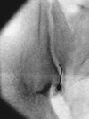
Try to get them lined up as straight on the table as you can. Positioning is step one in getting a good image. After that, I usually use a combination of rotating the table and the C arm to get my best picture. If you're not perfectly clear on what you need your picture to look like, reinforce that anatomical knowledge with a bookSounds like a basic question but sometimes when I use the wigwag I notice I have to go quite a bit in terms of the angulation which leads to the target being at the edge of the image so when I slide the base of the machine to get the target back in range of the source it seems all the work I put in with the wigwag went to waste as the image needs to be lined up again. Does anyone else have this issue or tips for this?
-
CycleTrack and SDN are teaming up to make medical school admissions more transparent and accessible! Read the announcement to learn how we’re supporting the future of CycleTrack’s free application-tracking tools .
You are using an out of date browser. It may not display this or other websites correctly.
You should upgrade or use an alternative browser.
Si joint injection
Started by PinchandBurn
Get help with your application
Use all the free resources available to you from SDN: articles, guides, expert advising, forums discussions, and school research.
Try to get the patient "straight." You have to guess and mess with optimizing wig-wag in the lateral, but once there, you don't need to go "back" to "Zero" when coming back to AP. Keep the machine where it is and the tech can just rotate the AP image on the screen so it looks plumb.Sounds like a basic question but sometimes when I use the wigwag I notice I have to go quite a bit in terms of the angulation which leads to the target being at the edge of the image so when I slide the base of the machine to get the target back in range of the source it seems all the work I put in with the wigwag went to waste as the image needs to be lined up again. Does anyone else have this issue or tips for this?
This doesn't help with the initial waste of time, but prevents repeating it.
thanks guys, its helpful. I only really have this issues on the occasion with very large patients and a S1 transforaminal when I try to line up the iliopectinal lines. I think I will have to be a bit more strict with them to try to lie as midline as possible and "evenly distribute" their extra body habitus when lying down 😛
Wigwag when it looks like my kypho is spot on in AP but inferior to the pedicle on lateral.
Don't care what anyone calls it. Just use your hands and make the motion of the direction you want it to go in.
Cranial tilt the ii (bring the top of the machine towards the head)
Caudal tilt the ii (bring the tube at the bottom of the machine toward the head)
Rotate the C towards me (ii towards me)
Rotate away (ii away from me)
Piston towards or away from me.
Wigwag: move the entire base of the machine to be parallel towards what we are looking at so we can eliminate parallax.
I aspire for my techs and I do have this kind of communication. If I lose some fingers, so be it.
Awesome! Just make sure it's your non-dominant hand and/or have good "own occupation" disability insurance.
He's actually PM&RAnother family practice pain surgeon
Really? Why’s he advertising sports med? Did he do a fellowship?He's actually PM&R
Believe it was a nonacgme but well reputed sports and spine fellowshipReally? Why’s he advertising sports med? Did he do a fellowship?
Image #2 here is what I was wondering if “forwho” was asking about when they said “are you tying to be coaxial or lateral to medial”, because I was a little confused at that image. Even if the detector is contralateral your needle there looks like you are trying to angle in towards the left… I think. The second series of images you posted below it looks more coaxial. I have tried your method four times and have gotten crazy contrast but have not checked lateral yet. Maybe that’s my problem. Will try that tomoro.Here are some images of an SI-J I did today.
This is on the right side - not a true AP. The fluorscope is tilted caudad (with reference to the detector, not the source) and contra-lateral. The caudad tilt brings the Posterior Superior Iliac Spine (PSIS) to (about) mid joint. Then, rotate contralateral (towards midline) until the two joint lines cross just under the PSIS. This will usually give a flashlight sign - meaning, you get a small square that lights up. This is your spot. It is extremely rare that if you line it up as such, that the needle doesn't slide in the joint.
View attachment 364962
The red line highlights the PSIS. The blue lines highlight the two joint lines as they cross right below the PSIS.
View attachment 364963
The lateral in this injection is not as far anterior as I would have liked to go. However, as mentioned above, doing these under CT has given me a much great appreciated for how these joint edges are - and sometimes, there is no way you can go further because of the jagged edges.
View attachment 364964
Had a great lateral today. AP and CLO as well. I don't always do this but sometimes I start inferior to inferior joint opening and advance cephalad into it. Posterior-inferior approach, hits capsule pretty deep, well past anterior margin of sacrum. Sometimes easier than messing around trying to figure out which joint line is anterior and posterior, lining them up, etc.
AP and CLO aren't great but you can see vertical flow. And that half-crescent shape that someone else mentioned usually means you're in. Yeah I was taught that way too, don't superimpose, go for the medial line. But medial isn't always posterior either.If I never did a lateral I would assume based on your AP that it was totally not in the joint. Also never did an injection without seeing clear separation of medial and lateral heads on AP. Was always taught the superimposed view was the the wrong view
If I never did a lateral I would assume based on your AP that it was totally not in the joint. Also never did an injection without seeing clear separation of medial and lateral heads on AP. Was always taught the superimposed view was the the wrong view
5% of the time the medial joint line is actually the ventral joint line. Benefit of superimposed lines is you don’t have to worry about that.
I did not know that. Citation or empirical? Though that is about the rate where I give up on my ipsilateral oblique approach separating the joint lines, pull out and just overlap them w clo then re-stick.5% of the time the medial joint line is actually the ventral joint line. Benefit of superimposed lines is you don’t have to worry about that.
I did not know that. Citation or empirical? Though that is about the rate where I give up on my ipsilateral oblique approach separating the joint lines, pull out and just overlap them w clo then re-stick.
Modified Fluoroscopy-Guided Sacroiliac Joint Injection: A Technical Report
AbstractBackground and Objective. Sacroiliac joint (SIJ) injection can occasionally be challenging. We describe our experience in using conventional techni
In this paper, first they overlapped the lines. When that failed, they obliqued to separate the lines. When the lines were separated, 1 out of 18 successful injections required entering the lateral line. Small N, but I think we all have been in those head scratcher moments where it looks wide open and then you hit bone very superficially. Not surprising given how varied these joints are from person to person.
D
deleted131481
5 degrees oblique and you'll know if medial is the ventral joint line.5% of the time the medial joint line is actually the ventral joint line. Benefit of superimposed lines is you don’t have to worry about that.
I was taught to superimpose. After enough struggling over my first year or so, I keep separation now. Less fluoro and higher success rate in my hands.
Edit: typo
Last edited by a moderator:
Can you explain that? You aim for medial line always?5 degrees oblique and you'll know if medial is the central joint line.
I was taught to superimpose. After enough struggling over my first year or so, I keep separation now. Less fluoro and higher success rate in my hands.
D
deleted131481
I had a typo, should have said ventral.Can you explain that? You aim for medial line always?
If you have straight AP you see medial and lateral joint lines. If you oblique 5° away from the side of the SI joint, and the joint lines are getting closer, then you know your medial line is posterior and your lateral line is anterior. If they're getting farther apart then the opposite is true which is rare. In my opinion it's not necessary to line them up.
Edit: another typo
Last edited by a moderator:
Anyone try entering at the superior portion of the joint? Had some attendings that did it in training from time to time but never really caught on. Seemed to give a higher rate of intraarticular from my limited exposure
Do you mean the superior part of the joint that is not a synovial joint but as a syndesmosis?Anyone try entering at the superior portion of the joint? Had some attendings that did it in training from time to time but never really caught on. Seemed to give a higher rate of intraarticular from my limited exposure
Yeah, I was a bit confused cause it went against what I read and was taught. Never really questioned it much in training because only a few people ever did it this way. Was able to find some pictures online that looked like what they did
SI-Joints are weird.
True, but a nice, clean crisp arthrogram like the one above is still so amazingly satisfyingSI-Joints are weird.
I could feel myself entering the capsule, then joint (you can see - red arrows - where the needle enters the joint and is deflected, and then the needle is again deflected within the joint).
Lateral shows my needle well within the joint.
I'm not sure what contrast will tell me at this point, other than give me a satisfying feeling of a cool contrast patern. If I get a crappy contrast pattern, what would I do since I already know I'm in the joint?
Also, nothing is free. If this happens to be the one patient that has a significant anaphylaxis reaction, how does my day go now?
Lateral shows my needle well within the joint.
I'm not sure what contrast will tell me at this point, other than give me a satisfying feeling of a cool contrast patern. If I get a crappy contrast pattern, what would I do since I already know I'm in the joint?
Also, nothing is free. If this happens to be the one patient that has a significant anaphylaxis reaction, how does my day go now?
Attachments
Patient with a contrast allergy so had to rely on imaging alone. My question is, do you tilt cephalad/caudad to line up the inferior edge of the joint (red arrow), or not needed? Thanks in advance
A New Sacroiliac Joint Injection Technique and Its Short-Term Effect on Chronic Sacroiliac Region Pain
Abstract. Objectives. Sacroiliac joint (SIJ) injections have been used to provide short-term relief of SIJ pain. In this study, the authors investigated a
@epidural man
Is this similar to your technique?
doi.org
Is this similar to your technique?
Far-Contralateral Oblique (FCO) Sacroiliac Joint Injection: Description of a Novel Technique
Sacroiliac (SI) joint arthropathy is the primary pain generator in approximately 15–25% of patients with axial low back pain and traditionally diagnosed with >50% pain reduction following an intra-articular injection localized to the inferior 1/3 of the SI joint. The conventional technique for...
Ple
Please dont post anything else from this shill unless it is to make fun of him.
His interacept talk at SIS a few years ago was embarrassing.
Well, I took a brief look and it does seem similar.@epidural man
Is this similar to your technique?
Far-Contralateral Oblique (FCO) Sacroiliac Joint Injection: Description of a Novel Technique
Sacroiliac (SI) joint arthropathy is the primary pain generator in approximately 15–25% of patients with axial low back pain and traditionally diagnosed with >50% pain reduction following an intra-articular injection localized to the inferior 1/3 of the SI joint. The conventional technique for...doi.org
I’ll take a deeper dive when I’m not at Indian Wells watching tennis.
Last edited:
So I changed my technique after reading through this thread. I had previously gone 15 degrees CLO and aimed for the Y point, but would rarely get a true arthrogram with that approach. You can see that it’s spreading along the joint, posteriorly, but not into the joint. Works fine in the younger patients who almost all seem to have ligament-based pain anyway, but especially with the Medicare changes I worry about efficacy if it’s not actually into their arthritic joint. Also I just like the look of a perfect arthrogram.
Today I did somewhere between straight AP and 5 degrees CLO, caudal tilt, aiming for the inferior cm or so of the joint. Got an arthrogram on 5/5 SI joints injections. The needle did not go very far into the joint itself, so without contrast I would not have known I was in.
Hate to break it to you, but she says that to all the boysmy rad techs tell me I have a way higher percentage of getting an arthrogram than the rest of my colleagues
This looks really nice. Sometimes I get that dark black spread that you have in the bottom of the screen around the 5oclock position despite having a arthrogram. Any idea what that is?Inferomedial to joint line. Come in at a 1 o'clock orientation. I did this shot 20 min ago.
View attachment 368095
Anyone do cranial tilt (with respect to top part of the machine, the detector) and CLO and go in bottom centimeter of joint?
I usually do caudalAnyone do cranial tilt (with respect to top part of the machine, the detector) and CLO and go in bottom centimeter of joint?