You are using an out of date browser. It may not display this or other websites correctly.
You should upgrade or use an alternative browser.
You should upgrade or use an alternative browser.
SIJ injection tips
- Thread starter needledox
- Start date

- Joined
- Aug 4, 2017
- Messages
- 695
- Reaction score
- 633
- Points
- 3,111
Contralateral oblique to open up the joint space. I aim for inferior medial border. Use 25 g. Confirm Contrast in the joint. Don’t use too much volume. I then inject 2 cc - kenalog 40 and marcaine 0.25%
- Joined
- Apr 13, 2016
- Messages
- 3,558
- Reaction score
- 3,698
- Points
- 6,696
- Location
- California
- Attending Physician
I use a 22g for better tactile feedback. Start 15 degrees oblique contralateral to the joint but oblique back and forth a little until the inferior margin opens up. Once you feel like it’s in a good spot, get a lateral to confirm.I’ve been struggling with these a bit as of late, what are some of your tips? Do you oblique the fluoro for better visualization?
- Joined
- Jan 26, 2017
- Messages
- 1,537
- Reaction score
- 489
- Points
- 3,356
Do you put any caudal or cranial in?
- Joined
- Nov 11, 2012
- Messages
- 2,352
- Reaction score
- 1,626
- Points
- 5,726
- Attending Physician
Do you put any caudal or cranial in?
Caudal ~20-30 degrees along with contralateral oblique.
- Joined
- Aug 18, 2011
- Messages
- 501
- Reaction score
- 136
- Points
- 4,896
- Attending Physician
You can also use 2 needle technique until you get back to more success.
- Joined
- Jan 26, 2017
- Messages
- 1,537
- Reaction score
- 489
- Points
- 3,356
Straight AP. Aim for more medial line. Since I started doing this I’ve been more successful with getting intraarticular.
So you don’t line up the two lines?
- Joined
- Jan 9, 2010
- Messages
- 14,272
- Reaction score
- 8,132
- Points
- 8,286
- Attending Physician
For me it depends how far they are apart. If they're right next to each other, I'm not sure which is posterior, so I line them up. If they're far apart, I stay AP and go for the medial line.So you don’t line up the two lines?
Advertisement - Members don't see this ad
If you have access to CT lumbar, or abdominal/pelvis CT the axial cuts will give you the perfect trajectory. It's amazing how variable they are, and how often an osteophyte blocks off part of the joint.
If no access to these images I agree with generally going slightly contralateral, but will take pics at multiple angles from slight ipsilateral to contra looking for crisp joint lines and joint to open.
If no access to these images I agree with generally going slightly contralateral, but will take pics at multiple angles from slight ipsilateral to contra looking for crisp joint lines and joint to open.
- Joined
- Mar 30, 2003
- Messages
- 3,411
- Reaction score
- 1,272
- Points
- 5,881
- Location
- Northeast
- Attending Physician
My technique:
Bent 23g needle.
Start 15° contralateral oblique with enough caudal tilt to get the PSIS away from the joint line.
Aim somewhere below the PSIS along the joint line.
Land at what looks like the joint line. You will probably land short of the joint.
Swing back to straight AP.
Redirect, and advance along the sacrum until you're wiggling into the joint.
Inject dye. Not every joint is going to give you the perfect arthrogram.
For extra credit, check a lateral to make sure you are cephalad enough.
Last edited:
- Joined
- Jan 26, 2017
- Messages
- 1,537
- Reaction score
- 489
- Points
- 3,356
“For extra credit, check a lateral to make sure you are cephalad enough.”My technique:
Bent 23g needle.
Start 15° contralateral oblique with enough caudal tilt to get the PSIS away from the joint line.
Aim somewhere below the PSIS along the joint line.
Land at what looks like the joint line. You will probably land short of the joint.
Swing back to straight AP.
Redirect, and advance along the sacrum until you're wiggling into the joint.
Inject dye. Not every joint is going to give you the perfect arthrogram.
For extra credit, check a lateral to make sure you are cephalad enough.
What does this mean?
- Joined
- Mar 30, 2003
- Messages
- 3,411
- Reaction score
- 1,272
- Points
- 5,881
- Location
- Northeast
- Attending Physician
“For extra credit, check a lateral to make sure you are cephalad enough.”
What does this mean?
Maybe not the best choice of words. Unless you look lateral, it's pretty easy to go through and through the joint, or place the needle in the caudad aspect of the joint and not get enough flow toward the superior aspect of the joint. In lateral you can redirect in a cephalad direction.
- Joined
- May 8, 2004
- Messages
- 4,275
- Reaction score
- 2,008
- Points
- 5,796
- Attending Physician
So you don’t line up the two lines?
Not anymore. I think I read about doing it this way on this forum and have found more success.
- Joined
- Aug 16, 2007
- Messages
- 7,422
- Reaction score
- 4,375
- Points
- 6,611
- Attending Physician
Exactly!Straight AP. Aim for more medial line. Since I started doing this I’ve been more successful with getting intraarticular.
- Joined
- Jun 20, 2012
- Messages
- 276
- Reaction score
- 380
- Points
- 4,846
- Attending Physician
Straight AP. Aim for more medial line. Since I started doing this I’ve been more successful with getting intraarticular.
What he said. That whole CLO stuff is ridiculous and often times if you go AP you see you're nowhere close.
- Joined
- Dec 13, 2005
- Messages
- 7,039
- Reaction score
- 5,782
- Points
- 6,481
- Attending Physician
Yep, straight AP and go for the medial line. Ralph Justiz taught me that one. Can numb up both sides using one fluoro shot. Saving time and rad exposure over a career.
- Joined
- Jul 27, 2017
- Messages
- 191
- Reaction score
- 271
- Points
- 2,781
- Attending Physician
Ipsilateral oblique was a game changer for me, hit joint that moves medial.
- Joined
- Apr 20, 2018
- Messages
- 2,168
- Reaction score
- 1,659
- Points
- 2,156
- Attending Physician
Ipsilateral oblique. Medial joint line is posterior. Go to the inferior aspect.
Advertisement - Members don't see this ad
- Joined
- Jun 3, 2007
- Messages
- 4,912
- Reaction score
- 3,319
- Points
- 6,651
- Age
- 56
- Location
- San Diego
- Attending Physician
My technique:
Bent 23g needle.
Start 15° contralateral oblique with enough caudal tilt to get the PSIS away from the joint line.
Aim somewhere below the PSIS along the joint line.
Land at what looks like the joint line. You will probably land short of the joint.
Swing back to straight AP.
Redirect, and advance along the sacrum until you're wiggling into the joint.
Inject dye. Not every joint is going to give you the perfect arthrogram.
For extra credit, check a lateral to make sure you are cephalad enough.
I do this too.
Identify the PSIS - tilt cameral caudal. (It's interesting to note that if you go straight AP and don't make a point to identify the PSIS - you will never notice that often it is sitting right where you think you need to go).
Once the PSIS is halfway up the joint (or so...), move the collector back and forth (most often is contralateral) until both joint lines cross RIGHT UNDER the PSIS (it will form a white box). Enter the box.
I no longer use contrast. You can feel yourself drop into the joint. I use a lateral to make sure I am in the joint, and far enough anterior.
The problem with not lining up the lines - is despite the joints USUALLY tilting medial to lateral (from a posterior to anterior direction), I have done enough of these injections under CT to notice that the joint tilt is not the same on everyone.
- Joined
- Aug 18, 2011
- Messages
- 501
- Reaction score
- 136
- Points
- 4,896
- Attending Physician
I do this too.
Identify the PSIS - tilt cameral caudal. (It's interesting to note that if you go straight AP and don't make a point to identify the PSIS - you will never notice that often it is sitting right where you think you need to go).
Agree. This is specially true when some literature recommends cephalad tilt leading to PSIS obstructing your target.
But after, you have tried and failed, take solace in this...
In 2007, Murakami et al studied whether intra-articular or periarticular injections were more effective at relieving SI joint pain. [48] They initially gave patients intra-articular SI joint injections and then performed additional periarticular injections in the patients who experienced no improvement from the intra-articular injection. The improvement from the periarticular injections was significantly higher than that from the intra-articular injections. This result could have various causes, including the following:
- The interosseous membrane and the surrounding ligaments have nociceptive fibers, and this may be part of the reason why the injection is effective even if it is periarticular
- The additional quantity of steroid may also be responsible for the improvement with the second injection
- The patients may have been initially misdiagnosed; if they were, in fact, experiencing pain from soft tissue dysfunction, their symptoms may have been improved by the extra-articular injection
In a 2016 study of 113 fluoroscopically guided SI joint injections in 99 patients, Nacey et al found that after adjustments were made for age, sex, preinjection pain score, time of year, and indication for injection, there was no significant difference between intra-articular injection and periarticular injection in terms of the degree of pain relief achieved. [49]
- Joined
- Oct 23, 2005
- Messages
- 8,317
- Reaction score
- 6,057
- Points
- 7,396
- Attending Physician
bringing this topic back up.
One thing I find odd, is that several procedural atlases, will recommend moving the C-arm cranial and several atlases recommend moving the C-arm caudal. Several atlases recommend moving C-arm ipsilateral oblique and several recommend contralateral oblique. Same thing with the posts on this thread.
I'm inclined to start using Doctors Jays/Bob Barkers SIJ approach since a straight AP is the quickest and most direct.
Such a weird procedure in that several experienced pain physicians who posted in this thread above, all gave exactly opposite recommendations regarding cranial vs caudal and ips vs contra oblique?
Last edited:
- Joined
- Aug 16, 2007
- Messages
- 7,422
- Reaction score
- 4,375
- Points
- 6,611
- Attending Physician
I agree with straight AP. I go straight AP and aim towards the inferior medial joint line. I then jimmy the needle and use a little muscle if need be but have increased my successful arthrogram rate to 90% using this method, no exaggerationbringing this topic back up.
One thing I find odd, is that several procedural atlases, will recommend moving the C-arm cranial and several atlases recommend moving the C-arm caudal. Several atlases recommend moving C-arm ipsilateral oblique and several recommend contralateral oblique. Same thing with the posts on this thread.
I'm inclined to start using Doctors Jays/Bob Barkers SIJ approach since a straight AP is the quickest and most direct.
Such a weird procedure in that several experienced pain physicians who posted in this thread above, all gave exactly opposite recommendations s to cranial vs caudal and ips vs contra oblique?
The other guys in my practice use the contralateral approach lining up the joint lines. I rarely see them get good arthrograms. One doesn’t even inject contrast anymore bc I think he knows he’ll just get a blobogram.
- Joined
- May 8, 2004
- Messages
- 4,275
- Reaction score
- 2,008
- Points
- 5,796
- Attending Physician
bringing this topic back up.
One thing I find odd, is that several procedural atlases, will recommend moving the C-arm cranial and several atlases recommend moving the C-arm caudal. Several atlases recommend moving C-arm ipsilateral oblique and several recommend contralateral oblique. Same thing with the posts on this thread.
I'm inclined to start using Doctors Jays/Bob Barkers SIJ approach since a straight AP is the quickest and most direct.
Such a weird procedure in that several experienced pain physicians who posted in this thread above, all gave exactly opposite recommendations s to cranial vs caudal and ips vs contra oblique?
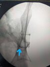
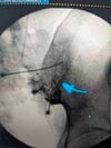
Skin marker over the target, no skin numbing, 25 gauge 3.5” needle, drop into joint, contrast, inject, done.
Edited by Lobel: Placed thumbnail showing needle trajectory medial to lateral as first picture and then arthrogram without needle as second picture.
Last edited:
- Joined
- Oct 28, 2009
- Messages
- 759
- Reaction score
- 536
- Points
- 4,831
- Attending Physician
Skin marker over the target, no skin numbing, 25 gauge 3.5” needle, drop into joint, contrast, inject, done.
Edited by Lobel: Placed thumbnail showing needle trajectory medial to lateral as first picture and then arthrogram without needle as second picture
So AP of the sacrum (with sacrum lined up (typically caudal tilt)) or AP just no tilt and whatever you see under there you go for. If the picture doesn't look like what you posted do you start tilting until you see the joints separated
- Joined
- May 8, 2004
- Messages
- 4,275
- Reaction score
- 2,008
- Points
- 5,796
- Attending Physician
typically just AP without any tilt or oblique. If I can see a medial line that's what I go for and walk in lateral a bit or the needle will bend that way. very rarely I'll ask for some oblique to see that medial line better.
- Joined
- May 30, 2010
- Messages
- 2,422
- Reaction score
- 2,296
- Points
- 5,626
- Attending Physician
A slight medial to lateral approach helps with getting intraarticular in most patients as well. The ilium is superficial to the sacrum, so this needle trajectory helps slide under it and into the joint.

- Joined
- Apr 28, 2010
- Messages
- 1,770
- Reaction score
- 1,902
- Points
- 6,296
I start with AP no oblique, no tilt. Most the time I get it going very inferior tip, medial line.
If spread isn't linear, I CLO 10-15 deg, try again.
If spread still isn't linear, I do far CLO mid-joint. David Lee technique paper.
 www.ncbi.nlm.nih.gov
It took going far CLO for me to get this one.
If spread isn't linear, I CLO 10-15 deg, try again.
If spread still isn't linear, I do far CLO mid-joint. David Lee technique paper.
Far-Contralateral Oblique (FCO) Sacroiliac Joint Injection: Description of a Novel Technique - PMC
Sacroiliac (SI) joint arthropathy is the primary pain generator in approximately 15–25% of patients with axial low back pain and traditionally diagnosed with >50% pain reduction following an intra-articular injection localized to the inferior 1/3 of ...
www.ncbi.nlm.nih.gov
It took going far CLO for me to get this one.
Attachments

- Joined
- Nov 28, 2011
- Messages
- 2,722
- Reaction score
- 1,748
- Points
- 6,761
- Location
- New Jersey
- Attending Physician
Apologies if this point has already been made.
A Tim Maus (Mayo Radiology/ IPSIS past President) pearl is to search chart for abdomen/ pelvis CT at any time in the past. It’s surprising how many patients, especially females, have had this study. Look at axial images at about the level of S3 foramen and you can see exact trajectory to posterior aspect of joint. You will also see if joint ankylosed, osteophyte overlying path to joint, etc. You will be surprised how often a straight AP approach is best as well as how often a lateral to medial approach is warranted. Granted, finding these studies is easiest if you are in a health system where there are no independent radiological practices.
A Tim Maus (Mayo Radiology/ IPSIS past President) pearl is to search chart for abdomen/ pelvis CT at any time in the past. It’s surprising how many patients, especially females, have had this study. Look at axial images at about the level of S3 foramen and you can see exact trajectory to posterior aspect of joint. You will also see if joint ankylosed, osteophyte overlying path to joint, etc. You will be surprised how often a straight AP approach is best as well as how often a lateral to medial approach is warranted. Granted, finding these studies is easiest if you are in a health system where there are no independent radiological practices.
Advertisement - Members don't see this ad