TFESI Troubles

This forum made possible through the generous support of SDN members, donors, and sponsors. Thank you.

jwalker12

Full Member
15+ Year Member
Joined
Aug 25, 2008
Messages
189
Reaction score
46
Hello again everyone!

I have some questions about TFESIs that have been causing me stress.
1.) I go typically at the neck of the scotty dog and try to walk off. Typically line up end plate and oblique 20-25 on average. Some how on AP I am frequently ending up too medial, past 6'o'clock on pedicle. What am I doing wrong?
2.) What techniques do you use if there is significant arthritis preventing you from entering in the technique mentioned above? Do you go infraneural?
3.) What is best way to have success at L5-S1, I find this area often has significant arthritis etc and prevents me from entering correctly
4.) If a patient has a sacaralized L5, can you enter as if it was a traditional S1? and if a Lumbarized sacrum do you enter as a traditional lumbar TFESI? Or would you just do a Interlaminar at that level as the in theory the ligament should be fine. Thanks so much everyone.
dont go AT the neck. that is why you end up too medial. should be right underneath the middle of the jaw

sometimes L5-S1 needs a little tilt
 
My technique:

1. I usually go around 25* and touch os at the neck as well. You're likely just targeting too medial a spot on the pedicle.
2. Infraneural, or keep on obliquing until things open up
3. For L5-S1, I usually utilize at least some cephalad tilt (maybe don't need it 25-50% of the time). Most often 5* is enough, but sometimes it's 10-20*. Usually use 25* oblique, but sometimes have to take 5* out to visualize the triangle around the medial aspect of the ilium
4) Yes, treat sacralized L5-S1 as S1 TFESI, and can do lumbarized S1 as L5-S1 ILESI or TFESI
 
I do this technique now exclusively.

Caudal tilt until pedicle shadow is below end plate.
Oblique until you can see VB underneath pedicle
Go coaxial under pedicle until you hit VB
Put dye in
 
-agree neck too medial, go under 6 o'clock pedicle on Scotty, or a bit more lateral than that
-i probably oblique 20-40 from upper to lower, even more than that if big facets
-with big facets, just oblique more. With osteophytes off TP, caudal tilt a bit or start lower and angle up. More bend in needle, 22 ga for steerability. Sometimes you just need to go through a facet joint capsule, essentially LOR technique with contrast going through capsule to epidural
 
Hello again everyone!

I have some questions about TFESIs that have been causing me stress.
1.) I go typically at the neck of the scotty dog and try to walk off. Typically line up end plate and oblique 20-25 on average. Some how on AP I am frequently ending up too medial, past 6'o'clock on pedicle. What am I doing wrong?
2.) What techniques do you use if there is significant arthritis preventing you from entering in the technique mentioned above? Do you go infraneural?
3.) What is best way to have success at L5-S1, I find this area often has significant arthritis etc and prevents me from entering correctly
4.) If a patient has a sacaralized L5, can you enter as if it was a traditional S1? and if a Lumbarized sacrum do you enter as a traditional lumbar TFESI? Or would you just do a Interlaminar at that level as the in theory the ligament should be fine. Thanks so much everyone.

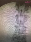
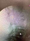
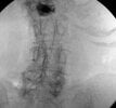
1. Line up SEP
2. 15 degree ipsi oblique to get the scotty dog.
3. start at the lateral inferior edge of the VB border.
4. move superiomedially towards the neck of the scotty dog. Alternatively, coaxial aim to pedicle. then walk off inferiorly and slip into the foramen.
5. contrast.
 
1. Line up SEP
2. 15 degree ipsi oblique to get the scotty dog.
3. start at the lateral inferior edge of the VB border.
4. move superiomedially towards the neck of the scotty dog. Alternatively, coaxial aim to pedicle. then walk off inferiorly and slip into the foramen.
5. contrast.
This is the way. Should be near subpedicular in the AP view and clearly in the foramen on lateral. Being too posterior in the foramen on lateral is typically the problem with contrast only outlining the spinal nerve. I usually have to advance a few mm to get that cephalad epidural spread.
 
1) get pedicle under end plate- usually caudal tilt
2) oblique until you see VB. Hit from inferior to superior mid pedicle
3) get AP, slide under pedicle but face away from nerve. Hit contrast
4) lateral
 

Attachments

  • image.jpg
    image.jpg
    228.6 KB · Views: 252
  • image.jpg
    image.jpg
    246.2 KB · Views: 249
  • image.jpg
    image.jpg
    239.9 KB · Views: 244
  • image.jpg
    image.jpg
    178.6 KB · Views: 247
1) get pedicle under end plate- usually caudal tilt
2) oblique until you see VB. Hit from inferior to superior mid pedicle
3) get AP, slide under pedicle but face away from nerve. Hit contrast
4) lateral
Would have been perfect if you had a way to upload to pacs and then SNIP images rather than cellphone camera on 60hz screen.
 
Here's another example
Perfect everytime if you go anterior above nerve (true supraneural)
Hitting VB is fine (don't have significant issue of "spearing nerve" as long as you don't get paresthesia, which when you align pedicle underneath VB endplate, you're usually coming at an angle that should prevent. Obviously don't continue to advance if you get paresthesia.
In this example, I had caudal tilt of 15 to get pedicle close to being underneath endplate
 

Attachments

  • 2.CaudalTilt.GetPedicle.jpg
    2.CaudalTilt.GetPedicle.jpg
    21 KB · Views: 254
  • 1.APwithoutcaudaltilt.jpg
    1.APwithoutcaudaltilt.jpg
    24.5 KB · Views: 245
  • 3.Startinferior.tosuperior.jpg
    3.Startinferior.tosuperior.jpg
    21.7 KB · Views: 247
  • 4.Hitlowerpedicleshadow.jpg
    4.Hitlowerpedicleshadow.jpg
    22.1 KB · Views: 257
  • 5.Swoopunderneathpedicle.jpg
    5.Swoopunderneathpedicle.jpg
    23.4 KB · Views: 252
  • 6.GetLateral.jpg
    6.GetLateral.jpg
    18.3 KB · Views: 264
Last edited:
Here's another example
Perfect everytime if you go anterior above nerve (true supraneural)
Hitting VB is fine (don't have significant issue of "spearing nerve" as long as you don't get paresthesia, which when you align pedicle underneath VB endplate, you're usually coming at an angle that should prevent. Obviously don't continue to advance if you get paresthesia.
In this example, I had caudal tilt of 15 to get pedicle close to being underneath endplate
that's perty. Picks like that get me excited. However if I posted a pic like that everyone would be yelling at me to collimate. Just sayin
 
that's perty. Picks like that get me excited. However if I posted a pic like that everyone would be yelling at me to collimate. Just sayin
Aside from significant foraminal stenosis or recess narrowing (at same level), this technique has always given me textbook supraneural pics

Collimation depends in my practice on skill of rad tech. Some days I do it and some days I can’t
 
Here's another example
Perfect everytime if you go anterior above nerve (true supraneural)
Hitting VB is fine (don't have significant issue of "spearing nerve" as long as you don't get paresthesia, which when you align pedicle underneath VB endplate, you're usually coming at an angle that should prevent. Obviously don't continue to advance if you get paresthesia.
In this example, I had caudal tilt of 15 to get pedicle close to being underneath endplate
Great share. Thanks so much. Thanks everyone for the help as well.
 
Aside from significant foraminal stenosis or recess narrowing (at same level), this technique has always given me textbook supraneural pics

Collimation depends in my practice on skill of rad tech. Some days I do it and some days I can’t
Pretty common and reasonable approach on a ‘virgin’ foramen… although some prefer to hit the pedicle for depth and walk inferior into the foramen. Both options may not work however on fusion/pedicle screw patients where a far lateral approach maybe easiest to access the foramen and avoid the BMP and osteophytes/post surgical overgrowth . Lobel has described this approach in the past …
 
Pretty common and reasonable approach on a ‘virgin’ foramen… although some prefer to hit the pedicle for depth and walk inferior into the foramen. Both options may not work however on fusion/pedicle screw patients where a far lateral approach maybe easiest to access the foramen and avoid the BMP and osteophytes/post surgical overgrowth . Lobel has described this approach in the past …
Would you mind posting some pics of difficult TFESI and sharing them.
Also, please view other posts
 
Pretty common and reasonable approach on a ‘virgin’ foramen… although some prefer to hit the pedicle for depth and walk inferior into the foramen. Both options may not work however on fusion/pedicle screw patients where a far lateral approach maybe easiest to access the foramen and avoid the BMP and osteophytes/post surgical overgrowth . Lobel has described this approach in the past …

Would you mind posting some pics of difficult TFESI and sharing them.
Also, please view other posts
Would love to see approaches described here. maybe some photos. fusion/pedicle screws always give me trouble and i find myself having to do >60 degree obliques to clear the obstruction.
 
Heresy I know, but I get better and longer lasting pain relief in this population doing a quick S1 TFESI with depo, than I do with a longer , complicated approach doing a TFESI at the exact pathology level using dex.
 
Here's another example
Perfect everytime if you go anterior above nerve (true supraneural)
Hitting VB is fine (don't have significant issue of "spearing nerve" as long as you don't get paresthesia, which when you align pedicle underneath VB endplate, you're usually coming at an angle that should prevent. Obviously don't continue to advance if you get paresthesia.
In this example, I had caudal tilt of 15 to get pedicle close to being underneath endplate
Nice pics. Curious if you get #5 pic with good spread, any benefit in obtaining lateral at that point?
 
Heresy I know, but I get better and longer lasting pain relief in this population doing a quick S1 TFESI with depo, than I do with a longer , complicated approach doing a TFESI at the exact pathology level using dex.
Hmm been debating this exact thing whether to switch to depo for my s1 tfesi. There's nothing there of concern.

Anyone else doing this?
 
Heresy I know, but I get better and longer lasting pain relief in this population doing a quick S1 TFESI with depo, than I do with a longer , complicated approach doing a TFESI at the exact pathology level using dex.
youre getting depo from S1 to L3? At that point, why not just do a caudal?
 
Heresy I know, but I get better and longer lasting pain relief in this population doing a quick S1 TFESI with depo, than I do with a longer , complicated approach doing a TFESI at the exact pathology level using dex.

Any tips on how to reliably identify S1 quickly?
 
Any tips on how to reliably identify S1 quickly?
Average S1 foramen obliquity is 25 degrees, so after cephalad tilt to square endplate, oblique 25. It's a big foramen and angle can vary, that's why AP can work or 15 degrees can work but usually 25 you'll see a big round hole.
 
S1 is the easiest thing we do.

Look at a model of the sacrum. Curved cranio-caudal as well as lateral to medial. So you will always need ipsilateral oblique and cranial tilt. It also almost always lines up with the pedicles of L4 and L5 vertically.

 
Look at Ferguson view, identify Charlie’s line (pedicle line)

Average S1 foramen obliquity is 25 degrees, so after cephalad tilt to square endplate, oblique 25. It's a big foramen and angle can vary, that's why AP can work or 15 degrees can work but usually 25 you'll see a big round hole.

S1 is the easiest thing we do.

Look at a model of the sacrum. Curved cranio-caudal as well as lateral to medial. So you will always need ipsilateral oblique and cranial tilt. It also almost always lines up with the pedicles of L4 and L5 vertically.


Thank you. I don't think I was going oblique aggressively enough. And I had too much fun with that 3d sacrum model.
 
Top