Confusion about location of malate-aspartate shuttle

This forum made possible through the generous support of SDN members, donors, and sponsors. Thank you.
D

deleted647690

In Kaplan biochem, they show cytosolic OAA reduced to malate so that it can cross the inner mitochondrial membrane from the cytosol into the mitochondrial matrix.

I'm confused by this, because isn't the mitochondrial matrix separated from the cytosol by both an inner and outer mitochondrial membrane? The diagram in Kaplan makes it look like there is only one membrane between the cytosol and matrix.


This diagram on wikipedia seems more accurate. However, it shows OAA as existing in the intermembrane space, contrary to what Kaplan says (Kaplan says it exists in the cytosol)
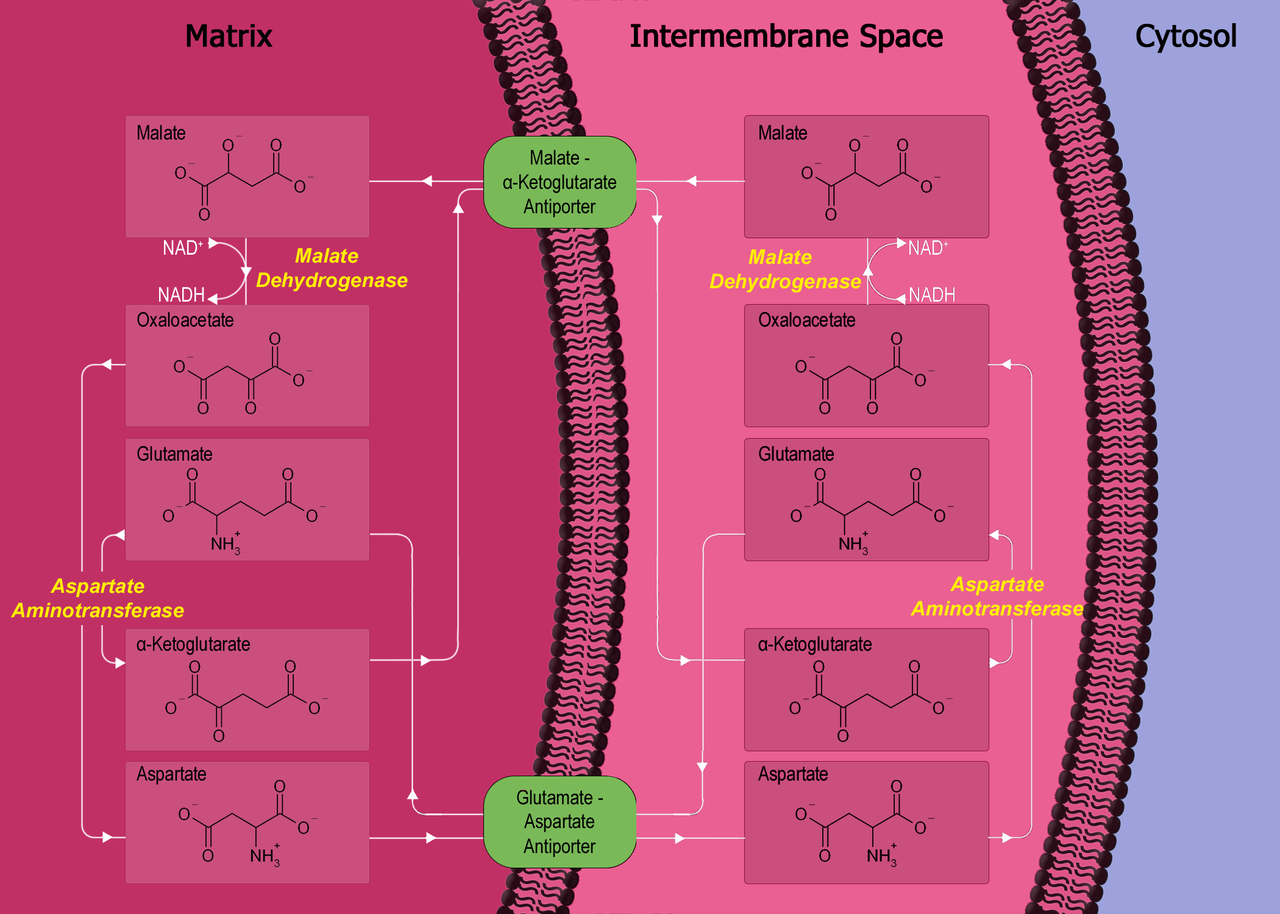
1280px-Malate-aspartate_shuttle.png


So, what is correct here?


Here is the diagram from Kaplan, which I'm not sure is accurate.

image227.jpg

Members don't see this ad.
 
There are two membranes, but the outer membrane is likely more porous. The takeaway is that oxaloacetate crosses the inner membrane through some kind of specific transport mechanism in it's reduced form as malate.

The point of reducing oxaloacetate -> malate isn't just so it can cross the membrane, it's so that malate can be oxidized inside the matrix, ultimately allowing for 2 electrons to be transferred from the cytosol to the matrix. Remember that the original two electrons were carried by NADH in the cytosol.


So OAA crosses both the inner and outer membrane after being reduced to malate? And thus it starts off in the cytosol?

Thank you
 
So OAA crosses both the inner and outer membrane after being reduced to malate? And thus it starts off in the cytosol?

Thank you

Let's think about a scenario like glycolysis. During glycolysis (in the cytosol) we take D-glucose and convert it to pyruvate. The net ATP output is 2 ATP and we also get 2 NADH. The 2 NADH carry high-energy electrons, but we need to get these electrons into the mitochondrial matrix for them to be useful. They can't help us by being carried in the cytosol.

So we need a way to transport high-energy electrons from the cytosol to the matrix- we use a shuttling system.

By reducing oxaloacetate -> malate in the cytosol, we have put 2 high-energy electrons on malate from NADH. Malate then crosses into the matrix and gets reoxidized to oxaloacetate (last step of the Kreb's cycle). In this last step, the high-energy electrons are now carried by NADH in the matrix, which can drop off the electrons at complex I of the ETC.

Note that the malate-aspartate shuttle system is specific to certain tissues like the liver, heart, and kidney. The brain and muscles use the g3p shuttle system (also important to know about).
 
  • Like
Reactions: 1 user
Members don't see this ad :)
Let's think about a scenario like glycolysis. During glycolysis (in the cytosol) we take D-glucose and convert it to pyruvate. The net ATP output is 2 ATP and we also get 2 NADH. The 2 NADH carry high-energy electrons, but we need to get these electrons into the mitochondrial matrix for them to be useful. They can't help us by being carried in the cytosol.

So we need a way to transport high-energy electrons from the cytosol to the matrix- we use a shuttling system.

By reducing oxaloacetate -> malate in the cytosol, we have put 2 high-energy electrons on malate from NADH. Malate then crosses into the matrix and gets reoxidized to oxaloacetate (last step of the Kreb's cycle). In this last step, the high-energy electrons are now carried by NADH in the matrix, which can drop off the electrons at complex I of the ETC.

Note that the malate-aspartate shuttle system is specific to certain tissues like the liver, heart, and kidney. The brain and muscles use the g3p shuttle system (also important to know about).


Thank you very much for the reply. I was mainly confused because the diagram from wiki shows OAA as being able to cross the outer mitochondrial membrane into the intermembrane space, where the reduction to malate occurs, which can then cross into the matrix.

On the other hand, kaplan shows OAA as not able to cross the outer mitochondrial membrane, thus it is oxidized to malate within the cytosol, but not in the intermembrane space.

So both diagrams are showing different locations for parts of the process


Also, I'm curious, you guys answering questions on here are probably extremely knowledgeable. When you answer questions, are you able to just come up with the answers on your own like these you have presented here? Just from memory and understanding from your own studies? Or do you consult resources? I'm assuming you have to consult resources since you most likely don't remember all of these tiny details.
 
I can only speak for myself, but the biochem knowledge above I have ingrained in me. Sometimes I look up an equation or a concept to explain it better, but for my test I knew pretty much everything that I could in terms of concepts and formulas.

Aim for deep understanding of the material as well :)
 
Top