Patient not dying?

In all seriousness clinical elements can be very helpful. If the sat improves, the heart rate stabilizes, color improves, etc. Im encouraged.
Second would be a good waveform capnograph.
Now all of these things get tricky especially with coding patients. I will say that I frequently have patients who are crumping, I tube them, they get worse, drop sats, brady down, I look and see that the tube is good, good sounds, no bounceback on the syringe (I like the esophageal detector device too
) and thats just how it goes. With a bad PE or an MI in cardiogenic shock an so on you can certainly have a good tube and a bad outcome.
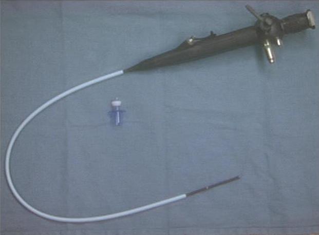
The gold standard is accepted as fiberoptic bronchoscopy. Obviously that isn't always an available option, but when it's there it takes 2 seconds to slip the bronch in and see some sort of lung anatomy. We've established difficult airway carts (at great expense) and distributed them across the hospital, including the ER. They have a full complement of fiberoptic scopes and ENT intruments, along of surgical airway kits.
I agree that clinical improvement helps add confidence. The benefit of a nonparalyzed patient is that they still have spontaneous ventilaton. So even if the tube is not in the trachea, these patients rarely suffer hypoxic brain injuries (related to the esophageal intubation).
Gold standard? Three of them - direct visualization (either outside with the eye or a camera or inside the tube with bronch), CT/MRI, and autopsy. Go for the first two.
Yes, but of the three you've listed, only bronch is reasonable realtime evidence. But we're talking about prehospital intubation, and I recognize that FOB isn't an option. What is available is capnography and esophageal detector devices. Sustained capnography, is probably the best option to establish intubation. But it doesn't help with mainstem placement or when the tip of the tube is above the cords (a scenario that I think happens very frequently in the field). Like usual, the pathologist is late to the party.
Even a combination of everything isn't 100% fool proof, but I think end-tidal CO2 detection (except in cardiac arrests), direct visualization, breath sounds, and oxygen saturation of 95+% (except in cardiac arrest) is required to confirm placement. The esophageal detector device should be used in cardiac arrests as an additional measure.
I am carefully reviewing intubations performed by paramedics where I am medical director. If there is a problem with undetected esophageal intubations, then I can easily revoke their airway privileges. Most studies I am familiar with don't show an improvement in mortality with patients tubed in the field.
I like the syringe version of the esophageal detector, but both the bulb and syringe have been shown to have false positive (esophageal) results from plugging in pulmonary edema/aspiration scenarios. As you guys have discussed the bad thing about capnography is that it depends on a reasonable cardiac output to produce and exchange gas.
As for the physical exam, I think it's much less reliable. I've heard breath sounds transmitted across the chest (plus we all know how noisy the diesel busses are). Chest rise is something that not easy to see. I'm a fan of ballotting the pilot balloon (high specificity, low sensitivity) in the suprasternal notch.
Back to the field, I think the most reasonable approach of confirming placement, understanding the limitations, would be
1) Direct visualization via laryngoscopy
2) Use of a syringe esophageal detector device for initial placement, with physical exam including ballottment of the balloon.
3) Continuous capnography
My argument that the tube doesn't commonly jump out of the trachea to the esophagus on transferring patients still stands.