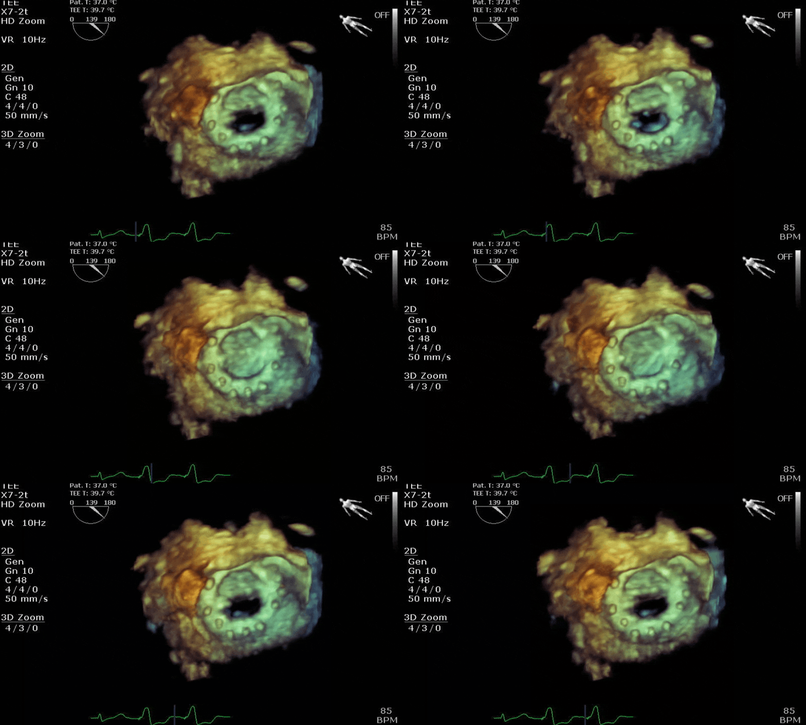
Let's do some echo:

This forum made possible through the generous support of SDN members, donors, and sponsors. Thank you.
Like @Hork Bajir was saying I was thinking pre-operatively. To evaluate someone's cardiac function and risk in preparation for a surgery (major or otherwise) what would you rather see? If a cath has been done, is there any point to getting a TEE? Or vice versa?

(This is all theoretical, I am taking oral boards soon and was wondering if I could argue for a simple TEE on said test)
Echo provides more qualitative and quantitative function of LV, RV, and valves. But preop, as a resting TTE/TEE it doesn’t really provide info regarding at risk myocardium. A stress echo sure.

LHC provides rudimentary LV function typically, and coronary disease which implies at risk myocardium.

You can have an echo that shows normal EF and no significant valvular pathology yet be at risk for a periop MI due to unknown CAD.

Two different tests.

Edit: I would study and understand the ACC/AHA guidelines. There’s a simple algorithm for preop testing. Know it. A pharmacological stress test is the study you’re looking for based on those guidelines/your question.

Members don't see this ad.
 
Last edited:
  • Like
Reactions: 3 users
Like @Hork Bajir was saying I was thinking pre-operatively. To evaluate someone's cardiac function and risk in preparation for a surgery (major or otherwise) what would you rather see? If a cath has been done, is there any point to getting a TEE? Or vice versa?

(This is all theoretical, I am taking oral boards soon and was wondering if I could argue for a simple TEE on said test)
It's good to know the ACC/AHA guidelines, RCRI, and the various tests available for pre-op stratification, but just FYI, 9 times out of 10 the examiner is looking for you to hold your ground and proceed with surgery without unnecessary tests and consults (assuming the pt can do 4 mets)
 
  • Like
Reactions: 1 users
But if you do need a test


Screenshot_20220305-230926_Opera.jpg

 
  • Like
Reactions: 1 user
Members don't see this ad :)
Anyone recognize this structure? I’ve never gotten such a nice picture of it before…

1647644398818.gif

(Hint, it’s not a cardiac structure. If you turn the probe to the right you’ll see the aorta, which is not seen in this image)
 
Anyone recognize this structure? I’ve never gotten such a nice picture of it before…

View attachment 351997
(Hint, it’s not a cardiac structure. If you turn the probe to the right you’ll see the aorta, which is not seen in this image)
tiny image...thoracic vertebra and sc.
 
  • Like
Reactions: 2 users
Anyone recognize this structure? I’ve never gotten such a nice picture of it before…

View attachment 351997
(Hint, it’s not a cardiac structure. If you turn the probe to the right you’ll see the aorta, which is not seen in this image)
One of my co-fellows recorded a really great TEE loop of a thoracic epidural being bolused.

Ooohs and aaaahs all around.

I'm pretty sure there was an image of the spine on my echo board exam too. Just the sort of thing they'd do to mess with us.
 
  • Like
Reactions: 1 users
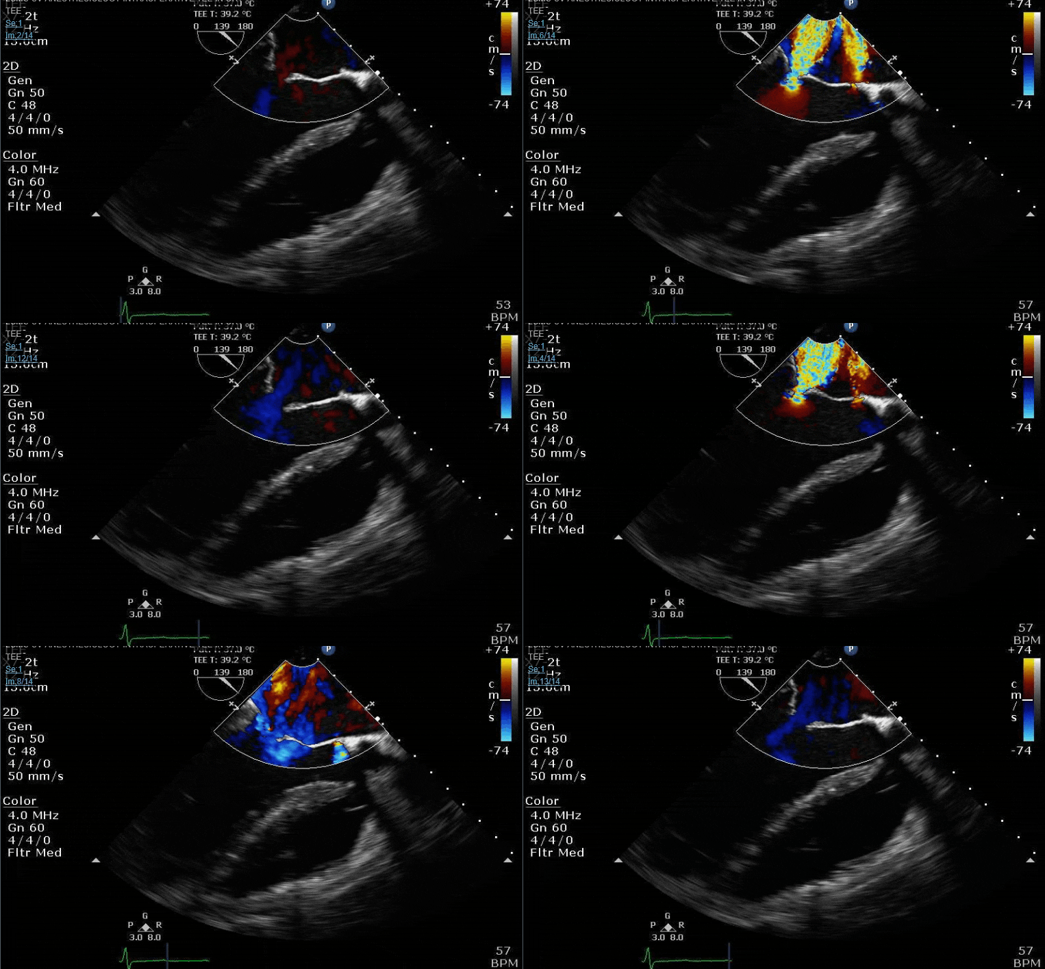
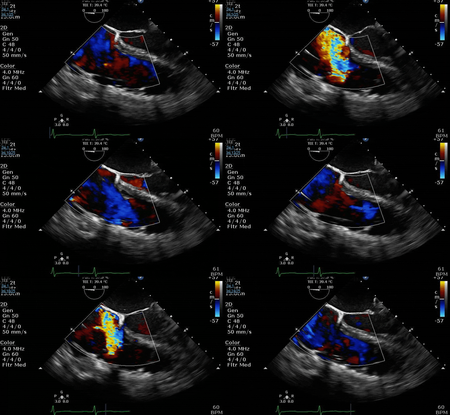
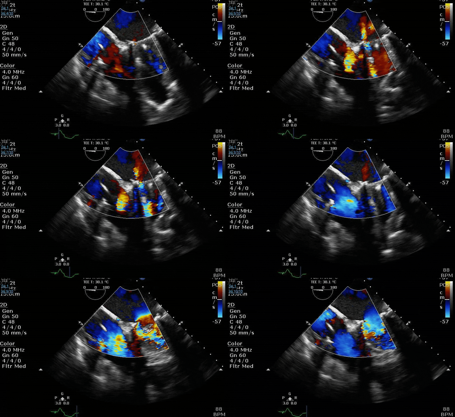
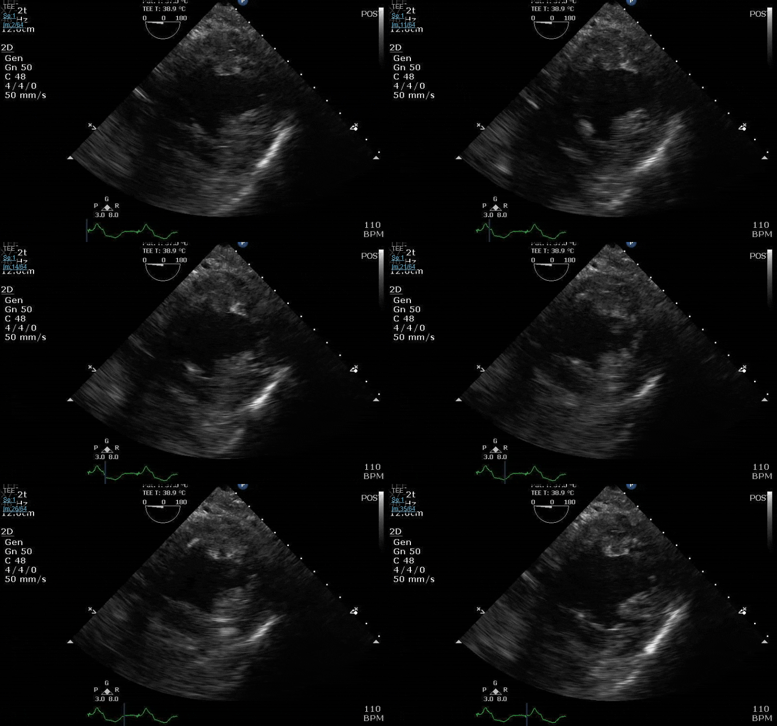
I love this series of clips- particularly the last picture, which nicely illustrates one of the (many) fundamental flaws of PISA, despite apparent agreement with 3D VCA in this case. Any current fellows want to explain what I’m getting at, particularly with the last picture? Do you think the “real” MR severity is more or less than what I’ve calculated here?

1652270200422.jpeg

1652270474994.jpeg

1652270314423.jpeg


1652270381820.jpeg
 
  • Like
Reactions: 1 user
I love this series of clips- particularly the last picture, which nicely illustrates one of the (many) fundamental flaws of PISA, despite apparent agreement with 3D VCA in this case. Any current fellows want to explain what I’m getting at, particularly with the last picture? Do you think the “real” MR severity is more or less than what I’ve calculated here?

Beautiful pictures!

I'm not an ACTA fellow, but I'm studying for my basic so I'll take a stab (also I love this thread). PISA makes the assumption that your MR jet is holosystolic. I think that your last photo is showing color / aliasing over time for your MR jet showing that it actually does vary (looks like it's higher at beginning and end systole).

My suspicion is this is overestimating your MR, since it's assuming the PISA you measured is consistent throughout but your photo shows that's clearly not true.
 
  • Like
Reactions: 1 users
Beautiful pictures!

I'm not an ACTA fellow, but I'm studying for my basic so I'll take a stab (also I love this thread). PISA makes the assumption that your MR jet is holosystolic. I think that your last photo is showing color / aliasing over time for your MR jet showing that it actually does vary (looks like it's higher at beginning and end systole).

My suspicion is this is overestimating your MR, since it's assuming the PISA you measured is consistent throughout but your photo shows that's clearly not true.
Bingo! You can actually measure the “area under the curve” in the last color M-mode shot and integrate that value to get a time-weighted “average” PISA radius over the course of systole, but you need special software to do this offline (and frankly it’s not really worth it outside of maybe some research settings).

You can also predict based on the color M-mode picture that this was functional MR, which is worse in early and late systole (since this is when the LV volume is larger, and therefore leaflet tethering is greatest). Primary MR (as with a flail leaflet) usually has an accelerating color M-mode signature that shows the MR ramping up and reaching maximum severity in mid-late systole when the ventricle is smallest, and therefore the excursion of any prolapsing leaflet tissue is maximal

Nerdy stuff, but I find it fascinating
 
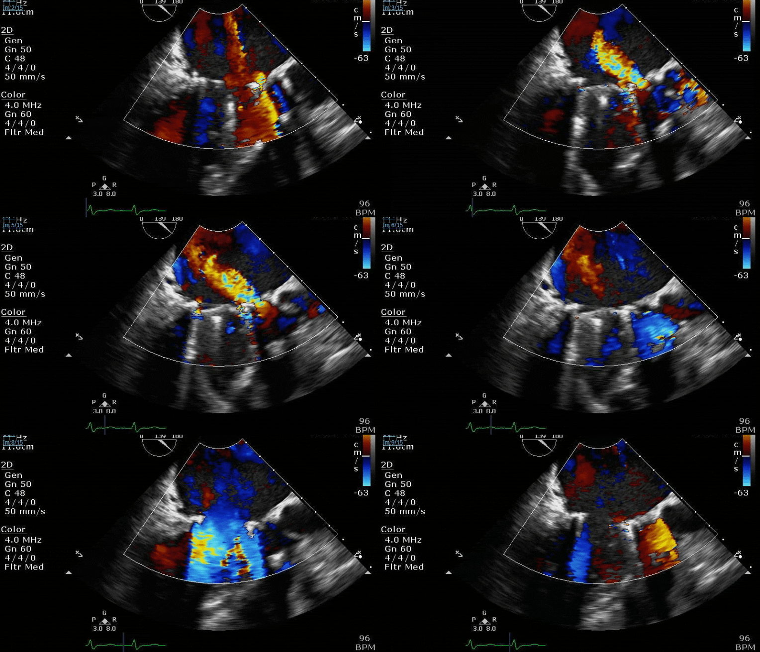
Some (post-bypass) clips to keep the thread alive:


7cuY1kN.gif


foqiZOg.gif


HTQYRQW.gif
Looks like there was a BOGO on mechanical mitrals for this patient. PTE masters has taught me that this is “incredibly rare” but apparently not THAT rare. A lot of aliasing through the functional side, curious what the gradient was.
 
Members don't see this ad :)
Some (post-bypass) clips to keep the thread alive:


7cuY1kN.gif


foqiZOg.gif


HTQYRQW.gif
Nice case. Was this taken immediately after coming off pump? Were you happy w loading conditions? I’d take a good look at the MV from the LV side and look for obstruction due to suture or subvalvular structures interfering with proper functioning of the valve. Early thrombosis or bad valve is rare.
 
Looks like there was a BOGO on mechanical mitrals for this patient. PTE masters has taught me that this is “incredibly rare” but apparently not THAT rare. A lot of aliasing through the functional side, curious what the gradient was.
Not as rare as you may think… 😉
AD4F3D1D-48EE-4E08-8F85-9BB8264E9214.jpeg
 
  • Like
Reactions: 1 user
Nice case. Was this taken immediately after coming off pump? Were you happy w loading conditions? I’d take a good look at the MV from the LV side and look for obstruction due to suture or subvalvular structures interfering with proper functioning of the valve. Early thrombosis or bad valve is rare.
No, I wasn’t particularly happy with the loading conditions at this point. However, if everyone would like to direct their attention to screen right of the clips you’ll notice that there is also a mechanical AVR in place as well. This guy had severe AS, moderate to severe MS and MR, moderate MAC and severe AV calcification preop…… with the AV calcification extending into the LVOT with chunks right under the intervalvular fibrosa. Surgeon did not debride calcium far enough into the LVOT, so what I think we’re seeing here is the anteriorly oriented occluder disc trying to open but getting stuck on one of those chunks. Unfortunately, pt was not the best protoplasm and this was not a particularly short pump run either. Surgeon was pretty adamant that “it is what it is” and that we weren’t going back on pump, which really wasn’t all that unreasonable a position considering how long a second run would take and the inability to do an impella given the mech AVR.

Luckily for this guy, kept getting volume resuscitated and I guess it was enough to change the geometry so that the disc was no longer stuck on the f/u echo. Either that or the calcium broke off and he managed to dodge a stroke or other organ ischemia. *shrug*
 
  • Like
Reactions: 4 users
I have had this on more than one occasion.
Vasoplegia, lack of proper loading conditions, etc do have this result sometimes. Almost always resolves as you adjust hemodynamics.
 
S/p MI. Clearly something bad happening. What is somewhat unique about this pathology (hint: the transgastric 2-chamber is most helpful for this question)?


07.12 Severe MR 3.PNG


  • Like
Reactions: 1 user
S/p MI. Clearly something bad happening. What is somewhat unique about this pathology (hint: the transgastric 2-chamber is most helpful for this question)?


View attachment 358861


Cool. Typically you think of just a chordal rupture with post MI MR, but a whole chunk of pap muscle tore off and is flopping in the wind here. Presumably post medial given single blood supply going to that pap muscle from RCA.

I see the defect in the inferior wall that @pocafx is referring to in the 2 chamber view (which further speaks to RCA infarct/post-med pap rupture), but I’m not seeing enough to call it as a VSD on that view alone. Though I am looking at it on a cell phone. I’d want to interrogate further before I could definitively call it for my surgeon.

Also, at risk of sounding pedantic, it’s technically flail, not prolapse.
 
Last edited:
Cool. Typically you think of just a chordal rupture with post MI MR, but a whole chunk of pap muscle tore off and is flopping in the wind here. Presumably post medial given single blood supply going to that pap muscle from RCA.

I see the defect in the inferior wall that @pocafx is referring to in the 2 chamber view (which further speaks to RCA infarct/post-med pap rupture), but I’m not seeing enough to call it as a VSD on that view alone. Though I am looking at it on a cell phone. I’d want to interrogate further before I could definitively call it for my surgeon.

Also, at risk of sounding pedantic, it’s technically flail, not prolapse.

If you look at the 3d it really looks like the flail segments are lateral in the A1/A2 region. Same goes for the ME 5 chamber which should be picking up A1. Lends credence to not being a PM pap rupture, although that would be irregular given the blood supply...
 
  • Like
Reactions: 1 users
If you look at the 3d it really looks like the flail segments are lateral in the A1/A2 region. Same goes for the ME 5 chamber which should be picking up A1. Lends credence to not being a PM pap rupture, although that would be irregular given the blood supply...

Yea, you’re right, flail segment appears to be more AL. I think I saw MI/ruptured pap and got tunnel vision. Would have to be either atypical solo blood supply to the AL pap or it has an accessory pap with solo blood supply and that’s what infarcted/ruptured. It’s unlikely the patient would be alive long enough after the left main went down for that to be the reason for AL pap rupture.
 
Yea, you’re right, flail segment appears to be more AL. I think I saw MI/ruptured pap and got tunnel vision. Would have to be either atypical solo blood supply to the AL pap or it has an accessory pap with solo blood supply and that’s what infarcted/ruptured. It’s unlikely the patient would be alive long enough after the left main went down for that to be the reason for AL pap rupture.
It is indeed the anterolateral papillary muscle that's ruptured. PM is 8-12 x more common from what I have read, so it was a reasonable initial thought anyway.

This guy spent a week in the ICU with a balloon pump in place prior to OR. He was exceptionally unwell.

He underwent CABG + MVR with a bioprosthetic valve. Pictures to follow. Last I saw, he got a trach and was still in the ICU.
 
  • Like
Reactions: 1 user
It is indeed the anterolateral papillary muscle that's ruptured. PM is 8-12 x more common from what I have read, so it was a reasonable initial thought anyway.

This guy spent a week in the ICU with a balloon pump in place prior to OR. He was exceptionally unwell.

He underwent CABG + MVR with a bioprosthetic valve. Pictures to follow. Last I saw, he got a trach and was still in the ICU.

How did the LHC/cors look? Curious to know the disease distribution
 
  • Like
Reactions: 1 users
Honestly, I don't recall the LHC, but I'll look it and get back to you. Here are the post MVR images. Mo betta.


Pre:

HM5zMtL.gif


2UezR7c.gif


Post:

EyfR8tV.gif


SmZg3LV.gif


GXf0cfM.gif

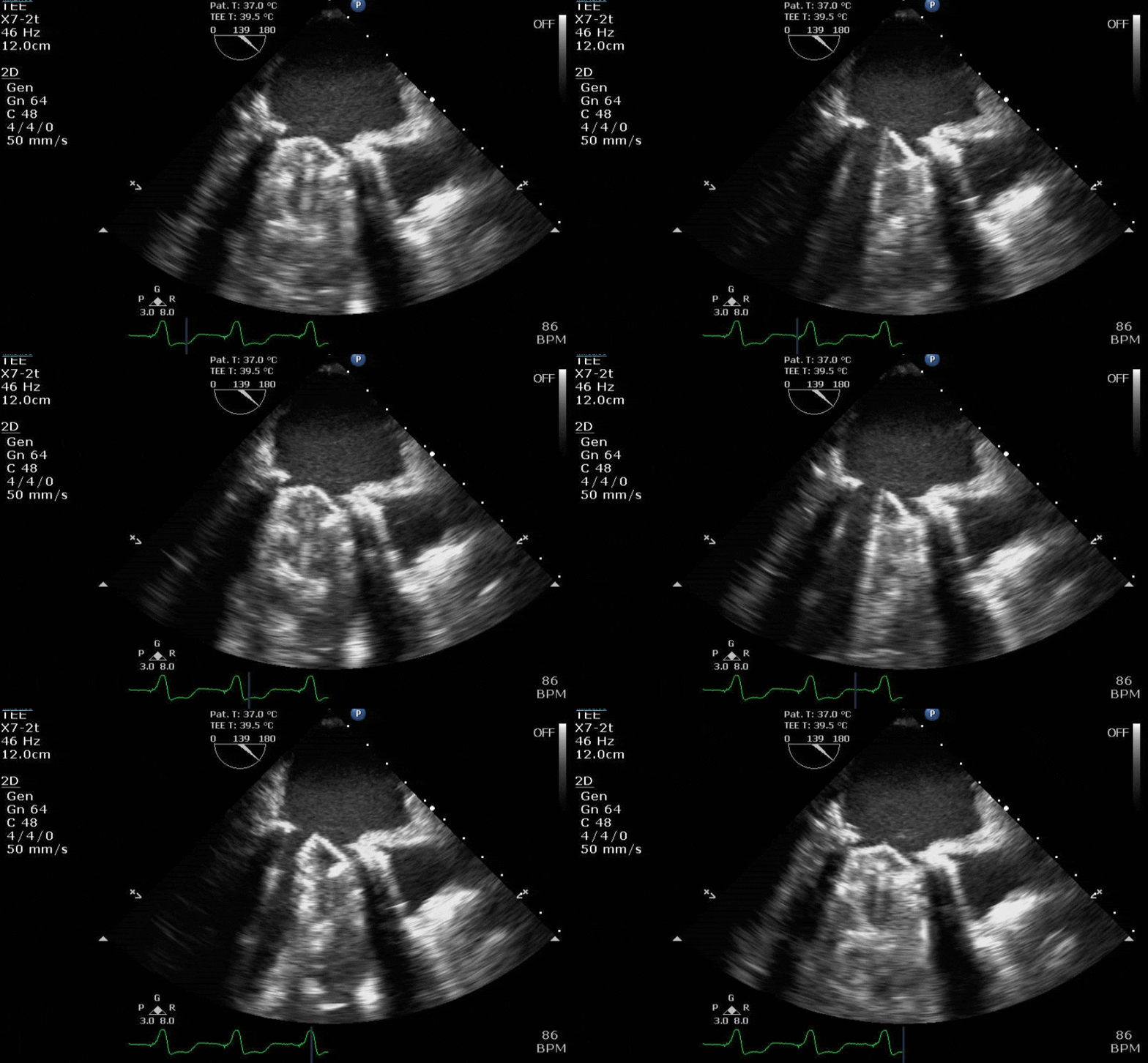
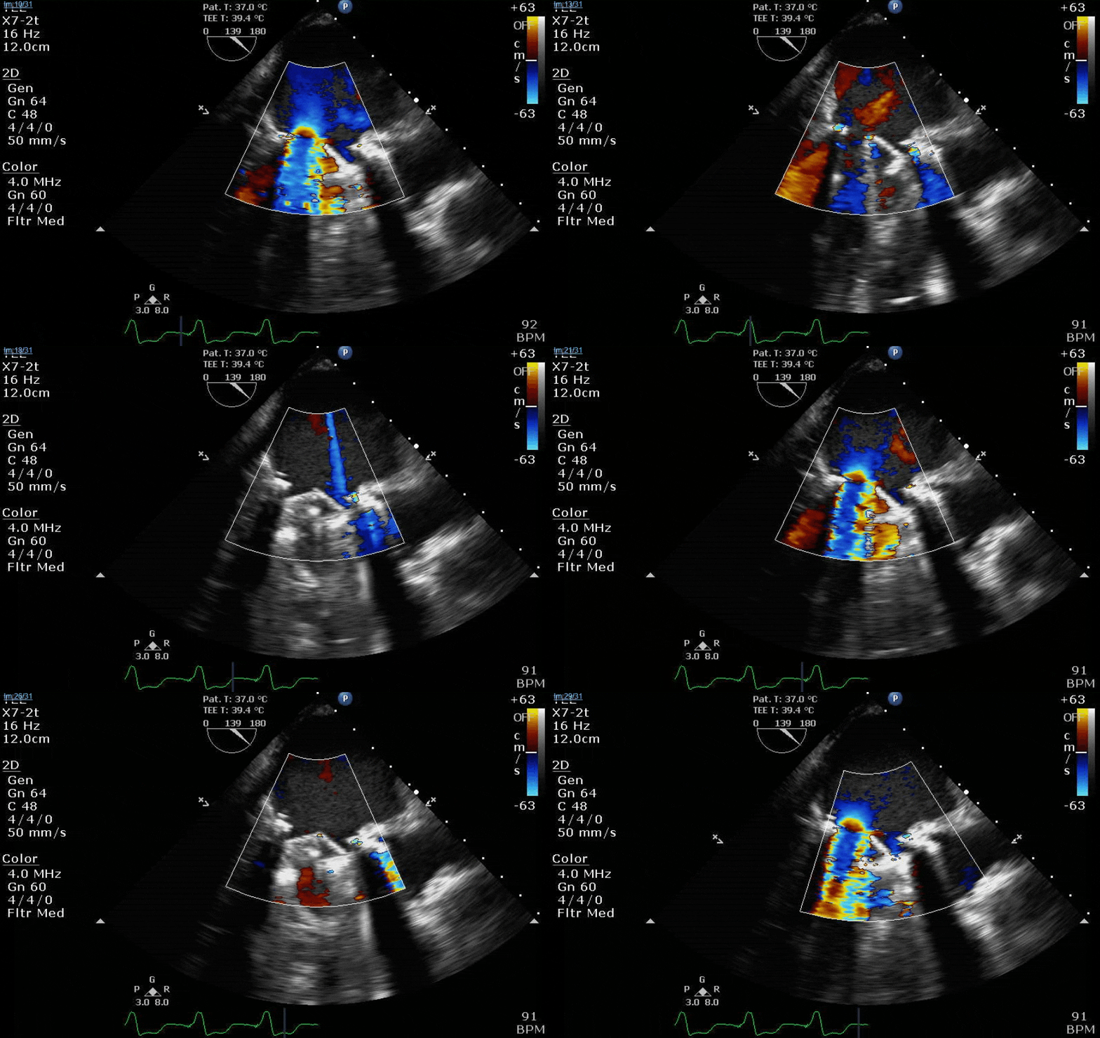
Lots happening here!

Looks like a perforation/hole from the LVOT to the LA that was made worse after pulling the MVR (I'd guess it's just from tissue distortion and tension from sutures).

Severe TR. And I feel like there's also a VSD, but my eyes may be deceiving me.
 
Lots happening here!

Looks like a perforation/hole from the LVOT to the LA that was made worse after pulling the MVR (I'd guess it's just from tissue distortion and tension from sutures).

Severe TR. And I feel like there's also a VSD, but my eyes may be deceiving me.

Was this done by a heart surgeon, or someone from EVS who stayed at a holiday inn express? Yikes.

Yikes… Was this the result of a (very) misguided attempt at chordal-sparing MVR, or what?

Yep, lot to unpack here. But first, some clinical history and context ( because of course it's easier to judge the result when all you have to go by are the clips).

Pt is a 47yo F. She's 360 lbs. This is her 3rd sternotomy with the prior two being for LA myxoma excisions. She is booked for an MVR/TVr/LAA clip/MAZE. She has a pre-op CMR with her LVEF being listed as 36% and her RVEF at 29%. This is obviously severe biventricular dysfunction given her regurgitant lesions.

Of course anyone in their right mind would get wires into the groin before sternotomy on this pt. Lead surgeon here gets frustrated though because her femoral vessels are buried under a million miles of fat and they can't access them even with ultrasound. Instead of doing a cutdown (or calling for fluoro and help from IC), he proceeds to sternotomy with the oscillating saw (of note, in 5 years I've never run into a [major] problem with this surgeon during redo sternotomies).

And as you can guess, this time, even with being really careful near the posterior table of the sternum, he saws right through the main PA and probably through some of the innominate vein. She immediately starts pouring blood and we start transfusing some prbc and she stabilizes. Lead surgeon is able to get enough exposure so that the surgery resident can hold manual pressure. Lead surgeon and 2nd surgeon then start working on groin dissection. Which takes FOREVER because they're super deep and not where they initially made incision. At this point we've been asleep for 2 hrs, but finally fems are cannulated and we go on bypass.

But having to do difficult redo dissections on pump is the worst. Just the worst. The patient is getting all the downsides of cardiopulmonary bypass without the surgeon actually making substantive progress on repairing the lesions for which they came to surgery. She's amazingly fat and the working space in her mediastinum is tiny, so stopping the bleeding and doing enough dissection just to be able to centrally cannulate takes another 2 hrs on pump. And she's so adhesed that doing the MAZE and clip is noted to be impossible anyway.

So 4 hrs into the case, we're clamped and pleged and lead surgeon can finally begin the actual valve replacement. Of course we discover that in addition to being surrounded by fat and deep in the chest, the adhesions have rotated her heart so that the "surgeon's view" of the mitral through a transseptal approach is dog****. Surgeon cannot really visualize the intervalvular fibrosa that well even though we know there is a perforation in that region (but if he could see it, it does raise questions of how to spare the chords, leaflets or how to do a transposition technique, etc. Leaflet/chord sparing is important, doubly so when LV function is down). He does the best he can and gets the MVR in. TVr goes fine as well.

Clamp off, we start pacing and deair, fill up a bit and at this point you see the big old anterior PVL where that prior perforation was. And the TR is still high mild to moderate because the septal leaflet is probably still tethered. In regard to the mitral, you know the replacement is not adequate, but the flip side is, at this point, the clamp time has been almost 2 hrs and the bypass time is over 4 hrs in a pt with severe biventricular dysfunction. Furthermore, the ICs who typically do impellas or help with VA ecmo (both of which we do pretty infrequently) are unavailable at the moment. Further furthermore, she is just profusely bleeding from everywhere. So even with MCS, unless you're gonna do it heparin free she is gonna bleed to death. Lead surgeon says the PVL is what it is and that there's no way he's clamping and pleging again. We manage to limp off on some high dose epi, milrinone, nitric, levo, vaso. Vac the chest, start a mini-MTP, she makes it to the unit.

Pt had her ups and downs (bedside washout for hemorrhage POD 0), but we managed to close the chest POD 3, extubate POD 9, weaned off all inotropes POD 10, and she managed to escape with no other organ failure. Future hemolysis or reop MVR notwithstanding, I'm still calling it a win.
 
  • Like
  • Wow
Reactions: 8 users
Yep, lot to unpack here. But first, some clinical history and context ( because of course it's easier to judge the result when all you have to go by are the clips).

Pt is a 47yo F. She's 360 lbs. This is her 3rd sternotomy with the prior two being for LA myxoma excisions. She is booked for an MVR/TVr/LAA clip/MAZE. She has a pre-op CMR with her LVEF being listed as 36% and her RVEF at 29%. This is obviously severe biventricular dysfunction given her regurgitant lesions.

Of course anyone in their right mind would get wires into the groin before sternotomy on this pt. Lead surgeon here gets frustrated though because her femoral vessels are buried under a million miles of fat and they can't access them even with ultrasound. Instead of doing a cutdown (or calling for fluoro and help from IC), he proceeds to sternotomy with the oscillating saw (of note, in 5 years I've never run into a [major] problem with this surgeon during redo sternotomies).

And as you can guess, this time, even with being really careful near the posterior table of the sternum, he saws right through the main PA and probably through some of the innominate vein. She immediately starts pouring blood and we start transfusing some prbc and she stabilizes. Lead surgeon is able to get enough exposure so that the surgery resident can hold manual pressure. Lead surgeon and 2nd surgeon then start working on groin dissection. Which takes FOREVER because they're super deep and not where they initially made incision. At this point we've been asleep for 2 hrs, but finally fems are cannulated and we go on bypass.

But having to do difficult redo dissections on pump is the worst. Just the worst. The patient is getting all the downsides of cardiopulmonary bypass without the surgeon actually making substantive progress on repairing the lesions for which they came to surgery. She's amazingly fat and the working space in her mediastinum is tiny, so stopping the bleeding and doing enough dissection just to be able to centrally cannulate takes another 2 hrs on pump. And she's so adhesed that doing the MAZE and clip is noted to be impossible anyway.

So 4 hrs into the case, we're clamped and pleged and lead surgeon can finally begin the actual valve replacement. Of course we discover that in addition to being surrounded by fat and deep in the chest, the adhesions have rotated her heart so that the "surgeon's view" of the mitral through a transseptal approach is dog****. Surgeon cannot really visualize the intervalvular fibrosa that well even though we know there is a perforation in that region (but if he could see it, it does raise questions of how to spare the chords, leaflets or how to do a transposition technique, etc. Leaflet/chord sparing is important, doubly so when LV function is down). He does the best he can and gets the MVR in. TVr goes fine as well.

Clamp off, we start pacing and deair, fill up a bit and at this point you see the big old anterior PVL where that prior perforation was. And the TR is still high mild to moderate because the septal leaflet is probably still tethered. In regard to the mitral, you know the replacement is not adequate, but the flip side is, at this point, the clamp time has been almost 2 hrs and the bypass time is over 4 hrs in a pt with severe biventricular dysfunction. Furthermore, the ICs who typically do impellas or help with VA ecmo (both of which we do pretty infrequently) are unavailable at the moment. Further furthermore, she is just profusely bleeding from everywhere. So even with MCS, unless you're gonna do it heparin free she is gonna bleed to death. Lead surgeon says the PVL is what it is and that there's no way he's clamping and pleging again. We manage to limp off on some high dose epi, milrinone, nitric, levo, vaso. Vac the chest, start a mini-MTP, she makes it to the unit.

Pt had her ups and downs (bedside washout for hemorrhage POD 0), but we managed to close the chest POD 3, extubate POD 9, weaned off all inotropes POD 10, and she managed to escape with no other organ failure. Future hemolysis or reop MVR notwithstanding, I'm still calling it a win.
Wow incredible stuff...
How she is still alive in incredible. Well done
I guess she is still young and that counts in her favor.
I would imagine this is her last sternotomy. Maybe an amplatzer and or tricuspid clip if someone is brave enough...
 
  • Like
Reactions: 2 users
Yep, lot to unpack here. But first, some clinical history and context ( because of course it's easier to judge the result when all you have to go by are the clips).

Pt is a 47yo F. She's 360 lbs. This is her 3rd sternotomy with the prior two being for LA myxoma excisions. She is booked for an MVR/TVr/LAA clip/MAZE. She has a pre-op CMR with her LVEF being listed as 36% and her RVEF at 29%. This is obviously severe biventricular dysfunction given her regurgitant lesions.

Of course anyone in their right mind would get wires into the groin before sternotomy on this pt. Lead surgeon here gets frustrated though because her femoral vessels are buried under a million miles of fat and they can't access them even with ultrasound. Instead of doing a cutdown (or calling for fluoro and help from IC), he proceeds to sternotomy with the oscillating saw (of note, in 5 years I've never run into a [major] problem with this surgeon during redo sternotomies).

And as you can guess, this time, even with being really careful near the posterior table of the sternum, he saws right through the main PA and probably through some of the innominate vein. She immediately starts pouring blood and we start transfusing some prbc and she stabilizes. Lead surgeon is able to get enough exposure so that the surgery resident can hold manual pressure. Lead surgeon and 2nd surgeon then start working on groin dissection. Which takes FOREVER because they're super deep and not where they initially made incision. At this point we've been asleep for 2 hrs, but finally fems are cannulated and we go on bypass.

But having to do difficult redo dissections on pump is the worst. Just the worst. The patient is getting all the downsides of cardiopulmonary bypass without the surgeon actually making substantive progress on repairing the lesions for which they came to surgery. She's amazingly fat and the working space in her mediastinum is tiny, so stopping the bleeding and doing enough dissection just to be able to centrally cannulate takes another 2 hrs on pump. And she's so adhesed that doing the MAZE and clip is noted to be impossible anyway.

So 4 hrs into the case, we're clamped and pleged and lead surgeon can finally begin the actual valve replacement. Of course we discover that in addition to being surrounded by fat and deep in the chest, the adhesions have rotated her heart so that the "surgeon's view" of the mitral through a transseptal approach is dog****. Surgeon cannot really visualize the intervalvular fibrosa that well even though we know there is a perforation in that region (but if he could see it, it does raise questions of how to spare the chords, leaflets or how to do a transposition technique, etc. Leaflet/chord sparing is important, doubly so when LV function is down). He does the best he can and gets the MVR in. TVr goes fine as well.

Clamp off, we start pacing and deair, fill up a bit and at this point you see the big old anterior PVL where that prior perforation was. And the TR is still high mild to moderate because the septal leaflet is probably still tethered. In regard to the mitral, you know the replacement is not adequate, but the flip side is, at this point, the clamp time has been almost 2 hrs and the bypass time is over 4 hrs in a pt with severe biventricular dysfunction. Furthermore, the ICs who typically do impellas or help with VA ecmo (both of which we do pretty infrequently) are unavailable at the moment. Further furthermore, she is just profusely bleeding from everywhere. So even with MCS, unless you're gonna do it heparin free she is gonna bleed to death. Lead surgeon says the PVL is what it is and that there's no way he's clamping and pleging again. We manage to limp off on some high dose epi, milrinone, nitric, levo, vaso. Vac the chest, start a mini-MTP, she makes it to the unit.

Pt had her ups and downs (bedside washout for hemorrhage POD 0), but we managed to close the chest POD 3, extubate POD 9, weaned off all inotropes POD 10, and she managed to escape with no other organ failure. Future hemolysis or reop MVR notwithstanding, I'm still calling it a win.
Very impressive case.

Although the PVT (peri valvular Torrent) is not optimal, it may be a good popoff valve for her struggling LV!??!

I wonder what the discussion was like during the M&M
 
Well done getting her out of the OR. It's easy to criticize the echo result but yeah sometimes the surgeon just cannot get good visualization and you get what you get. Perhaps a thoracotomy approach would've been better for visualization (and adhesions, sternotomy risk) but it's impossible to know. Veering away from just echo discussion though -- my opinion is that if your surgeon is comfortable doing 3x redo sternotomy double valves on severe BiV dysfunction, they also need to be comfortable in mechanical support. Although she survived (impressive), in my experience an Impella 5.5 can be a lifesaver in situations like this. Direct aortic implant is possible through a chimney that allows chest closure and eventual bedside removal. There are places that don't run anticoagulation, although that's controversial. We'd reverse the heparin in OR, attempt hemostasis, then anticoagulate when more stable in the ICU.

Amplatz may be a future option...but the perforation seems quite close to the AV so it could be tough to fix without impinging on AV cusps or the underside of the mechanical mitral leaflet.
 
  • Like
Reactions: 4 users
if your surgeon is comfortable doing 3x redo sternotomy double valves on severe BiV dysfunction, they also need to be comfortable in mechanical support
This x1000.

…Of course recognizing that you need not just surgeon comfort, but comfort amongst the nurses/RTs/perfusionists/physical therapists/etc etc etc in order to have a successful MCS program

@itwasalladream at my institution we’re doing central impellas, but still coming back to the OR for removal (oversewing the end of the graft and leaving a short stump, so no XC needed). Would love some more info/technical details on how you’re doing with bedside removal? Does your surgeon just pull it out and staple the graft off at the skin or something?
 
  • Like
Reactions: 1 user
This x1000.

…Of course recognizing that you need not just surgeon comfort, but comfort amongst the nurses/RTs/perfusionists/physical therapists/etc etc etc in order to have a successful MCS program

@itwasalladream at my institution we’re doing central impellas, but still coming back to the OR for removal (oversewing the end of the graft and leaving a short stump, so no XC needed). Would love some more info/technical details on how you’re doing with bedside removal? Does your surgeon just pull it out and staple the graft off at the skin or something?
Our surgeon pulls the dacron graft up a little, clamps and ties off with silk. Then trims/tucks it and closes skin. Exit site is just above clavicle, so no tunneling between the ribs or anything is needed. They just do it under local in the ICU so we aren't involved. It's a long stump but we've had no clot propagation issues that I'm aware of. We do a similar thing with central RVAD (femoral venous drainage -> centrimag -> end-to-side dacron graft off the main PA, tunneled through skin). It's nice not to have to leave their chest open, or take them back to the OR for decannulation/weaning.
 
  • Like
Reactions: 1 user
Wow incredible stuff...
How she is still alive in incredible. Well done
I guess she is still young and that counts in her favor.
I would imagine this is her last sternotomy. Maybe an amplatzer and or tricuspid clip if someone is brave enough...

Very impressive case.

Although the PVT (peri valvular Torrent) is not optimal, it may be a good popoff valve for her struggling LV!??!

I wonder what the discussion was like during the M&M

Well done getting her out of the OR. It's easy to criticize the echo result but yeah sometimes the surgeon just cannot get good visualization and you get what you get. Perhaps a thoracotomy approach would've been better for visualization (and adhesions, sternotomy risk) but it's impossible to know. Veering away from just echo discussion though -- my opinion is that if your surgeon is comfortable doing 3x redo sternotomy double valves on severe BiV dysfunction, they also need to be comfortable in mechanical support. Although she survived (impressive), in my experience an Impella 5.5 can be a lifesaver in situations like this. Direct aortic implant is possible through a chimney that allows chest closure and eventual bedside removal. There are places that don't run anticoagulation, although that's controversial. We'd reverse the heparin in OR, attempt hemostasis, then anticoagulate when more stable in the ICU.

Amplatz may be a future option...but the perforation seems quite close to the AV so it could be tough to fix without impinging on AV cusps or the underside of the mechanical mitral leaflet.

I spoke with IC after the initial surgery and given the extremely anterior location of the leak they were pretty hesitant about trying to plug it. Not to mention, as @dchz said, having a bit of a popoff probably helped her hemodynamics in the initial post-op period. She will probably need a follow up TEE and we'll see whether with good GDMT for HFrEF / MR and not being coagulopathic if the leak severity has lessened. If she's not hemolyzing and the leak is mild or mild-moderate then they may just leave well enough alone.

As far as MCS, we're in a weird spot with this CT surgery group. They've been out for a long time and have pretty much done it all (except transplant) at some point during their careers. The lead surgeon in this case, for instance, used to do Heartmate IIs before one of the other hospitals that he covers lost their AHF/MCS cardiologists. They could do MCS if they wanted, but given the number of hospitals they cover and the stage they're at in their careers, they just don't want the headache.

Also, our ICU is not built for this kind of patient because it's a low volume cardiac center. There is no dedicated CTICU. There are no real cardiac intensivists. The surgeons pretty much manage their pts post-op by themselves along with the surgery resident and a perfunctory cards consult. Say for instance they were putting in their own Impella 5.5s, maybe some of the calls would go to the cards fellow or cards attending on consults covering "CCU," but odds are the nurses who are in over their heads would simply hammer page them about one issue after the next. Don't get me wrong, they have called in IC for the occasional Impella 5.5, Impella RP, or Protek Duo when an IABP isn't enough, but it's pretty rare. And this is the same reason that when we get type A dissections with arch involvement, they go out of their way to avoid hemiarch/total arch + DHCA. If they can graft the ascending, seal off the ascending entry tear, and establish good flow in the true lumen then that's what they do, even if arch repair is probably warranted. They're more than capable of doing the full surgery or even debranching, but dealing with the post-op vasoplegic, LCOS, coagulopathic total arch pt.... is just something they don't want to do.

This kind of situation is a good cautionary tale for the current ACTA fellows who are joining community practices in centers which are way different from the quaternary mothership hospitals in which they're training. In ACTA fellowship you take care of lot of deathly sick people, but there's also a mental ease that comes with them because literally every CT surgeon you work with is comfortable doing central VA-ECMO cannulation, slapping on the Centrimag, venting the LV, and then hauling it to the ICU....upon where you're met by an army of residents, ICU/CT surg fellows, and dual trained CT/CCM attendings. Out in the community, you will run into occasional sick as **** pts....but without all the toys and the deep bench of support people. Definitely increases the pucker factor when the initial wean from pump isn't looking so hot.
 
  • Like
Reactions: 4 users
They could do MCS if they wanted, but given the number of hospitals they cover and the stage they're at in their careers, they just don't want the headache.
Also, our ICU is not built for this kind of patient because it's a low volume cardiac center. There is no dedicated CTICU. There are no real cardiac intensivists. The surgeons pretty much manage their pts post-op by themselves along with the surgery resident and a perfunctory cards consult.

I’d argue your surgeons probably shouldn’t be bringing cases like this to your hospital if this is the case. Not a commentary on you/your partners or your surgeons ability. But if you’re doing sick patients like this with any regularity, you really need robust, ready access to MCS and ICU care/support staff capable of managing it.

Having great cardiac surgeons and anesthesiologists who have seen and done it all and can take care of anyone is only half the battle. These patients need experienced, high level ICU care to get them the rest of the way home.
 
  • Like
Reactions: 6 users
I’d argue your surgeons probably shouldn’t be bringing cases like this to your hospital if this is the case. Not a commentary on you/your partners or your surgeons ability. But if you’re doing sick patients like this with any regularity, you really need robust, ready access to MCS and ICU care/support staff capable of managing it.

Having great cardiac surgeons and anesthesiologists who have seen and done it all and can take care of anyone is only half the battle. These patients need experienced, high level ICU care to get them the rest of the way home.
Yeah these kind of patients aren’t a regular occurrence since we mostly do b&b CABG and valves. And I (and the surgeons) are in total agreement with your sentiment, so much so to the point that not infrequently they turn down pts thinking the risk is too high even when I think the pt is doable. But really, most of these pts of the highest complexity get referred elsewhere……assuming they have Medicare or private insurance. For charity care and/or Medicaid cases like our lady in question, my shop is all they get if the big private center across town turns them down based on ability to pay, not to mention they were also slightly more inclined to do this one given her young age.
 
  • Like
Reactions: 2 users
I’d argue your surgeons probably shouldn’t be bringing cases like this to your hospital if this is the case. Not a commentary on you/your partners or your surgeons ability. But if you’re doing sick patients like this with any regularity, you really need robust, ready access to MCS and ICU care/support staff capable of managing it.

Having great cardiac surgeons and anesthesiologists who have seen and done it all and can take care of anyone is only half the battle. These patients need experienced, high level ICU care to get them the rest of the way home.

We're a big referral zone for all the CVTS crashing/burning patients that get done at smaller, outside facilities (and some not-so-small). We still have issues with high turnover/burnout in our ICUs with new travel ICU nurses who "know it all" every single week trying to kill our sicker-than-sick patients. That being said, our MCS team is pretty good at quickly cannulating and prolonging suffering/delaying death.
 
  • Like
Reactions: 1 user
Butterfly clip on a fresh post-op mitral. It's the worst of the handhelds but you really can't beat it for 2 grand

ezgif-2-6ae005c957.gif
 
  • Like
Reactions: 2 users
mitrezgif-4-8ecd04809f.gif


Here's another fresh sternotomy. Caveat is that these two pts I've posted are (atypically for us) pretty damn skinny. This lady does have COPD though so I'm surprised the windows were this good
 
Ostium primum with some mitral annulus involvement? Endocardial cushion defect? Trisomy 21 kiddo?
 
Ostium primum with some mitral annulus involvement? Endocardial cushion defect? Trisomy 21 kiddo?

Nope, not a kiddo, not congenital.

I can see though how from the first clip one could get the idea that flow is crossing the septum, however you can see on the non-color 4ch that the IAS and ventricular septi are intact, so that color you see in the RA is probably artifact/RA inflow.
 
Pre:

MR-12-22-1.gif

MR-12-22-2.gif

MR-12-22-3.gif


Post:

MR-12-22-4.gif

MR-12-22-5.gif

MR-12-22-6.gif

MR-12-22-7.gif
Ill try. 2nd bottle of wine open...

T2 carpentier, severe mr. Hard to assess precise mechanics given 1 shot only and not really in middle of LA so cant see much.

MVRt pretty good, maybe some PVL butnhave to assess it better. Lv is tanked after. Need ++mil, flolan epi to get thru. Big dilated ****e. Did he take a coronary too?
 
  • Like
Reactions: 1 users
Ill try. 2nd bottle of wine open...

T2 carpentier, severe mr. Hard to assess precise mechanics given 1 shot only and not really in middle of LA so cant see much.

MVRt pretty good, maybe some PVL butnhave to assess it better. Lv is tanked after. Need ++mil, flolan epi to get thru. Big dilated ****e. Did he take a coronary too?
Pretty much spot on. The gifs didn't capture the way I would've liked (they're kinda choppy), but if you squint real hard you'll notice in the 4ch color shot that there is a central jet and a highly, eccentric anteriorly directed jet. There ended up being a cleft plus myxomatous degeneration leading to both Carpentier type I and II mechanisms.

You'll also notice the LV function is pretty bad pre-pump, and absolutely atrocious post-pump. The RV function is also moderately to severely depressed post-pump. Both the clamp time and pump run were very short, MVR went without a hitch. I expressed to my resident that this case highlighted the difference between doing pump cases for revascularization in ischemic cardiomyopathy with viable myocardium vs doing valvular surgery for people with non-ischemic cardiomyopathy. In the literature, AMI cardiogenic shock does better than decompensated NICM cardiogenic shock, and I find that to be generally (and anecdotally) true in the OR as well when one has post-cardiotomy shock.

The post-pump LV clip with an EF of ~15% is with the pt on high dose epi, dobutamine, milrinone, iNO, levo, vaso and IABP with a barely acceptable MAP and CI. He went on to require a takeback to the cathlab a few hours after he arrived in the ICU for impella placement due to worsening shock, and that highlighted some of the system issues we face with having surgeons who do not routinely put in their own impellas, VA-ECMO, LV vents, etc. They tried to punt on this case (either for transfer or possible clip), but pressure from cards was heavy given this was a young man (about age 50) with good functional status.

Finally, these clips highlight that in some patients you are just not going to be able to get a classic valentine heart 4 chamber view. I tried every combination of omniplane and flexion but his cardiomegaly and rotation made it impossible get a clip where I wasn't severely obliquely cutting through the LA. People btch a lot about the quality of clips in the aPTE, but difficult views are not terribly infrequent IRL.
 
  • Like
Reactions: 3 users
RCARWMA.gif


Question for residents (and maybe fellows but might be too easy):

1. What coronary distribution typically supplies the wall with the regional wall motion abnormality?

2. How do you explain it if I told you an angiogram of the coronary that typically supplies that wall is normal?
 
  • Like
Reactions: 1 user
Top