Let's do some echo:

This forum made possible through the generous support of SDN members, donors, and sponsors. Thank you.

sevoflurane

Ride
20+ Year Member
Joined
Jul 16, 2003
Messages
6,032
Reaction score
3,820
Let's do some basic Echo:
This comes from my personal data base, so I hope you guys dig it. Med studs and residents first.

Name 1-7

0fd48cdc-3014-4e39-889d-80c88113ca83_zps0828fe70.jpg

Members don't see this ad.
 
  • Like
Reactions: 1 user
I'll get the ball rolling. This is a mid-esophageal AV LAX view. As I am echo boarded, I'll hold off and let the kids have their fun.
 
Rising MS-3 who just took step 1 last week (i.e., idiot here). Without googling "esophageal echo primer" I'll make a fool of myself and take a stab at it:

1: not sure (pulm vein?)
2: wall of proximal pulm art
3 & 4: cusps of pulm valve
5: left atrium
6: left pulm artery
7: right atrium? or bit of aorta? I don't know
 
Members don't see this ad :)
Rising MS-3 who just took step 1 last week (i.e., idiot here). Without googling "esophageal echo primer" I'll make a fool of myself and take a stab at it:

1: not sure (pulm vein?)
2: wall of proximal pulm art
3 & 4: cusps of pulm valve
5: left atrium
6: left pulm artery
7: right atrium? or bit of aorta? I don't know

I'll give this a 1/7, but an A+ for stepping up to the plate!
 
  • Like
Reactions: 1 users
Lol... I certainly wasn't very confident but was going off the assumption that the first thing the probe sees from the esophagus is left atrium.. is #5-LA the one I got right?
 
I'll take a crack at it. I'm putting down my original guesses, however, I did look it up to confirm what I thought I was seeing. I still don't have great labels for 3&4. And that's embarrassing.

1. PML
2. AML
3. Don't know what particular label you are looking for, but one of the aortic valve leaflets.
4. Ditto number 3.
5. Left atrium
6. Left ventricle
7. Right ventricle

SPOILER: Does this patient have a bicuspid aortic valve?

Pretty good! You can get a bit more specific on the mitral valve leaflets (at least in theory), though you'd want to confirm that with other views. I usually think of #7 as the RVOT, but I guess that is technically the right ventricle. Strong work!
 
  • Like
Reactions: 1 user
Ok I see it now.. wow, didn't realize the aortic valve and LVOT was oriented almost 90 degrees relative to the LA-LV axis
 
Ok I see it now.. wow, didn't realize the aortic valve and LVOT was oriented almost 90 degrees relative to the LA-LV axis
Remember, you are seeing a mm (or less) thick slice of the heart. So it is hard to appreciate the total orientation of these things. But yes, there is a pretty sharp turn the blood has to make from coming out of the mitral valve on its way out of the aortic valve.
 
It's a posterior leaflet prolapse at least. I wouldn't be able to confirm what the offending scallop was but I'd guess p2/3 and if a papillary muscle was responsible I'd blame the posterior pap.
 
  • Like
Reactions: 1 user
Thanks for jumping in B-bone. Kinda busy today.

To the rest of the gang here... there are NO wrong answers.

What are the 4 leaflets labeled 1, 2, 3, and 4? We'll get to that... but here is another pic... to keep things cooking.

0eaed0ec-1587-4334-95b7-ffd411b87c70_zps08cd4708.jpg


What the heck is going on there?
 
  • Like
Reactions: 1 user
It's a posterior leaflet prolapse at least. I wouldn't be able to confirm what the offending scallop was but I'd guess p2/3 and if a papillary muscle was responsible I'd blame the posterior pap.

Very nice sir. Good pickup. Which direction will the jet go?
 
  • Like
Reactions: 1 user
Members don't see this ad :)
For the intellectually curious among you, I will put in a plug for e-echocardiography. I found the subscription service very useful while preparing for the NBE exam, but the free portions (like echo of the day/case of the week) are great as well.

https://e-echocardiography.com/index.php
 
  • Like
Reactions: 1 user
You bet. :thumbup:
In the abscense of severe systemic hypertension or aortic stenosis, an eccentric/wall hugging MR jet tends to UNDERESTIMATE the severity of MR.

Here is the same case, but in a different view.

60a48d90-a614-451e-a845-f4f9e8937600_zps5eabb805.jpg


= Bigger Jet area.
 
  • Like
Reactions: 1 user
Red jet toward the LA?

You're right. It is kinda RED. Remember this: RED = towards the probe. Blue = away... and then there is flow convergence which mixes everything up.
The pnemonic that is commonly used is BART =Blue Away from the probe, Red Towards the probe.

You're also right in that mitral regurgitant flow heads from the LV to the LA. The direction of the JET is very telling about the pathology of the mitral valve:

Let me explain: There is this thing called the "Carpentier" grading system of mitral regurgitation. A "fail" leaflet is considered Type II and the direction of the mitral regurgitant jet is typically "away" from that flail leaflet. In this case it was a posterior flail leaflet with an anterior directed jet (away/opposite direction fo the flail leaflet). This is exactly what happens with this type of lesion. Perforated leaflets, say by vegetations, is typically very different looking.
FundTypenew.jpg
 
Last edited:
0e52977c-0e4f-4e51-bcad-ab7818ae7839_zps91e80b85.jpg


IMG_7261_zpsc985918e.jpg


Notice the "flail" leaflet and the direction of regurgitant flow = opposite that of the leaflet = Type 2 Carpentier
 
Probably systolic reversal of flow in the pulmonary veins but I've always just pulsed the flow and looked at the wave form.

Systolic reversal of flow = severe MR

What if I gave you this?

afd88606-68de-4ec0-8ee6-7e594b84b716_zpsfc0fc581.jpg
 
Flow pattern on the long axis is normal. There is another finding.

That's a dissected aorta. Based on the angle of the flap it looks like flow is in the true lumen. You'd also look at timing of the appearance of the flow (true lumen earlier).
 
What if I gave you this?

afd88606-68de-4ec0-8ee6-7e594b84b716_zpsfc0fc581.jpg

I'm not sure what you're pulsing but it doesn't show systolic reversal of flow in pulmonary veins. Generally the S and D waves are above the baseline and the AR wave is below. With systolic reversal, the S wave is also below.
 
I'm not sure what you're pulsing but it doesn't show systolic reversal of flow in pulmonary veins. Generally the S and D waves are above the baseline and the AR wave is below. With systolic reversal, the S wave is also below.

Long and short axis of descending thoracic aorta showing diastolic flow reversal. Bad AI

Yep. :thumbup:

Holodiastolic flow reversal in the aorta indicating an aortic disseciton that has caused severe AI.
 
That's a dissected aorta. Based on the angle of the flap it looks like flow is in the true lumen. You'd also look at timing of the appearance of the flow (true lumen earlier).

Yep. :thumbup:
 
Here is a great view to learn for med studs and residents. It is of particular importance because it shows you the distribution of the RCA, LAD and Circ.

What are structures labeled 1 and 2?

Name the correct heart vessel that supplies walls 3, 4, 5 and 6.

What does it mean to have a "right dominant" circulation?

c92c9a14-2977-499f-9526-2254da945e89_zps6d2e2113.jpg

1 left pap
2 right pap (posterior)
3 RCA
3/4 RCA & circ
4/5 lad and circ
5 lad
5/6 lad
6 lad and/or RCA
6/7 lad or/and RCA

Right dominant = PDA from RCA
Left dominant = PDA from lad
 
That's a dissected aorta. Based on the angle of the flap it looks like flow is in the true lumen. You'd also look at timing of the appearance of the flow (true lumen earlier).

This dissection is a little unusual in that the true lumen is the larger lumen. Usually the true lumen is smaller and lens-shaped while the false lumen is bigger and crescenty or Pac-Mannish.
 
Good, but we generally refer to the papillary muscles as anterolateral and posteromedial. Also, with the left dominant, the PDA is more commonly supplied by the Circumflex. For extra points, why is it relevant to know whether a patient is right or left dominant?

Great thread, I will have to dig up some of my images for the residents
 
upload_2014-5-15_19-40-14.png
upload_2014-5-15_19-36-47.png
[/ATTACH]

Two images. One is the way it should look, the other is potentially a problem. What is going on in this patient and how do we fix it?
 
  • Like
Reactions: 1 user
Nice. Just did one of these a couple weeks ago.
Thanks for jumping in!
 
There is more than one thing going on with sethco's echo.
 
Good, but we generally refer to the papillary muscles as anterolateral and posteromedial. Also, with the left dominant, the PDA is more commonly supplied by the Circumflex. For extra points, why is it relevant to know whether a patient is right or left dominant?

yes sir.
 
Are you a current or future CT fellow?

What else do you look for after the LVAD is in place?
 
Are you a current or future CT fellow?

What else do you look for after the LVAD is in place?
That the RV works
That the RV and LV have a reasonably balanced / "natural" geometry
That the AV is opening, hopefully at least every third beat
 
Excellent response.

What about volume status do we look for? What is the "suckdown" phenomenon?
What do we look for in the inflow and outflow cannulas?
Why is it bad if the patient has significant AI? What are our options?
 
RV is of paramount importance. If you can't get blood to the LV (via RV ejection), then the LVAD will not work. When you start adding 1-2 l/m to the systemic circulation, that is 1-2 l/m extra flow that the RV suddenly is confronted with. It doesn't have a lot of time to accommodate for such a change in hemodynamics. Therefore, inhaled vasodilators and RV augmentation with volume and inotropes may be necessary. If these maneuvers don't work this may cause the RV to go into failure. One thing to keep in mind is that the RCA (supplies the RV) is anterior. Air tends to go down the RCA when coming off bypass. Furthermore, CPB doesn't protect the RV nearly as well as the LV. This only adds insult to injury when dealing with LVAD placement. Many of the the RVADS that are placed in practice are actually due to an LVAD that overwhelmed the RV leading to RV failure.


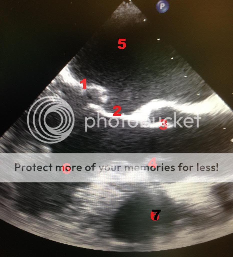
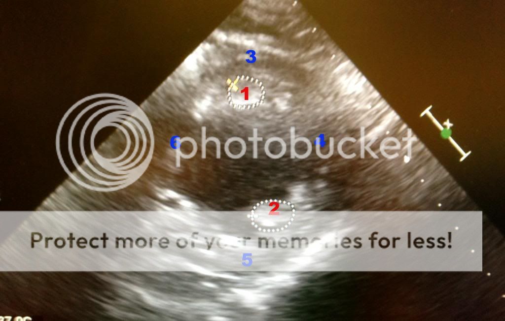
The "suckdown" phenomena is usually due to an underfiled LV causing the interventricular septum to get pulled towards the LV free wall. Doing this alters the geometry of the RV. Augmenting the geometry of the RV may equate to poor systolic performance AND tricuspid annulus dilatation leading to acute tricuspid regurgitation.


Here is a modified bicaval view showing this:

IMG_7335_zps10a137f9.jpg


If you see this coming off bypass you need to think: oh... oh... what can we do to make this better. :nurse:

The answer...? As weird as it sounds, reduce your flow rates on your LVAD and augment volume status. These maneuvers will return the RV to it's regular shape and hopefully increase RV unloading to the LV.

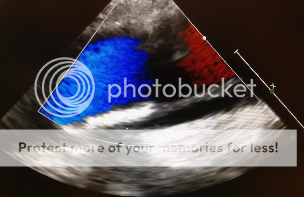
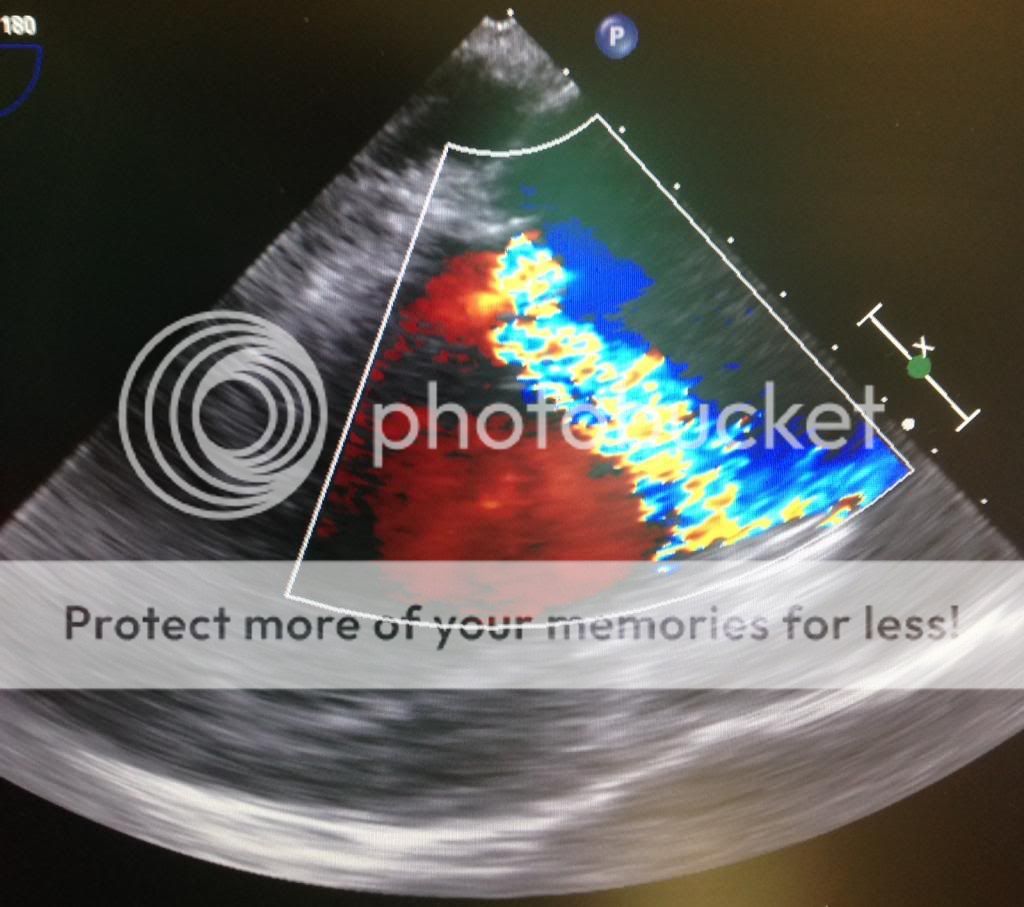
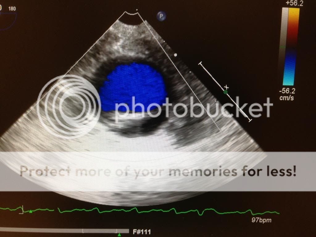
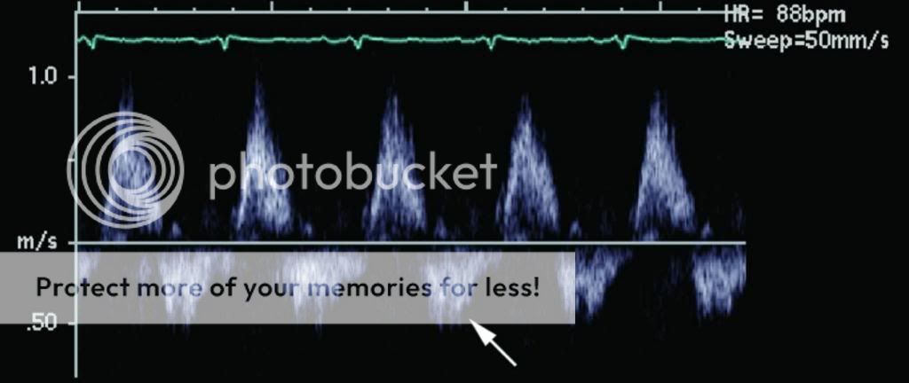
In addition to causing the suckdown phenomena, the inflow canulas can become obstructed. Check for this by using color Doppler and seeing laminar flow. You can also use CWD. Flows should be less than 2.5 m/s. Similarly, the outflow cannula (ascending aorta) should show slightly lower flows. 1-2 m/s.

AI is bad... LVAD worsens AI and therefore you may not achieve increased organ perfusion. You can create a partial closed loop from the ascending aorta back into the LV causing a volume overload problem. You need a competent valve. What are the choices?

1) Repair the valve.
2) Replace the valve and deal with higher morbidity/mortality. Never use mechanical valves. Always bioprosthetic ones.
3) Sounds crazy... but if it's destination therapy or bridge to transplant, you can surgically sew the aortic valve shut so that it doesn't open at all! :eek:


Good stuff.
 
Last edited:
  • Like
Reactions: 1 user
Top