Step 1 Complicated Concepts Thread

This forum made possible through the generous support of SDN members, donors, and sponsors. Thank you.

TheSeanieB

Full Member
10+ Year Member
Joined
Dec 17, 2010
Messages
263
Reaction score
1
ASK AND ANSWER TOUGH QUESTIONS RELATED TO STEP 1.

Starting with me:
physiologic chloride shift - When CO2 diffuses into a RBC, it quickly converts with H2O to H+ and HCO3- so that CO2 will continue to passively diffuse into the RBC. The HCO3- is then excreted into the plasma by a Cl-/HCO3- exchanger. When the RBC enters the pulmonary capillaries, the process reverses. HCO3- is taken up by exchange for a Cl-. It combines with H+ to creates CO2 +H2O. The CO2 then diffuses out of the RBC and ultimately into the alveoli. This process allows for maximal CO2 excretion by a RBC.

Members don't see this ad.
 
Last edited:
  • Like
Reactions: 1 user
Members don't see this ad :)
Can anyone explain why you get Pellegra-like symptoms with Hartnup disease?

You get pellagra because hartnup is a failure of resorbing Tryptophan. Less tryptophan = less niacin -> pellagra.

There are two other scenarios that also causes pellagra:

1. carcinoid syndrome -> increased tryptophan is used to make serotonin means less tryptophan can be used to make niacin -> pellagra.

2. using INH means less pyroxidine. b6 is required to make niacin so less niacin is made -> pellagra.
 
so, hepatic encephalopathy mainly comes from increased ammonia in the blood. Does this not mean the BUN goes up? I thought ammonia counted towards the nitrogenous products in the N of BUN?

I think the liver converts NH4+ to urea, but they're not the same thing. The excess ammonia causes encephalopathy, but if you have liver cirrhosis or if it's not functioning, then the BUN won't go up because you're not converting the ammonia to urea.
 
I think the liver converts NH4+ to urea, but they're not the same thing. The excess ammonia causes encephalopathy, but if you have liver cirrhosis or if it's not functioning, then the BUN won't go up because you're not converting the ammonia to urea.

ah, ok. I thought BUN measured all nitrogen containing products in the blood, not just urea.

thanks!
 
Here's a concept I'm still scratching my head over...
In First Aid it says that potassium shifts into cells in the context of hypo-osmolar states, and out of cells in hyperosmolar states. What's the reasoning/mechanism behind this? The only thing I can come up with is that K+ exchanges for Na+ to correct the extracellular osmolarity, but that doesn't quite add up since doesn't Na+ draw water with it? Would abnormal serum potassium be a clue to either SIADH or diabetes insipidus for this reason?

If my question even makes sense... anyone have some insight?
 
I came across a question saying a good first step in the management of cardiac tamponade would be IV fluids, and inotropic agents to increase preload and contractility of the compressed heart. Then you can perform pericardiocentesis. I didnt know that, and I thought id share.
 
I came across a question saying a good first step in the management of cardiac tamponade would be IV fluids, and inotropic agents to increase preload and contractility of the compressed heart. Then you can perform pericardiocentesis. I didnt know that, and I thought id share.

if there was a list out there for:

1. best initial work up in management
2. gold standard for screening
3. confirmatory test

for some of the high yield diseases...that would be:

iBXfiMvtklBMI.gif
 
I came across a question saying a good first step in the management of cardiac tamponade would be IV fluids, and inotropic agents to increase preload and contractility of the compressed heart. Then you can perform pericardiocentesis. I didnt know that, and I thought id share.

Interesting! Thanks for sharing.
 
I came across a question saying a good first step in the management of cardiac tamponade would be IV fluids, and inotropic agents to increase preload and contractility of the compressed heart. Then you can perform pericardiocentesis. I didnt know that, and I thought id share.

This... doesn't make any sense to me haha. I looked it up and apparently you give IV fluids to increase CO, especially if the patient is hypovolemic. Some papers said the inotropic agents won't work because the heart is already contracting maximally, but I guess it wouldn't hurt. Thanks for this. Word on the street is that Step 2CK material occasionally shows up...
 
This... doesn't make any sense to me haha. I looked it up and apparently you give IV fluids to increase CO, especially if the patient is hypovolemic. Some papers said the inotropic agents won't work because the heart is already contracting maximally, but I guess it wouldn't hurt. Thanks for this. Word on the street is that Step 2CK material occasionally shows up...

From current diagnosis and treatment Cardiology
Intravenous fluids and pressors can be administered as temporizing measures until the procedure can be performed.

In real life, you would do this if pericardiocentesis cannot be immediately performed.
On a test youd choose IV fluids if pericardiocentesis isnt an answer choice.
And the reason IV fluids help is because it increases the preload, which would be beneficial for a heart that is being compressed and cannot fill adequately.
 
Members don't see this ad :)
From current diagnosis and treatment Cardiology


In real life, you would do this if pericardiocentesis cannot be immediately performed.
On a test youd choose IV fluids if pericardiocentesis isnt an answer choice.
And the reason IV fluids help is because it increases the preload, which would be beneficial for a heart that is being compressed and cannot fill adequately.

Ah, that makes sense now, thanks. I guess in my mind, I was thinking that the patient already had problems with heart expansion, so giving more fluids would somehow overwork the heart.
 
I seem to recall learning that 2 types of bacterial meningitis cause CSF lymphocytosis. I think one is syphilis...thoughts?
 
pg.114 - It seems like the more you exercise the more aerobic metabolism takes over. I just thought before that it makes more sense that you switch to anaerobic as time goes on with using up oxygen and needing to get quick energy, and then you start forming lactic acid. But here it seems like aerobic is the main form of metabolism long term during exercise. Can someone explain?
 
And one more on pg.114-

Starvation 1-3d. "hepatic gluconeo... from adipose tissue glycerol and propionyl CoA (from odd chain ffa - the only triacylglycerol components that contribute to gluconeo)"

So is this saying that only odd chain ffa are used for gluconeogenesis? That confuses me as they put down propionyl coa there, which I think is 16 carbons? Do even chain ffa not contribute? I just don't know what they're trying to say in general w/ this statement.
 
Ah found it. Subacute meningitis: TB and syphilis

pg.114 - It seems like the more you exercise the more aerobic metabolism takes over. I just thought before that it makes more sense that you switch to anaerobic as time goes on with using up oxygen and needing to get quick energy, and then you start forming lactic acid. But here it seems like aerobic is the main form of metabolism long term during exercise. Can someone explain?

I think you start off by breaking down muscle glycogen, but then you run out so your body turns to fat which has to be metabolized in mitochondria and hence aerobic? At this point i'm thinking your adp is really high which turns on the TCA cycle. I think keeping fat reserves is the body's number one priority which explains anaerobic being used first

And yes, odd FA only
 
Nice thank you. And this might be paying way too much attention to details, but LPL and HL working on chylomicron remnants seems contrary to the definitions given for them in first aid. ANd if VLDL have B-100, why isn't it shown binding to the LDL receptor like IDL and LDL are? Should I just memorize this and move on lol
 
Anybody have a good way to tell the difference between hyper-PTH and vitamin D deficiency based on bone and lab findings, but obviously without a PTH or vitamin D level... Just got a uworld question with a bone description and labs that amount to high calcium and low phosphorus.
 
Anybody have a good way to tell the difference between hyper-PTH and vitamin D deficiency based on bone and lab findings, but obviously without a PTH or vitamin D level... Just got a uworld question with a bone description and labs that amount to high calcium and low phosphorus.

Honestly, I bet it's more based upon age and presentation.
 
Anybody have a good way to tell the difference between hyper-PTH and vitamin D deficiency based on bone and lab findings, but obviously without a PTH or vitamin D level... Just got a uworld question with a bone description and labs that amount to high calcium and low phosphorus.

You should have low calcium in vitamin D deficiency, no? Also, I've only heard of osteitis fibrosa cystica in regards to hyper-PTH.
 
Anybody have a good way to tell the difference between hyper-PTH and vitamin D deficiency based on bone and lab findings, but obviously without a PTH or vitamin D level... Just got a uworld question with a bone description and labs that amount to high calcium and low phosphorus.
This was explained nicely by Goljan...I think FA 2013 pg 293, 294, 300:

Low Ca stimulates PTH release with causes
1. Bone resportion to release more Ca.
2. Goes to the kidney and Stimulates reabsorption of Ca and inhibits Phosphate reabsorption
3. Stimulates 1,25 OH2D synthesis.
Vit D then increases absorption of dietary Ca and Phosphate.
Vit D also inhibits PTH synthesis.

I think from this you can workout what to expect from most situations.

Primary HyperPTH(adenoma, ): High Ca; Low PO4; High 1,25 Vit D(But it cant stop inhibit adenoma synth of PTH); High PTH; Cystic Bone Spaces(increase re-absorption of bone); Subperiosteal thinning: Radiograph shows Subperiosteal erosions in medial sides of 2nd and 3rd Phalanges of hand, Salt and pepper appearance of Calvarium.

Secondary HyperPTH(Renal Failure-->Hypovitaminosis D) : High PTH; Low Ca (Due to Low Vit D, and Kidneys not working to reabsorb Ca); Increase PO4 in Renal failure; Increase Alkaline Phosphatase;

Vit D Def ( No sunlight, Renal Failure): Rickets/ Osteomalacia; Low Ca and Low PO4

In summary:
Primary HPTH: High Ca, hypophosphatemia, High PTH
Cause: adenoma MCC, hyperplasia, cancer
S/S: renal stone, peptic ulcers, pancreatitis, hypertension, metastatic calcification; Cystic bone spaces
Secondary HPTH: Low Ca, High PTH;
Cause: hypovitaminosis D from renal failure


Vitamin D deficiency: Decreased mineralization; pathologic fractures; rickets children; osteomalacia adults
Causes: renal failure ; malabsorption; liver disease; low sunlight
Lab: Decrease Ca, Decrease phosphate (malabsorption), Increase phosphate renal failure, Increase PTH
 
Here is what it confuses me:

Predominant tone of arterioles is sympathetic and especially beta2---> vasodilation.

I've read that hexamethonium (nicotinic ganglion blocker) blocks predominant tone of arterioles (sympathetic) and of heart (parasympathetic). So it decreases BP and increases HR.
I get the HR. But I can't understand how decreased BP occurs from hexamethonium MOA..
 
FA says that with renal or liver damage, maintenance dose decreases but loading dose remains unchanged.
I get the former. As soon as CL decreases, maintenace dose decreases.
But I have a problem understanding the latter. If there is liver damage--> decreased protein production--> decreased binding to protein--> increased Vd --> increased loading dose.

Am I wrong here?
 
Here is what it confuses me:

Predominant tone of arterioles is sympathetic and especially beta2---> vasodilation.

I've read that hexamethonium (nicotinic ganglion blocker) blocks predominant tone of arterioles (sympathetic) and of heart (parasympathetic). So it decreases BP and increases HR.
I get the HR. But I can't understand how decreased BP occurs from hexamethonium MOA..

Well you said it yourself, hexamethonium removes sympathetic tone of arterioles and causes vasodilation. This reduces total peripheral resistance and hence blood pressure. The tachycardia is not enough to offset this

FA says that with renal or liver damage, maintenance dose decreases but loading dose remains unchanged.
I get the former. As soon as CL decreases, maintenace dose decreases.
But I have a problem understanding the latter. If there is liver damage--> decreased protein production--> decreased binding to protein--> increased Vd --> increased loading dose.

Am I wrong here?

I would think that the amount of drug given compared to binding proteins in the blood is so small that even if you were to greatly reduce plasma proteins, there would still be more than enough to bind the drug
 
Can someone explain what we're supposed to know about aminoglycoside mechanism of action? On the Wiki page it says exact mechanism is unknown but it's thought that it intereferes with proofreading and translocation, which is also what we were taught in our pharm course.

But, a World question I got said inhibition of translocation is only clindamycin and erythromycin and that aminoglycosides actually distort the 30S subunit structure and prevent synthesis from ever starting by blocking initiation. Anyone that can clear it up for me?

Yeah, inhibiting translocation is done by the macrolides and clindamycin only. AGs inhibit the formation of the initiation complex. The diagram on FA2013 p. 179 is pretty good, and it's accurate (they fixed it from the FA2012 version which was incorrect without the errata). I know because I actually missed a question on a lecture exam because I looked at the FA2012 diagram without fixing the errata. I'll never miss it again.
 
Can someone explain what we're supposed to know about aminoglycoside mechanism of action? On the Wiki page it says exact mechanism is unknown but it's thought that it intereferes with proofreading and translocation, which is also what we were taught in our pharm course.

But, a World question I got said inhibition of translocation is only clindamycin and erythromycin and that aminoglycosides actually distort the 30S subunit structure and prevent synthesis from ever starting by blocking initiation. Anyone that can clear it up for me?

All of the above are among the mechanisms of action for aminoglycosides:

(1) They inhibit the formation of the initiation complex by binding to 30S ribosomal subunit (this is the main one)
(2) They cause misreading of mRNA
(3) They block translocation
 
Yeah, inhibiting translocation is done by the macrolides and clindamycin only. AGs inhibit the formation of the initiation complex. The diagram on FA2013 p. 179 is pretty good, and it's accurate (they fixed it from the FA2012 version which was incorrect without the errata). I know because I actually missed a question on a lecture exam because I looked at the FA2012 diagram without fixing the errata. I'll never miss it again.

All of the above are among the mechanisms of action for aminoglycosides:

(1) They inhibit the formation of the initiation complex by binding to 30S ribosomal subunit (this is the main one)
(2) They cause misreading of mRNA
(3) They block translocation

I see, thanks guys
 
Because you guys are the hardest working most badass step 1 dominators I know, I figured I'd ask here. Any recommendations on whether to do NBME 11 vs. NBME 13? It'll most likely be my last practice test :scared: since it's doubtful I'll have time to do them both. Already done 12 and 15.
Edit: I heard from a friend 13 kind of sucks. Also, where the heck is 14?
 
Last edited:
Because you guys are the hardest working most badass step 1 dominators I know, I figured I'd ask here. Any recommendations on whether to do NBME 11 vs. NBME 13? It'll most likely be my last practice test :scared: since it's doubtful I'll have time to do them both. Already done 12 and 15.

Personally I would do 13 just because it is more recent. The NBME 11 discussion thread on here is pretty extensive, though, so if you do 11 you'll more than likely be able to find the correct answers to all of your missed questions (assuming you get extended feedback) so that's something to consider.
 
Personally I would do 13 just because it is more recent. The NBME 11 discussion thread on here is pretty extensive, though, so if you do 11 you'll more than likely be able to find the correct answers to all of your missed questions (assuming you get extended feedback) so that's something to consider.

I took 13 and corrected all of my incorrects, so I might be able to help with that a little bit.
 
Aren't there threads for 13 and 15? Sorry I'm at the beginning of the study period.. haven't really looked into the threads to see their completeness because I didn't want to accidentally see answers
 
I tookd 13 and did the same is coolforschool, so we could definitely work out any questions you guys might have!!

I have two questions on general concepts.

1) Nystagmus - there is a line in first aid that said that PPRF lesions your eyes point away from the lesion while frontal eye field lesions, your eyes point towards the lesion. That is all it says on the topic and I don't really understand it.

2) Glaucoma - I'm having trouble understanding what receptors affect closed-angle vs open angled glaucoma. The one thing I do know is that mydriasis precipitates closed angle glaucoma (so you have to be careful with atropine). But in general how does the ciliary muscle, pupillary constrictor, and pupillary dilator affect glaucoma? And do alpha agonists help glaucoma by centrally decreasing sympathetic activity? i don't recall first aid mentioning that alpha receptors directly decrease aqueous humor synthesis.

Thanks!!
 
Rh- women and Rh+ fetuses. I thought I understood this completely but I sat down with the First Aid QandA book and read a question that I don't understand the explanation to.

Basically there's a woman who's serum is sent in for an indirect coombs test to check for anti Rh. It's positive and the question says something along the lines of "it would be correct to conclude:..."

The first 3 options are clearly incorrect, but the last two seem to be saying the same thing to me.

D) the presence of anti-Rh in the serum suggests that she's been pregnant with an Rh+ fetus before. (I picked this one and it was the correct answer per the book)

but then there's this answer choice:

E) The results of the test cannot be used to determine the Rh status of a current fetus; the results only assess the presence of antibodies in the mother. To determine fetal Rh status, direct typing of fetal blood must be done.

What's incorrect about that answer? I just don't know. It looks to me like both of these answers could be correct...

Anyone have any insight here?
 
We had pretty extensive (i.e. way too much) lecture on blood type incompatibilities, and both of those sound right to me too

Edit: The only thing I can think of is that they're not using precise words when they say "direct typing of fetal blood". You can determine fetal Rh status and Rh antigen load using maternal serum, but that's due to the presence of fetal RBC's in maternal circulation

http://en.wikipedia.org/wiki/Kleihauer–Betke_test

Edit2: Actually, I'm not sure if you can do that test if the mom seroconverts b/c the fetal RBC's may be destroyed, so that could be a possibility
 
Rh- women and Rh+ fetuses. I thought I understood this completely but I sat down with the First Aid QandA book and read a question that I don't understand the explanation to.

Basically there's a woman who's serum is sent in for an indirect coombs test to check for anti Rh. It's positive and the question says something along the lines of "it would be correct to conclude:..."

The first 3 options are clearly incorrect, but the last two seem to be saying the same thing to me.

D) the presence of anti-Rh in the serum suggests that she's been pregnant with an Rh+ fetus before. (I picked this one and it was the correct answer per the book)

but then there's this answer choice:

E) The results of the test cannot be used to determine the Rh status of a current fetus; the results only assess the presence of antibodies in the mother. To determine fetal Rh status, direct typing of fetal blood must be done.

What's incorrect about that answer? I just don't know. It looks to me like both of these answers could be correct...

Anyone have any insight here?


I'm going to go out on a limb here... (E) sounds right, but it isn't as definite as (D)... D is definite in all cases, while E is not... the reason I'm thinking is that the woman's indirect combs could possibly be +ve as a result of blood mixing the current fetus (placental hemorrhage).. in the case this is her first kid (or no prior Rh+ baby)... that is why if a woman with any sort of bleeding in the 2nd of 3rd trimester (and an unknown father's Rh type) - they typically give Rhogam...

This is what I recall... from when I read this a while back...
 
A 64 year old man comes to the physician's office for a health maintainence examination. He has been unable to sustain erections during intercourse or masturbation for the past month. He does have spontaneous nocturnal and morning erections . He had a myocardila infarction 2 months ago. Which of the following is the most likely explanation for his current erectile disorder?

A) Adverse effect of antihypertensive medication.
B) Altered body image
C) Decreased libido
D) Decreased vascular perfusion
E) Fear of sudden death

Apparently the answer is E. Why is C incorrect? You would still have nocturnal erections with decreased libido right? I figure he isn't being turned on sexually for whatever reason

These questions are almost always designed to point to emotional distress. UWorld has a similar one.

You'd think if he's doing those other things, then his libido probably isn't the issue. If it were he'd be more or less asexual and the question would probably be about "T" levels or something.
 
I tookd 13 and did the same is coolforschool, so we could definitely work out any questions you guys might have!!

I have two questions on general concepts.

1) Nystagmus - there is a line in first aid that said that PPRF lesions your eyes point away from the lesion while frontal eye field lesions, your eyes point towards the lesion. That is all it says on the topic and I don't really understand it.

2) Glaucoma - I'm having trouble understanding what receptors affect closed-angle vs open angled glaucoma. The one thing I do know is that mydriasis precipitates closed angle glaucoma (so you have to be careful with atropine). But in general how does the ciliary muscle, pupillary constrictor, and pupillary dilator affect glaucoma? And do alpha agonists help glaucoma by centrally decreasing sympathetic activity? i don't recall first aid mentioning that alpha receptors directly decrease aqueous humor synthesis.

Thanks!!

I've only got an answer to the first one... I used to understand it perfectly but I had to go look it up again and re-teach myself just now. You can simplify frontal eye field vs. PPRF lesions as if they were UMN and LMN respectively. To initiate voluntary eye movement to one side or another (just in horizontal dimensions... vertical is a whole different beast) the frontal eye fields *kind of around BA areas 4/6/8, in the motor cortex; 'FEF' in the picture on the link: http://www.neurology.org/content/46/2/451/F2.large.jpg* will signal down to the CONTRALATERAL PPRF, which is near the CN 6/abducens nucleus. That PPRF will tell its ipsilateral CN6 to contract (lateral rectus), and use the MLF to also tell the contralateral CN3 to contract (medial rectus).

So now if we lesion the PPRF say on the left, you can't look left - the right frontal eye field will fire at the left PPRF, but the left lateral rectus and right medial rectus can't get the signal to contract. So you will instead look right, away from the lesion.

If you instead lesion the frontal eye field on the RIGHT, it can't fire at the left PPRF, so again no signal to the extraocular muscles to look left. You will still look right, just like with the PPRF example above, but in this case it is towards the lesion.

Edit: here's my source: http://www.dartmouth.edu/~rswenson/NeuroSci/chapter_8D.html

Another thing I used to understand perfectly but no longer do is the difference between these voluntary eye movements vs. pursuit movements... I may or may not try to refresh myself on that.

I'm with you on the glaucoma stuff... ugh... I wish there were maybe a YouTube video or something, illustrating how & when the aqueous humor flow gets blocked vs. opened up...

Edit 2: here's the best I could find for glaucoma and the pupillary dilation vs. constriction: http://www.youtube.com/watch?v=MQIF5__l5J0. Doesn't mention anything about alpha agonists, although I've seen it mentioned elsewhere that they help decrease aqueous humor synthesis by causing local a1-vasoconstriction. Since it also causes a1-pupillary dilation, it would still be bad for closed-angle glaucoma but ok for open-angle. Also, I'm still not sure how relevant the CILIARY muscle tone is to glaucoma, as opposed to PUPILLARY muscle tone or ciliary epithelial secretion of the aqueous humor.
 
Last edited:
my friends, can anyone help me out here?

X2604-C-07.png


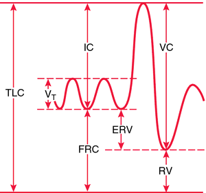
question asked at what point total pulmonary vascular resistance is lowest. I guessed at the RV. UW says it is at the end respiratory volume at the maximal FRC. I don't get the answer at all....

When lungs are totally empty after a maximal exhalation, there is positive pressure exerted on the lungs by the surrounding musculature, causing both airway and blood vessels to collapse?

Can any of your super intelligent homies explain this to me in terms I might understand?
 
A 64 year old man comes to the physician's office for a health maintainence examination. He has been unable to sustain erections during intercourse or masturbation for the past month. He does have spontaneous nocturnal and morning erections . He had a myocardila infarction 2 months ago. Which of the following is the most likely explanation for his current erectile disorder?

A) Adverse effect of antihypertensive medication.
B) Altered body image
C) Decreased libido
D) Decreased vascular perfusion
E) Fear of sudden death

Apparently the answer is E. Why is C incorrect? You would still have nocturnal erections with decreased libido right? I figure he isn't being turned on sexually for whatever reason

The USMLE wants us to know that the elderly (especially 64yo men) do NOT lose their sexual desires. The dude just had an MI 2 months ago so it's not surprising that he's scared for his life
 
my friends, can anyone help me out here?

X2604-C-07.png


question asked at what point total pulmonary vascular resistance is lowest. I guessed at the RV. UW says it is at the end respiratory volume at the maximal FRC. I don't get the answer at all....

When lungs are totally empty after a maximal exhalation, there is positive pressure exerted on the lungs by the surrounding musculature, causing both airway and blood vessels to collapse?

Can any of your super intelligent homies explain this to me in terms I might understand?

Haha not super-intelligent but I'll try, because I remember tripping over this question too...
So the way I see it, there's 2 opposing forces on the pulmonary vasculature when we breathe: the diaphragm versus the lungs. If we exhale hard all the way out (so just RV is left), we've got the diaphragm pushing and compressing the lungs and pulmonary vasculature, so that's not the point of lowest resistance against blood flow. If we start inhaling, we're decreasing intrathoracic pressure, pulling more air in the lungs and more blood in the vessels. However, the more we inhale, the more we get the lungs pushing on and compressing the vessels this time instead of the diaphragm, again increasing resistance.

So the point at which there is the least compressive force from both the diaphragm AND the lungs is at the end of a quiet respiration - top of FRC (and ERV).

Does that make sense? Hopefully someone else can either second this or correct me...
 
Top