Step 1 Complicated Concepts Thread

This forum made possible through the generous support of SDN members, donors, and sponsors. Thank you.

TheSeanieB

Full Member
10+ Year Member
Joined
Dec 17, 2010
Messages
263
Reaction score
1
ASK AND ANSWER TOUGH QUESTIONS RELATED TO STEP 1.

Starting with me:
physiologic chloride shift - When CO2 diffuses into a RBC, it quickly converts with H2O to H+ and HCO3- so that CO2 will continue to passively diffuse into the RBC. The HCO3- is then excreted into the plasma by a Cl-/HCO3- exchanger. When the RBC enters the pulmonary capillaries, the process reverses. HCO3- is taken up by exchange for a Cl-. It combines with H+ to creates CO2 +H2O. The CO2 then diffuses out of the RBC and ultimately into the alveoli. This process allows for maximal CO2 excretion by a RBC.

Members don't see this ad.
 
Last edited:
  • Like
Reactions: 1 user
Haha not super-intelligent but I'll try, because I remember tripping over this question too...
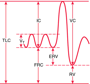
So the way I see it, there's 2 opposing forces on the pulmonary vasculature when we breathe: the diaphragm versus the lungs. If we exhale hard all the way out (so just RV is left), we've got the diaphragm pushing and compressing the lungs and pulmonary vasculature, so that's not the point of lowest resistance against blood flow. If we start inhaling, we're decreasing intrathoracic pressure, pulling more air in the lungs and more blood in the vessels. However, the more we inhale, the more we get the lungs pushing on and compressing the vessels this time instead of the diaphragm, again increasing resistance.

So the point at which there is the least compressive force from both the diaphragm AND the lungs is at the end of a quiet respiration - top of FRC (and ERV).

Does that make sense? Hopefully someone else can either second this or correct me...

I remember this questions from World and that was basically the same reasoning I took away from the explanation
 
my friends, can anyone help me out here?

X2604-C-07.png


question asked at what point total pulmonary vascular resistance is lowest. I guessed at the RV. UW says it is at the end respiratory volume at the maximal FRC. I don't get the answer at all....

When lungs are totally empty after a maximal exhalation, there is positive pressure exerted on the lungs by the surrounding musculature, causing both airway and blood vessels to collapse?

Can any of your super intelligent homies explain this to me in terms I might understand?

Why not post the qid?

edit: nevermind, found my notes. As usual, the explanation is spot on. I'm not sure how anyone on SDN could make it more clear.

During inhalation there is expansion pressure on the capillaries and during expiration there is compression pressure. The Goldilocks principle, the middle is "just right".
 
Last edited:
Members don't see this ad :)
Haha not super-intelligent but I'll try, because I remember tripping over this question too...
So the way I see it, there's 2 opposing forces on the pulmonary vasculature when we breathe: the diaphragm versus the lungs. If we exhale hard all the way out (so just RV is left), we've got the diaphragm pushing and compressing the lungs and pulmonary vasculature, so that's not the point of lowest resistance against blood flow. If we start inhaling, we're decreasing intrathoracic pressure, pulling more air in the lungs and more blood in the vessels. However, the more we inhale, the more we get the lungs pushing on and compressing the vessels this time instead of the diaphragm, again increasing resistance.

So the point at which there is the least compressive force from both the diaphragm AND the lungs is at the end of a quiet respiration - top of FRC (and ERV).

Does that make sense? Hopefully someone else can either second this or correct me...

Thanks! Great explanation!
 
One more q - For nAChR's, is it all of them or just the ones at NMJ's that require binding of 2 ACh to open? Also, how is this tested / what's applicable to the fact clinically? I feel like there was something (maybe stoichiometric binding of an irreversible blocking agent to nAChR's causing complete shutdown of all of them?)

Also, if someone could confirm that they do in fact require two ACh to open at all, I would appreciate it. Wiki says some contradictory stuff on the matter I don't remember every learning

TYIA
 
Is there a condition we'd be tested on in which the ventricles would be paced by purkinje cells? I think purkinje-pacing is also the point where you get QRS complex widening (His and above you maintain tight QRS), right?
 
Can anyone explain the difference between the following:

Aplastic anemia
Pancytopenia
Agranulocytosis
Bone marrow suppression

I kind of just jumble them all together and the drugs that cause them but I feel like they are pretty significantly different I'm just not sure how.

Thanks!
 
Why does CHF cause increased hydrostatic pressure? I would think it would be the opposite because of decreased CO.
 
Why does CHF cause increased hydrostatic pressure? I would think it would be the opposite because of decreased CO.

You're right! Decreased CO - the hydrostatic pressure is decreased, from the aorta and forward. But the left ventricle isn't pushing out everything that's coming in, so eventually there's a backlog... because of that, from the left atrium and back, the hydrostatic pressure is still increased.

Edit: What CrouchingLiger says, below :)
 
Last edited:
Why does CHF cause increased hydrostatic pressure? I would think it would be the opposite because of decreased CO.
Increased hydrostatic pressure is in the post-capillary venules, from blood backing up on the venous side of the heart. There is no increased hydrostatic pressure on the arterial side b/c of CHF as you said, but those vessels are thicker anyway and so wouldn't have much fluid filtration through them. These post-capillary venules are the main source of edema, because of the increased hydrostatic pressure and thus fluid filtration. They are thinner and allow for movement of water.
 
Members don't see this ad :)
Can anyone explain the difference between the following:

Aplastic anemia
Pancytopenia
Agranulocytosis
Bone marrow suppression

I kind of just jumble them all together and the drugs that cause them but I feel like they are pretty significantly different I'm just not sure how.

Thanks!

Still not too sure about all of this after looking it up, but I'll give it a shot: I think these are all similar in that they refer to decreased blood cell counts, some of them just more specifically refer to certain cell lineages. And I think they can all be caused by decreased hematopoiesis, if not other things like increased destruction.

Aplastic anemia: I consider this to only refer to rbc's. So the abnormality you'd find is purely a decrease in the cell counts, and I think it would be normocytic. Some causes... Parvo B19 (because it infects rbc progenitors specifically); felbamate (a newer anticonvulsant), and carbamazepine (drug of choice for partial & generalized tonic-clonic seizures).

Pancytopenia: All blood cell counts decreased! Not much else to say about that.

Agranulocytosis: Decreased granulocytes (neutrophils, basophils, eosinophils...). Some causes are clozapine (atypical antipsychotic), and again carbamazepine.

Bone marrow suppression: I think it's more of a blanket-term and drug side-effect buzzword, not sure if it can be used to describe specific blood cell lineage suppressions or if it has to mean all blood cells are suppressed (ie pancytopenia). But either way it means decreased synthesis... Some causes: antimetabolites (like MTX, 5-FU...), cyclophosphamide, sirolimus, rituximab, zidovudine.
 
Why not post the qid?

edit: nevermind, found my notes. As usual, the explanation is spot on. I'm not sure how anyone on SDN could make it more clear.

Obviously the UWorld explanation wasn't enough for me. Akr gave me an awesome explanation. If you could tone down the condensation in this subforum it would be great, John.

Are you actually taking Step 1 soon? Are you a second year? Just wondering....
 
Ok, so I have a hard time keeping aortic dissection aneurysm straight.

Can someone tell me if this is correct:

Aneurysm: will involve all three layers of vessel. Atherosclerosis and tertiary syphilis are a risk.

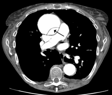
Dissection: involves intimal tear. Hypertension is the main risk factor. Can DX with image of double barrel aorta, which an aneurysm will not have:
220px-DissectionCT.png


Both: Will both look like a huge, swollen vessel on imaging from the exterior of the vessel, like so:
nature10145-f1.2.jpg
 
Quick question. beta-blockers are bad for diabetes because they mask hypoglycemic symptoms? or because they prevent beta-2 mediated insulin release?
 
Quick question. beta-blockers are bad for diabetes because they mask hypoglycemic symptoms? or because they prevent beta-2 mediated insulin release?

Insulin release is not majorly beta mediated. Beta 2 blockade can however inhibit hepatic gluconeogenesis and cause fasting hypoglycemia. More of an issue with type 1 diabetics I presume

Unless I'm wrong about the insulin thing
 
Quick question. beta-blockers are bad for diabetes because they mask hypoglycemic symptoms? or because they prevent beta-2 mediated insulin release?

I think it's more because they block hypoglycemic symptoms.

There are two stages of the hypoglycemic response:

1. adrenergic -> sweating, tremor, etc
2. central -> passing out, seizure, etc.

B-blockers masks stage 1 so people end up going straight into stage 2, which has much worse long term effects.

edit: page 288 of FA2013 goes over the B2/A1 receptor stuff. I think this stuff is a non-factor when prescribing, if someone needs a B-blocker (say for hypertension associated with an MI, to prevent heart remodeling or whatever) you just titrate the diabetes meds around it. the key here is that the patient has to be aware of the hidden hypoglycemia and must check their glucose levels vigilantly.
 
Last edited:
Ok, so I have a hard time keeping aortic dissection aneurysm straight.

Can someone tell me if this is correct:

Aneurysm: will involve all three layers of vessel. Atherosclerosis and tertiary syphilis are a risk.

Dissection: involves intimal tear. Hypertension is the main risk factor. Can DX with image of double barrel aorta, which an aneurysm will not have:

Don't forget that the location for the dissection will differ from that of the abdominal aneurysm. Haven't see a question asking the layers involved for aneurysm, but have for the dissection, which in case you're right - intimal tear.

There's a lot of other distinguishing features that'll be included in the vignette to help you differentiate the two.

thanks, Jonari, you're a boss dude
 
So Uworld constantly says that high lipid solubility of an inhaled anesthetic represents slow induction, NOT high potency.

Meanwhile, p453 of FA2013 clearly implies a direct relationship between high lipid solubility and high potency.

My take is that I should go by UWorld. Any thoughts?
 
So Uworld constantly says that high lipid solubility of an inhaled anesthetic represents slow induction, NOT high potency.

Meanwhile, p453 of FA2013 clearly implies a direct relationship between high lipid solubility and high potency.

My take is that I should go by UWorld. Any thoughts?

I ran into one of these questions last night. I just think of lipid solubility as relating to the MAC (and therefore potency). The speed of induction is not related to lipid solubility but blood solubility.

Lipid solubility = potency (MAC).
Blood solubility = speed of induction.

Keep potency and induction separate... it's still pretty confusing to me though, tbh.

yeah thats what i thought until i saw a question that asked which would correlate most with potency... and blood-lipid solubility coefficient and MAC were both answers. obviously MAC is the better answer, but i think uworld wouldn't usually put an answer choice like lipid solubility if it was close to being right
 
yeah thats what i thought until i saw a question that asked which would correlate most with potency... and blood-lipid solubility coefficient and MAC were both answers. obviously MAC is the better answer, but i think uworld wouldn't usually put an answer choice like lipid solubility if it was close to being right

It's UW, not the NBME haha. They would do that just to make sure you know that MAC is an actual measure of potency, and blood-lipid solubility is a property that is related to it. I get pretty angry with UW sometimes when they pull this stuff though.
 
So Uworld constantly says that high lipid solubility of an inhaled anesthetic represents slow induction, NOT high potency.

Meanwhile, p453 of FA2013 clearly implies a direct relationship between high lipid solubility and high potency.

My take is that I should go by UWorld. Any thoughts?

I ran into one of these questions last night. I just think of lipid solubility as relating to the MAC (and therefore potency). The speed of induction is not related to lipid solubility but blood solubility.

Lipid solubility = potency (MAC).
Blood solubility = speed of induction.

Keep potency and induction separate... it's still pretty confusing to me though, tbh.

Thanks for bringing this up, I had forgotten about how much this still frustrates me... so high blood solubility = slow induction, right? But then UWorld also says that high lipid solubility = slow induction, although it's better to think of lipid solubility as related to MAC/ potency, rather than speed of induction? It's kind of contradictory.

The only way I could make sense of high lipid solubility causing slow induction, would be to think of it as high solubility into non-target tissues that also have lipid in them. But I know that's probably not what's actually happening...
 
The only way I could make sense of high lipid solubility causing slow induction, would be to think of it as high solubility into non-target tissues that also have lipid in them. But I know that's probably not what's actually happening...

I think this is exactly what happens. Think of it like this:

bloodsteam must fill before interstituim does.
Interstituim(ECF) must fill before ICF does.
When the drug hits the ICF, the effect starts.
Equillibria.jpg


The more soluble(lipophillic) the compound is, the more of it dissolves into the bloodsteam. I believe this has to do with the high amount of fat carriers, such as albumin, that bind to the compounds. The more more lipophillic they are, the more binding to albumin there is, the more you need to dissolve to fill that compartment.

This is a super dumb analogy, but think of it like this:
a.baa-Waterfall-from-cups.jpg


Top cup(highest one) is the bloodsteam. Compound won't flow into the cup below it until the top cup overflows. Same goes for the ECF to ICF.
 
The more soluble(lipophillic) the compound is, the more of it dissolves into the bloodsteam.
Confused as crap now. I really liked your pictures, but isn't it hydrophilic things that dissolve better into blood? Or am I thinking about this totally wrong and should think about things binding to albumin like you said?
 
Can someone tell me what the answer to this concept is? If they show you a multinucleated giant cell and ask what was most important in it's formation, what's the answer? I feel like, on UWORLD, I've seen both IFN-y and TNF-a as the correct answer. It's kind of a perpetuating cycle between macrophages and T cells so I can see the logic in both, but...you know. I don't know.
 
Confused as crap now. I really liked your pictures, but isn't it hydrophilic things that dissolve better into blood? Or am I thinking about this totally wrong and should think about things binding to albumin like you said?

No you're correct, the more lipophilic things are the better they dissociate into tissue.
 
Can someone tell me what the answer to this concept is? If they show you a multinucleated giant cell and ask what was most important in it's formation, what's the answer? I feel like, on UWORLD, I've seen both IFN-y and TNF-a as the correct answer. It's kind of a perpetuating cycle between macrophages and T cells so I can see the logic in both, but...you know. I don't know.

Always look for IL1, IFN, or TNF

I'm assuming the answer will never including more than one of the above, because they all play a role.
 
I've always really loved how the enzyme deficient in acute intermittent porphyria has 3 different names that don't sound anything alike. ****ing scientists

Can someone tell me what the answer to this concept is? If they show you a multinucleated giant cell and ask what was most important in it's formation, what's the answer? I feel like, on UWORLD, I've seen both IFN-y and TNF-a as the correct answer. It's kind of a perpetuating cycle between macrophages and T cells so I can see the logic in both, but...you know. I don't know.

If they have both TNF-a and IFN-g, I would choose TNF-a. IFN-g activates macrophage cytotoxicity and cytokine production, TNF-a recruits monocytes and induces their differentiation into epithelioid histiocytes.

This is what I've gathered anyway
 
Confused as crap now. I really liked your pictures, but isn't it hydrophilic things that dissolve better into blood? Or am I thinking about this totally wrong and should think about things binding to albumin like you said?

No you're correct, the more lipophilic things are the better they dissociate into tissue.

Ok, there are two seperate ideas working here:


Here is a more detailed explanation from a someone else on SDN:

In general, there are two properties of general inhalation anesthetics that we should be familiar with. These are 1) the blood/gas partition coefficient and 2) the lipid/gas partition coefficient.

Blood/gas partition coefficient:
The blood/gas partition coefficient is a measure of the solubility of a gas in the blood and is what determines the SPEED OF ONSET of an inhalation anesthetic. If a gas is MORE soluble in the blood, the blood/gas partition coefficient will be higher. If a gas is LESS soluble in the blood, the blood/gas partition coefficient will be lower. In order for a gas to reach the brain, it must be inhaled. Once in the alveoli, the gas must equilibrate with the blood in the capillaries. The speed at which this occurs is determined by how soluble a gas is in the blood. If a gas is MORE soluble in the blood, more gas particles will exist in the blood and thus the blood/gas coefficient will be higher (ex. 200 particles in the blood and 1 particle in the gas suggests slower equilibration, so the coefficient is 200/1, or 200). In effect, it takes more gas particles dissolving in the blood to have the pressures equilibrate. Conversely, if a gas is LESS soluble in the blood, less gas particles will exist in the blood and the gas will have a lower blood/gas coefficient (ex. 1 particle in the blood and 200 particles in the gas suggests very rapid equilibration, or 1/200, or 0.005). In effect, less gas particles need to dissolve for the partial pressures to equilibrate. So, in summary, the speed at which a gas in the alveoli equilibrates with the blood in the capillaries is determined by its solubility in blood, and this then determines the speed of onset of an inhalation anesthetic.

Lipid/gas partition coefficient:
The lipid/gas partition coefficient is a measure of the lipid solubility of an inhalation anesthetic. The higher the lipid/gas coefficient, the more lipid soluble the gas and thus the easier the drug can cross the blood brain barrier. The lipid/gas coefficient is what determines the MAC. The Minimum Alveolar Concentration is defined as the amount of gas necessary to remove reaction to a noxious stimulus (i.e. surgical incision) in 50% of the population, and is actually a percentage of alveolar gas. For example, a gas with an MAC of 2, or 2%, means that its concentration in the alveoli must only be 2% of the total gas within the alveoli at that time in order to remove reaction to noxious stimulus in 50% of people. MAC also determines potency, and potency is expressed as the inverse of MAC, such that potency=1/MAC. So, a gas (Gas A) with a MAC of 2% will have a high potency, since potency=1/2, or 0.5. Gas B, which has a MAC of 75%, will have a low potency, since potency=1/75, or 0.013. So, in summary, the potency of a gas is determined by its MAC, and is expressed as 1/MAC.

It is absolutely possible to have a drug with high blood solubility and low lipid solubility, such that time of onset is slow and potency is low. The opposite (fast onset and high potency) is also true, and, as pointed out, is what makes for a favorable inhalation anesthetics. Hope this helps.
 
Ahhh maybe a source of confusion comes from blood:lipid partition coefficient (which would affect speed of onset) vs. lipid/gas partition coefficient (which would affect MAC/potency)?

Just noticed the post above mentions lipid/gas while the uworld question i was originally referring to mentions blood/lipid

Yeah, I don't think FA even mentions blood/lipid as I had to write that in
 
Ahhh maybe a source of confusion comes from blood:lipid partition coefficient (which would affect speed of onset) vs. lipid/gas partition coefficient (which would affect MAC/potency)?

Just noticed the post above mentions lipid/gas while the uworld question i was originally referring to mentions blood/lipid

yeah, exactly.

The principals behind potency and induction are similar but different and easy to confuse.

It doesn't help when you add in how albumin plays a factor, haha.
 
yeah, exactly.

The principals behind potency and induction are similar but different and easy to confuse.

It doesn't help when you add in how albumin plays a factor, haha.

Blood:lipid... Argh, the things I ignore in question stems!! Thanks for the cup analogy too, I'm sure that guy understands the principles of inhaled anesthetics very well...
 
Anyone know off the top of their heads what is the difference between neurofibrillary tangles and pick bodies? I know they are found in alzheimers and pick's disease, respectively, but why do they have different names? Aren't they all Tau proteins?
 
Anyone know off the top of their heads what is the difference between neurofibrillary tangles and pick bodies? I know they are found in alzheimers and pick's disease, respectively, but why do they have different names? Aren't they all Tau proteins?

Pick bodies are round whereas the tangles are flame shaped.. Think there was something else Sattar said... Can't remember for sure. It's in the CNS chapter dementia disorders
 
Just got the stupid ass UW question with blood/lipid partition coefficient. Funny how I can't find this term used anywhere other than UW or people asking about it on step forums. I think I'm just going to ignore what they have to say and go on whatever I remember from class
 
Just got the stupid ass UW question with blood/lipid partition coefficient. Funny how I can't find this term used anywhere other than UW or people asking about it on step forums. I think I'm just going to ignore what they have to say and go on whatever I remember from class

I ink its in big lippincotts if you want clarification, but yeah, probably not a bad idea to use class notes on this one
 
I ink its in big lippincotts if you want clarification, but yeah, probably not a bad idea to use class notes on this one

Yeah, I looked in G&G and class notes and didn't see it. Think I found something in Katzung though. What UW calls "blood/lipid partition coefficient" should actually be "tissue/blood partition coefficient". A high tissue/blood partition coefficient slows onset of drug by increasing the alveolar-venous partial pressure gradient and decreases elimination rate.

I think that's more or less right anyway
 
Can anyone summarize potassium and proton handling? Particularly in the collecting ducts I keep stumbling upon things that seem contradictory. To be specific

1) On essentially all cells throughout the body, there are K/H exchangers that can react to transient changes in pH or potassium status

2) In the collecting duct, aldosterone promotes potassium secretion and proton secretion

3) On intercalated cells, do you have a K/H exchanger, or is it an H+ ATPase with nothing for potassium?

4) The means by which alkalosis stimulates renal excretion of potassium is...?
 
I realized it was lame starting a bunch of new threads....regarding the notion of "Dyspnea in left CHF"...ultimately you get pulmonary edema....now the physiology concept that comes to mind when I think of pulmonary edema is diffusion-limited exchange...Uworld hits at the decreased in lung compliance part....is it pretty much BOTH diffusion limited exchange and decreased compliance that affect adequate gas exchange, hence = dyspnea?
 
Found a pathoma/first aid discrepancy: Pathoma says aortic stenosis causes mIcroangiopathic hemolytic anemia, FA says it's mAcroangiopathic. What do you guys think? Does it even matter since the end result is essentially the same?
 
that would be mAcroangiopathic hemolytic anemia. I think micro or macro refers to what is causing the RBC to be destroyed (Valve = large, macro vs thrombi = small, micro)
 
I've always really loved how the enzyme deficient in acute intermittent porphyria has 3 different names that don't sound anything alike. ****ing scientists

Correct me if I'm wrong but my general rule with the 2 porphyrias is that they are more or less alphabetical. AIP corresponds to HMB synthase (or whatever you call it) slash porphosomething deaminase slash uroporphowhatever 1.

In PCT, the deficient enzyme is only called uroporphosomething 3. P (as in PCT) comes after A (as in AIP), 3 comes after 1, and all the other AIP enzyme name synonyms start with letters that come before "U" as well.

It's also nice because the interruption for AIP occurs before the interruption for PCT. Once again, alphabetical order.

The substrate that builds up for AIP starts with the letter P, while the substrate that builds up for PCT starts with the letter U. Alphabetical as well.

If you somehow managed to follow that, I promise it's super helpful.
 
I remember seeing a question on food poisoning and attack rate or something. Can someone refer me to an example to explain how to figure out what food made people sick when given a table of numbers?
 
Top