Biochemistry, cell biology, and genetics question thread

This forum made possible through the generous support of SDN members, donors, and sponsors. Thank you.

Nutmeg

Green globule
Lifetime Donor
20+ Year Member
Joined
Aug 18, 2003
Messages
30,458
Reaction score
21,945
All users may post questions about MCAT, DAT, OAT, or PCAT cell/molecular biology, genetics, and biochemistry here. Anatomy, physiology, development, embryology, and evolution questions should be posted in the other biology thread. We will answer the questions as soon as we reasonably can. If you would like to know what biology topics appear on the MCAT, you should check the MCAT Student Manual (http://www.aamc.org/students/mcat/s...anual/start.htm)

Acceptable topics:
-general, MCAT-level biology.
-particular MCAT-level biology problems, whether your own or from study material
-what you need to know about biology for the MCAT
-how best to approach to MCAT biology passages
-how best to study MCAT biology
-how best to tackle the MCAT biological sciences section

Unacceptable topics:
-actual MCAT questions or passages, or close paraphrasings thereof
-anything you know to be beyond the scope of the MCAT

*********

If you really know your cell/molecular biology, I can use your help. If you are willing to help answer questions on this thread, please let me know. Here are the current members of the Cell/Molecular Biology Team:

-Nutmeg (thread moderator): My background is in neurobiology. Please note that I am nocturnal, and generally only post between the hours of 10pm and 8am PST.

I'm going to make this thread a bit different than the others, because the material covered in the BS section is a bit different. With o-chem, gen-chem, and physics, there are a number of core concepts to understand. While there is also a lot of that in the BS, there is also a great deal of specific knowledge involved in this section (relative to the others). Test questions often introduce an experimental set-up, asking for either expected results or the interpretation of results. As such, passages might relate to advanced concepts that you are not expected to know coming into the test, and that they will explain in the passages. Any familiarity that you have with these concepts will make the test easier.

While in general this forum is designed for people studying for the MCAT, I welcome any questions relating to molecular biology, even though they might be beyond the scope of the MCAT. I know some people also like to use these threads to get help on homework questions, and I welcome that, too.

-LT2: LT2 is finishing her MS in microbiology.

Members don't see this ad.
 
ooo. That helps a lot. Kaplan's stuff didn't help whatsoever.
So what determines whether it's independent or non-independent? Is one just ideal and the other non-ideal?


1) Genes that are on DIFFERENT chromosomes always assort independently (they have an equal chance of going to the same gamete or to different gametes).

2) Genes on the SAME chromosome that are far enough apart from each other could assort independently.

3) Genes on the SAME chromosome that are close to each other (i.e. linked) don't assort independently.
 
I can't seem to get this,
whenever a questions gives me the a sequecence and asks me for the complementary sequence like if i'm given
mrna sequence " 5-CGAUC-3 and ask to find the complementary rna, i mean i know the sequence is read 5 to 3' , and synthesized 3 to 5', but how do i write this? help?:confused:

First you write 3-GCUAG-5, and then you flip it to 5-GAUCG-3 since the standard way of writing sequences is 5' to 3' (it's still the same sequence). The MCAT might break that convention, who knows.
 
Members don't see this ad :)
a genetics question... It is from Kaplan lesson book, page #160

Question: What is the likelyhood that the offspring is a carrier?

(empty square) x (empty sircle) --> empty square.

The answer is 67%.

After drawing the pennett squares, i got 25% RR, 50%Rr, and 25% rr. So the carrier will be 50%. I do not understand how it is 67%?

Since the offspring is an empty square the offspring is not affected (i.e he doesn't have the disease), which is why we can throw away rr because we know for a fact that the offspring is not affected. So 2/3 = 67%.

You just forgot to account for this piece of information and you were doing 2/4 = 50%.

HTH
 
It still does not make sense logically. You see you get 3 different genotypes: RR, Rr, and rr. The probability The offspring has an equal probability to become RR or rr. The way the question is worded "what's the likelyhood of the offspring", it is 25% likely to be rr, 25% RR, and 50% Rr. I mean if we are talking about probabilities, it does not matter what the picture shows. "rr" is still probable, no matter what. I thought about calling Kaplan tech support about this, I still do not understand.
 
It still does not make sense logically. You see you get 3 different genotypes: RR, Rr, and rr. The probability The offspring has an equal probability to become RR or rr. The way the question is worded "what's the likelyhood of the offspring", it is 25% likely to be rr, 25% RR, and 50% Rr. I mean if we are talking about probabilities, it does not matter what the picture shows. "rr" is still probable, no matter what. I thought about calling Kaplan tech support about this, I still do not understand.


empty square) x (empty sircle) --> empty square.


If the child is an empty square, the child is a male (because of the "square" part) and the child doesn't have the disease (because of the "empty") part. So rr is ruled out. The child has a zero chance of being rr.
 
Okay, so the parents are both heterozygous -> you have the correct genotypic frequencies if we didn't know anything about the outcome already. However, this trait is homozygous recessive so a child with rr would be afflicted, but in this example the child is either a carrier or free of allele since his square is not filled in. From this we know he is either RR or Rr and since the original frequencies were 25% and 50%, respectively, we can conclude that he has a 66.66...% of being a carrier using these two percentages.

I don't have any kaplan stuff, but that is how I would try to explain it

yeah, this is what I was trying to say...
 
thanks guys. I think this problem would've made more sense if "rr" was an some embryon. lethal allele or something like that. I think that sometimes test prep companies are incosistent and sometimes they do not shade the last generation.
 
Prior to infecting a bacterium, a bacteriophage must:

A) reproduce, making copies of the phage chromosome
B) integrate its genome into bacterial chromosome
C) penetrate the bacterial cell wall completely
D) attach to a receptor on the bacterial cell membrane

I ruled out A and B obviously, and was about to select D until I thought:
The cell wall is the outermost enclosure, so it would have to interact with that first, choosing C.

I'm guessing this is wrong and that there is another membrane outside of the cell wall? Is this where bacteria has its receptors?
 
The phage needs to bind to a specific host cell molecule outside the cell wall (LPS, LTA, or some other protein receptor) first before it can penetrate the cell wall and deliver its genetic material.

I would make sure you know the diff. b/t gram - and gram + cellular morphologies for the mcat...it's pretty simple, but I know a lot of people that continually mix them up
 
Quick question:

In immunohistochemistry, why must the secondary anitbody be of a different organism than the primary antibody? Thanks.
 
Members don't see this ad :)
Quick question:

In immunohistochemistry, why must the secondary anitbody be of a different organism than the primary antibody? Thanks.

Remember that an antibody is created when you place a foreign substance into an organism and it is not compatible. For example, if you give someone with A+ blood, B- blood, and you get an immune response.

In immunohistochemistry, you want to be as specific as possible. So the primary antibody is created in an animal like a mouse. that means that the antibody used in the primary reaction is a mouse antibody. If you then put the primary antibody back into the mouse, no other antibodies will be created because it is not considered foreign. Therefor you will never get your secondary antibody. However, if you place the same primary mouse antibody into a rabbit, then you will illicit an immune response from the rabbit because its body has detected a foreign substance.

Now you have the secondary antibody that will bind to the primary antibody because it is specific.

Do you understand? The purpose is to create antibodies to illicit an immune response from the host animal. In order to do this, you need a forein substance to create this response.

I hope this helps.
 
Why are sex linked traits never associated with the Y-chromosome. I know that the genes for colorblindness and hemophelia (the two classic examples) are located on the X chromosome, so males that are positive for those conditions can only pass it to female offspring. However, are there any traits associated with the genes on the Y-chromosome so that males can only pass the trait to male offspring. Does this have to do with the Y-chromosome not containing as much genetic material as the X-chromosome? Or are any Y-linked genetic defects eliminated through evolution due to the fact that carriers that do not express the trait are not possible.
Thanks

No vital genes reside only on the Y chromosome, since 50% of humans do not have Y chromosomes. The only well-defined human disease linked to a defect on the Y chromosome is defective testicular development (due to deletion or deleterious mutation of SRY. This results in sex reversal, so that a person with an XY karyotype has a female phenotype (i.e., is born a female). The lack of the second X results in infertility.

However, it is possible for an abnormal number (aneuploidy) of Y chromosomes to result in problems.

47,XYY syndrome is caused by the presence of a single extra copy of the Y chromosome in each of a male's cells. Males with 47,XYY syndrome have one X chromosome and two Y chromosomes, for a total of 47 chromosomes per cell. Researchers are not yet certain why an extra copy of the Y chromosome is associated with tall stature and learning problems in some boys and men. These effects are variable and often minimal or undetectable. When chromosome surveys were first done in the 1960s, it was reported that a higher than expected number of men in prisons were found to have an extra Y chromosome, so that for a while it was thought to predispose a boy to antisocial behavior (and was dubbed the "criminal karyotype"). Better population surveys have since demonstrated that the association was simply that boys and men with learning problems are more likely statistically to spend time in prison and that there is no other independent statistical association with extra Y. The "criminal karyotype" concept is inaccurate and obsolete.

Greater degrees of Y chromosome polysomy (e.g., XYYYY) are very rare. Rarely, males may have more than one extra copy of the Y chromosome in every cell (polysomy Y). The extra genetic material in these cases can lead to skeletal abnormalities, decreased IQ, and delayed development, but the features of these conditions are variable.

There are also problems that arise from having an incomplete Y chromosome: the usual karyotype in these cases is 46X, plus a fragment of Y. This usually results in defective testicular development, such that the infant may or may not have fully formed male genitalia internally or externally. The full range of ambiguity of structure may occur, especially if mosaicism is present. When the Y fragment is minimal and nonfunctional, the child usually is a girl with the features of Turner syndrome but a risk of malignancy. (wikipedia).

There is a y-linked autoimmune disease
 
why is phosphate group on the DNA acidic?
it sayd that histones are basic and that DNA phosphate group are acidic
but i coulnt know why since phosphate group on the DNA helix have O-
...wouldnt this mean that they are basic?

also, where in the respiration does protein enter? fatty acids enter in TCA cycle...but i couldnt find protein

The defintion of an acid is a hydrogen ion donator, because the phosphate group releases the hydrogen, the phosphate group is considered acidic, hence the name, Deoxyribose nucleic ACID.

In respirations, you have many enzymes that catalyze the release of ATP and the synthesis of ATP from ADP, as well as NADH, etc. These enzymes are the proteins that are in the cycle. For example, G-6PD (glucose 6-phosphate dehydrogenase).

Does this clarify things a bit better?
 
is it DNA ligase or telomerase that removed RNA primer and replaces with DNA during DNA replication?

i know in prokaryotes, it is DNA polI that removes primers, but in
eukaryotes?

im getting conflicting information from my books..help!!
 
is it DNA ligase or telomerase that removed RNA primer and replaces with DNA during DNA replication?

i know in prokaryotes, it is DNA polI that removes primers, but in
eukaryotes?

im getting conflicting information from my books..help!!

i'm copying and pasting from my school's website:

http://www.biosci.ohio-state.edu/~mgonzalez/Micro521/04.html


DNA polymerase I - the function of this polymerase during replication is to remove the RNA primers (after they have done their job of initiating DNA replication) using a 5'--->3' exonuclease activity. It then uses its 5'--->3' polymerase activity to fill in the resulting gaps.

Hope this helps:luck:
 
I am having a hard time with Pedigree questions. Can someone write a review or summary of pedigrees and how to distinguish autosomal vs sex linked and the methodology of how to distinguish between dominant or recessive?

Thanks
 
Can I say that F1 is a recombinant of the parents if F1's phenotype is different from those of the parents? That is, does this rule work for sex-linked traits (or any other linked genes)? I would say yes and it makes sense but I wanted to make sure I am on the right track. Thank you! :)
 
Can I say that F1 is a recombinant of the parents if F1's phenotype is different from those of the parents? That is, does this rule work for sex-linked traits (or any other linked genes)? I would say yes and it makes sense but I wanted to make sure I am on the right track. Thank you! :)

Recombination has to do with crossovers during meiosis. F1's phenotype being different from parental phenotype is a different thing.
 
So the easiest way to test if something is a recombinant of the parents is to look at the gametes inherited from the parents and see if they match or not? Are there any easier/quicker way to know if it's a recombinant genotype in the MCAT?
 
So the easiest way to test if something is a recombinant of the parents is to look at the gametes inherited from the parents and see if they match or not? Are there any easier/quicker way to know if it's a recombinant genotype in the MCAT?

Take a look at the first few slides here. They show well what is meant by
"parentals" and what is meant "recombinants"

www.siue.edu/~smccomm/220/Genetic%20Maps%20III.ppt
 
is it DNA ligase or telomerase that removed RNA primer and replaces with DNA during DNA replication?

i know in prokaryotes, it is DNA polI that removes primers, but in
eukaryotes?

im getting conflicting information from my books..help!!

does telomerase also removed RNA primers at the end of the chromosomes during DNA replication?
 
How does decreased filtrate osmolarity in distal tubule indicate a reduced filtration rate? Shouldn't the concentration of the filtrate the same no matter how large hydrostatic pressure (or GFR) of the afferent arteriole is? It's the amount, not the concentration (or osmolarity), of the filtrate that should change depending on the filtration rate. osmolarity just means that that are more solutes than usual in a given solvent (water). So this means that either there's less solute or more solvent. More solvent here means more water so GFR should be higher than normal?
 
How does decreased filtrate osmolarity in distal tubule indicate a reduced filtration rate? Shouldn't the concentration of the filtrate the same no matter how large hydrostatic pressure (or GFR) of the afferent arteriole is? It's the amount, not the concentration (or osmolarity), of the filtrate that should change depending on the filtration rate. osmolarity just means that that are more solutes than usual in a given solvent (water). So this means that either there's less solute or more solvent. More solvent here means more water so GFR should be higher than normal?

Try these:

http://en.wikipedia.org/wiki/Juxtaglomerular_apparatus

http://www.lib.mcg.edu/edu/eshuphysio/program/section7/7ch03/7ch03p17.htm

http://www.wisc-online.com/objects/AP2204/AP2204.swf

http://www.cf.ac.uk/biosi/staff/jacob/teaching/jga1.html
 
Hey, I have a quick question.

Can someone go over Epinephrine and Norepinephrine - their functions and where they are secreted? Are there similarities/differences between them?

For some reason I remember that Adrenal medulla only secreted one of the two, but I was talking with someone who said that it secretes both. So now I'm a bit confused about this topic. Thanks for the help.

Hey!

Epinephrine: aka adrenaline; released by the adrenal gland (adrenal medulla) in response to danger (fight or flight response). It is a hormone so it is secreted into the bloodstream. Some of the responses: increase heart rate, pupil dilation, elevation of blood sugar (glycogen to glucose), arteriole constriction in the skin.

Norepinephrine: aka noradrenaline; released by the adrenal medulla; also increases the heart rate and release of glucose; released in response to stress. It is also a neurotransmitter in the CNS.

I hope this helps and good :luck:.
 
So apparently the Gold Standard says that for maternal-fetal circulations, some stuff like CO2, O2, wastes are transferred, which is all good and dandy. But it also states that certain antibodies also cross over.

If someone knows the detail about this, could you please explain this further?

I thought the blood barrier shouldn't allow that b/c the mother's immune system around the placenta should have been suppressed to not allow it to transfer stuff (such as antibodies) over that can potentially kill the "foreign" fetus?

I don't know how transferring Abs would help the growth of the baby b/c it would most likely lead to inflammation somewhere, which would be baaad. Plus, it's not like Abs can train T/B cells either since I doubt their MHC complex would match.

Does anyone else have any insight for this?
 
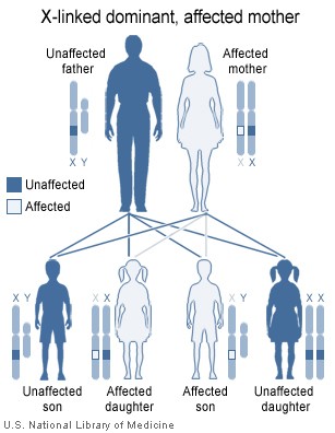
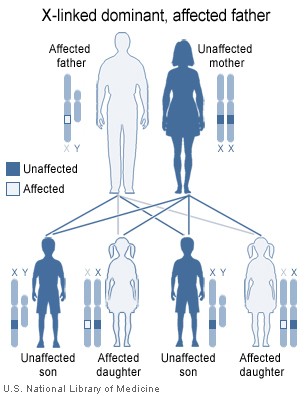
Another question guys. This one on the topic of genetic disorders.

I'm sure most of us are familiar with Autosomal Dominant/Recessive diseases. As well as X-linked disease / Y-linked disease.

The question I have now is that are there X-linked dominant diseases? Is that a possibility? Generally speaking, when we discuss x-linked disease, we automatically think of X-linked recessive because females become carriers (whose disease are masked).

But, in X-linked dominant (if this is actually a real thing), this would mean that a carrier would actually have the disease.
 
http://www.uic.edu/classes/bms/bms655/lesson6.html

Xlink_dominant_mother.jpg
X-link_dominant_father.jpg
 
Can someone explain in detail how the whole carbonic annyhdrase/bicarbonate ion process take place:

All I know is and increase in CO2 and acidity cause a rightward shift in the O2 hemoglobin curve..

I don't want to simply memorize this.. so can someone explain step by step if possible, what happens.. how to relate the whole bicarbonate ion process with decreased O2 affinity and increased acidity, and CO2?

the prep books don't seem to do a good job with this. Also can you explain if CO2 is present in CO2 forms in the cells or is it converted inside the blood?
 
In humans, how does cell shuttle the NADH from cytoplasm to mitrochondria.
Kaplan book tells that all eukaryotic cell only produce 36ATP/glucose, and use a shuttle system(no name given) that give only 4ATP from 2 NADH produced during glycolysis. However, I learned in my biology class that there is Glycerolphosphate shuttle that give 36 ATP/glucose and there is maltate-Aspartate that gives 38 ATP/glucose. So which one we use?
 
We used the malate-aspartate shuttle system in our biochem class since NADH itself doesn't actually cross the membrane, rather its reducing equivalents do. It is a proposed model so I think you could get away with just knowing 36 atp/glucose...they wouldn't ask "is it 36 or 38 atp per glucose fully oxidized".
 
Can someone explain in detail how the whole carbonic annyhdrase/bicarbonate ion process take place:

All I know is and increase in CO2 and acidity cause a rightward shift in the O2 hemoglobin curve..

I don't want to simply memorize this.. so can someone explain step by step if possible, what happens.. how to relate the whole bicarbonate ion process with decreased O2 affinity and increased acidity, and CO2?

the prep books don't seem to do a good job with this. Also can you explain if CO2 is present in CO2 forms in the cells or is it converted inside the blood?

I'll try to be succinct here because the total explanation, in detail, is very long.

CO2 moves from cells(which is from metabolism) into the blood down a pressure difference gradient. The CO2 moves into the red blood cell and will react with the RBC's carbonic anhydratase to form carbonic acid which reversibly converts into HCO3- and H+. The HCO3- will move out into the blood plasma(since there is a conc. diff. of which HCO3- is greater in the RBC) while a chloride ion moves into the RBC for charge conservation. When the blood gets to the lungs the alveolar PCO2 is lower than the blood so the reverse reactions all occur(Cl-moves out RBC & HCO3- moves into RBC where it converts back to carbonic acid and on to water and CO2) while the CO2 is unloaded to the alveoli to be expired.

If someone is exercising, their PCO2 in their cells will increase due to increase cellular activities like metabolism which gives off CO2 as an end product. This in crease in PCO2 between the cell and blood at the capillaries will cause an increase in the CO2 diffusing across which leads to a drop in pH. Your blood has its own inherent buffer system, but carotid chemoreceptors also detect this drop in pH to send a message to the medullary inspiratory center to increase ventilation to remove the CO2 to get the pH level back to normal. When looking at the O2 dissociation curve you notice the rightward shift at low pH/increased CO2 levels. This allows for more O2 to be liberated from Hb at a given PO2 and this occurs so that your body will give your cells, that are working extra hard from that exercise, more O2 than normal so they don't go into anaerobic metabolism and keep up with the required metabolic needs of the cell.

Thanks a lot for the explanation..

but I always thought a leftward shift was "good" or favorable due to increases in O2 affinity (long term for example.. when living at high altitudes) and that a rightward shift is "negative", except when exercising. Is this enough for the MCAT? Is it really necessary to reason everything out.. like why theres a rightward shift during exercise?
 
Oxyhaemoglobin_dissociation_curve.png


If there is a left-ward shift in a normal person that that person may not deliver enough oxygen to cells. The affinity, in this context, is how strong Hb will hold onto each O2 molecule; so at high affinity it will be releasing less O2 at a given PO2 than a lower affinity. From that graph you can see when there is a left-ward shift and it mainly occurs because the cells aren't very active and don't need much O2 so it helps prevent oxygen toxicity from occurring. One thing not noted on this graph is that fetal Hb has a higher affinity/greater left-ward shift than the maternal Hb which allows for the fetus to basically "grab" O2 from the mothers blood.

There was no question on the curve on my exam and I haven't seen a question dealing with it, but you can never know what will show up. I think you should know what makes it shift right and what makes it shift left because they could give a scenario and you might have to guess which way it will shift...
 
Hi folks,
Does anyone know the ATP cost of shuttling NADH from glycolysis into the mitochondrial matrix?

I know pyruvate shuttling costs 1 ATP/pyruvate...isn't NADH also carried actively across membrane??

Is this ATP cost accounted for in the 36ATP/Gluc molecule-- or needs to be further subtracted?

Along these same lines, do we attribute 3ATP to each NADH (and only 2 to FADH2) based on the number of H+ they push into intermembrane space? I'm not sure why NADH from glycolysis only contributes 2ATP, while NADH from Krebs contributes 3. Is this the cost of shuttling?

Thanks a lot.

**Nevermind, unless someone has further elaboration than corbis' above.
 
So apparently the Gold Standard says that for maternal-fetal circulations, some stuff like CO2, O2, wastes are transferred, which is all good and dandy. But it also states that certain antibodies also cross over.

If someone knows the detail about this, could you please explain this further?

I thought the blood barrier shouldn't allow that b/c the mother's immune system around the placenta should have been suppressed to not allow it to transfer stuff (such as antibodies) over that can potentially kill the "foreign" fetus?

I don't know how transferring Abs would help the growth of the baby b/c it would most likely lead to inflammation somewhere, which would be baaad. Plus, it's not like Abs can train T/B cells either since I doubt their MHC complex would match.

Does anyone else have any insight for this?

tennis, we learned tetanus vaccine should be administered to pregnant women in the third trimester, the point at which Abs DO cross over the placenta. This is for protection of infant when umbilical cord care puts infants at high risk for infection. Also, live measles vaccine is not given until 9 mos +- b/c maternal anti-measles Abs are circulating in infant until this point; then passive immunity breaks down, and attenuated measles strain is innoculated so child can develop active immune response.

I can't generalize, but do know that some Abs are surely crossing over...I think this is one reason an infant's HIV serostatus is unclear until six months or so, when the presence of anti-HIV Abs will clear (if they were simply passed from mother) or remain if the child is mounting its own immune response to live virus.

Maybe others can clarify further...
 
IgG crosses the placenta while the mother can pass on secretory IgA via her colostrum (a type of milk from her breast) to provide some protection to the newborn's mucosal membranes.
 
Top