CRPS consult

This forum made possible through the generous support of SDN members, donors, and sponsors. Thank you.

Crybaby

Full Member
7+ Year Member
Joined
Dec 4, 2014
Messages
321
Reaction score
161
Snow has scared away half my clinic so thought I would post :)

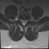
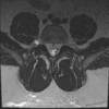
Had a CRPS consult for severe right foot pain after ankle sprain. Really didn't seem like CRPS to me so MRI L spine and found what looks like a lateral cyst. Radiologist calls it a facet cyst, I find this particular case interesting as I have not seen one this far lateral before and I can not see good communication with joint.
So, next move do I drain this sucker? What approach would you guys use? My first thought was to approach like a TFESI, however that nerve is just anterior.
Has anyone encountered a case like this?
l spine cyst sag.png

Members don't see this ad.
 

Attachments

  • l spine ax cyst.png
    l spine ax cyst.png
    200 KB · Views: 143
  • l spine ax.png
    l spine ax.png
    203.1 KB · Views: 138
Personally have a poor success rate with cyst drainage, but at least some temporary relief would help diagnostically. Take measurements on position and angle relative to bony landmarks and plot your approach trajectory. If you get a paresthesia just pull back a little...
 
Umm. No.

Not even close to the facet and completely in the wrong location for a facet cyst. L5 has a perineural cyst. Ok to get with and without contrast. And get a 2nd Rad to look at it.
 
  • Like
Reactions: 7 users
Members don't see this ad :)
Saw a grand rounds on fibrin sealant inj ct guided for these. Anyone have experience w that?
 
Saw a grand rounds on fibrin sealant inj ct guided for these. Anyone have experience w that?

That's crazy and no
 
TFESI +/- intra articular facet and try to drain it. That could be a facet cyst
 
Umm. No.

Not even close to the facet and completely in the wrong location for a facet cyst. L5 has a perineural cyst. Ok to get with and without contrast. And get a 2nd Rad to look at it.
agreed.
 
if you want to prove it is not a facet cyst, put a needle in the nearest facet joint
and inject some contrast. if it is connected to the joint it will take up contrast.
 
Umm. No.

Not even close to the facet and completely in the wrong location for a facet cyst. L5 has a perineural cyst. Ok to get with and without contrast. And get a 2nd Rad to look at it.


Agreed- that is a peri-neural cyst and not a facet cyst. Send to neurosurg.
 
Members don't see this ad :)
TFESI +/- intra articular facet and try to drain it. That could be a facet cyst

Ummmm………………… no. That is a perineural cyst and should be sent to neurosurg for eval. Do not be sticking needles in perineural cysts.
 
It could be an asymptomatic or incidental perineural cyst. Does the patients exam meet clinical criteria for CRPS? Consider three phase scan if the exam suggest sympathetic mediated pain. Consider EMG if you think the ankle pain is a radic.

The history doesn’t suggest a spontaneous perinieral cyst exacerbation...
 
so what about the ankle? You said ankle sprain and foot pain initially, but we seem to have found a tangent. Doesnt look like the typical facet cyst, so i wouldnt touch it until you treat the ankle. We might not have the full history so it is hard to say what comes next.
 
Ummmm………………… no. That is a perineural cyst and should be sent to neurosurg for eval. Do not be sticking needles in perineural cysts.

when did i say to stick a needle in the cyst?

we dont have all the MRI slices, so that definitely could be a facet cyst. it may not be, but i have seen some pretty funky looking ones.
 
what happens if u stick a needle in a perineural cyst?
 
  • Like
Reactions: 1 user
I had an Ortho spine surgeon take a peek, and he didn't think it was perineural, he agreed no communication seen with facet. he has never seen facet cyst like that or perineural cyst quite like that.
He suggested tfesi and contrast into joint as well, to see pattern if it is perhaps cyst. If it is a facet cyst he doesn't think that it will be able to be drained as doesn't think the needle would penetrate.
Does not fit clinical criteria at all for CRPS. Pain is present, no dysthesias, skin, hair, and nail growth all normal. Temp normal. EMG is good idea, thank you.
 
  • Like
Reactions: 1 user
I think a TFESI will more likely turn into a transforaminal intracyst steroid injection. I don't think injecting contrast or other fluid into that thing is going to make it any better.
 
  • Like
Reactions: 1 user
I had an Ortho spine surgeon take a peek, and he didn't think it was perineural, he agreed no communication seen with facet. he has never seen facet cyst like that or perineural cyst quite like that.
He suggested tfesi and contrast into joint as well, to see pattern if it is perhaps cyst. If it is a facet cyst he doesn't think that it will be able to be drained as doesn't think the needle would penetrate.
Does not fit clinical criteria at all for CRPS. Pain is present, no dysthesias, skin, hair, and nail growth all normal. Temp normal. EMG is good idea, thank you.
Definitely get the EMG before you inject around or into that cystic structure .
 
  • Like
Reactions: 1 user
I would try a simpler and safer far lateral interlaminar LESI aimed at that neuroforamen, contrast should show you whether that cyst is wrapped in dural sheath or not.

The paper above shows good results with perineural cyst drainage and fibrin injection, but I wouldn't do that myself
 
  • Like
Reactions: 1 users
Thank you guys, I really appreciate all the feed back. I though this was a pretty interesting case.
 
Top