Advertisement - Members don't see this ad
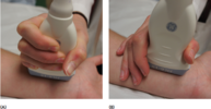
Soon to be third year anesthesiology resident here, I think im doing decent in most things but lately I have been struggling with regional anesthesia, I feel too clumsy holding the us probe and because of that the needlee often drifts out of plane, any helpful tips from the regional wizards around here would be greatly appreciated.