High Yield Anatomy/Neuroanatomy thread

This forum made possible through the generous support of SDN members, donors, and sponsors. Thank you.

sswang00

MS4
10+ Year Member
Joined
Feb 20, 2010
Messages
144
Reaction score
5
Anatomy is one of those subjects most of us don't want to devote too much time to because it isn't as high yield as some of the other subjects on the Step 1.
So how about we quiz each other with high yield anatomy facts? I'll get the ball rolling.

Question: A woman comes in with an ovarian mass and is scheduled for surgery to remove the tumor. During the surgey, which ligament should be ligated to prevent excess bleeding?

Members don't see this ad.
 
Last edited:
  • Like
Reactions: 1 user
Keep this thread going.

What's the basal rhythm frequency of the stomach, duodenum, and ileum?
 
Keep this thread going.

What's the basal rhythm frequency of the stomach, duodenum, and ileum?

S- 3
D-12
I- 9


What arteries are at greatest risk of damage in:
-Gastric Ulcer?
-Duodenal ulcer due to h.Pylori?


-If you have an ulcer in the lower duodenum, what should you suspect?


And i agree, lets keep this thread alive..maybe even throw in some high yield CT/MRI's
 
IMG10_fs.jpg


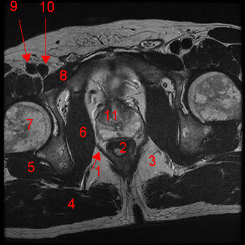
Patient comes in with back pain sx of fequent urination blablabla..
during rectal exam you feel a hard mass. where is it?
 
Members don't see this ad :)
S- 3
D-12
I- 9


What arteries are at greatest risk of damage in:
-Gastric Ulcer?
-Duodenal ulcer due to h.Pylori?


-If you have an ulcer in the lower duodenum, what should you suspect?


And i agree, lets keep this thread alive..maybe even throw in some high yield CT/MRI's

Left Gastric
Gastroduodenal
Lower duodenum ulcer indicates zollinger ellison!! I remember this from Uworld. Good q!

What artery is damaged if the posterior wall of the stomach is ulcerated?


What are the pacemaker cells of the gut?


IMG10_fs.jpg


Patient comes in with back pain sx of fequent urination blablabla..
during rectal exam you feel a hard mass. where is it?


is 11 the prostate? :xf:
 
yessir you got it :thumbup:

-pacemaker cells of stomach are the Cajal Cells
-Posterior wall of the stomach...im guessing splenic a? im picturing that see thru artery behind the stomach in FA lol


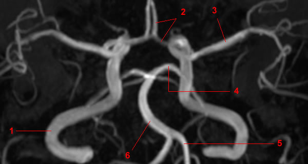
Question: Patient has bitemporal hemianopsia. Wheres the lesion?
polygone-willis-face.0004_fs.jpg
 
Whats in the red, whats in the green?

Because yeah..they actually ask this. and people like me get it wrong!
2001_cat_scan_annotated.jpg
 
Shouldn't it be anterior communicating, but only anterior cerebral is labeled #2.

Red - maxillary sinus
Green - frontal sinus?


Also, aren't you surprised cajal cells aren't mentioned in the FA GI section?
 
Where in the nephron is:

Urine most concentrated (w/ ADH):
Urine most dilute (w/ ADH):

Urine most concentrated (w/o ADH):
Urine most dilute (w/o ADH):
 
Where in the nephron is:

Urine most concentrated (w/ ADH):
Urine most dilute (w/ ADH):

Urine most concentrated (w/o ADH):
Urine most dilute (w/o ADH):

the frontal sinuses would be on the forehead, which are gray'd out on that CT

what are the answers to your nephron question? i suck at that stuff.

and my question is: which kidney do you take in transplant, what side does it go on when you transplant it, and what does the renal artery connect to
 
Members don't see this ad :)
Where in the nephron is:

Urine most concentrated (w/ ADH): At the bottom of the Thick descending loop of Henle
Urine most dilute (w/ ADH): At the end when it comes out of the collecting duct.

Urine most concentrated (w/o ADH): At the bottom of Thick descending LOH
Urine most dilute (w/o ADH): At the start of the distal convoluted tubule and stays the same till the end out of the Collecting ducts.
 
the frontal sinuses would be on the forehead, which are gray'd out on that CT

what are the answers to your nephron question? i suck at that stuff.

and my question is: which kidney do you take in transplant, what side does it go on when you transplant it, and what does the renal artery connect to

Left kidney, because longer renal vein.

Not sure about the rest, but I remember something about it being lower down in the abdomen on the right maybe? What do you connect it to?

Answers are:

Most concentrated w/o ADH: Junction between descending and ascending loop of henle (end of concentrating segment)

Most concentrated w/ ADH: End of Collecting duct (after absorption of H2O and Urea)

Most dilute w/ ADH: Junction of TALH and DCT (end of diluting segment)

Most dilute w/o ADH: End of collecting duct (no water absorption)


What vessels do you use for making an artificial porto-caval shunt in severe portal hypertension? (2 possibilities/4 vessels total)
 
Last edited:
the frontal sinuses would be on the forehead, which are gray'd out on that CT

what are the answers to your nephron question? i suck at that stuff.

and my question is: which kidney do you take in transplant, what side does it go on when you transplant it, and what does the renal artery connect to

You take the left kidney since it has a longer renal vein.

I believe you can transplant it on either side but you leave it in the pelvic region and you actually connect it to a branch of the Aorta (External Iliac or Internal Iliac). That's what I remember learning in school.
 
Left kidney, because longer renal vein.

Not sure about the rest, but I remember something about it being lower down in the abdomen on the right maybe? What do you connect it to?

Answers are:

Most concentrated w/o ADH: Junction between descending and ascending loop of henle (end of concentrating segment)

Most concentrated w/ ADH: End of Collecting duct (after absorption of H2O and Urea)

Most dilute w/o ADH: Junction of TALH and DCT (end of diluting segment)

Most dilute w/ ADH: End of collecting duct (no water absorption)


What vessels do you use for making an artificial porto-caval shunt in severe portal hypertension? (2 possibilities/4 vessels total)
1) Splenic Vein to the Renal Vein.
2) Portal Vein to the IVC.

My Question : Which nerve is mostly to be injured in a car bumper injury and what would be the findings (sensory and motor). :rolleyes:


Last edited:
I'm going to assume after the bumper injury, the car keeps going and runs him over resulting in the following pathogenesis: Crush injury -> Bone fracture -> Fat emboli -> Pulmonary Saddle Embolism -> Sudden death (Every nerve dysfunctional, permanent flaccid paralysis)

I have no idea haha
 
I'm going to assume after the bumper injury, the car keeps going and runs him over resulting in the following pathogenesis: Crush injury -> Bone fracture -> Fat emboli -> Pulmonary Saddle Embolism -> Sudden death (Every nerve dysfunctional, permanent flaccid paralysis)

I have no idea haha

Forgot to add the person was crossing the street and the bar bumper hit them laterally?

Comeon Knuckles you can get this?
 
1) Splenic Vein to the Renal Vein.
2) Portal Vein to the IVC.

My Question : Which nerve is mostly to be injured in a car bumper injury and what would be the findings (sensory and motor). :rolleyes:


i know that first aid says portal vein to IVC but i learned it in school that the TIPS procedure hooks up the portal vein to the HEPATIC vein (yeah, it ends up draining into IVC so im being technical here), so not exactly sure but keep that in mind
 
I'm going to assume after the bumper injury, the car keeps going and runs him over resulting in the following pathogenesis: Crush injury -> Bone fracture -> Fat emboli -> Pulmonary Saddle Embolism -> Sudden death (Every nerve dysfunctional, permanent flaccid paralysis)

I have no idea haha


Fine - cannot hold the answer any longer.

- Common fibular nerve
.o .Damage to fibular neck (car bumper injury)
§ Foot drop (loss of anterior and lateral of leg) and loss of sensation over most of anterio-lateral leg and dorsum of the foot. ==> Superficial fibular nerve
§ between Digits 1 and 2 sensation = deep fibular nerve loss of sensation
Don't forget loss of eversion too :p

I have never heard of that between digit 1 and 2 bs. F that haha
 
Don't forget loss of eversion too :p

I have never heard of that between digit 1 and 2 bs. F that haha

haha yeah that is part of it but screw that too, lol.

hey guys, i have this written in my notes but don't know if its true. the most common place in the stomach for a peptic ulcer to form is the lesser curvature?

i though it'd be the antrum for h. pylori but have no idea
 
haha yeah that is part of it but screw that too, lol.

hey guys, i have this written in my notes but don't know if its true. the most common place in the stomach for a peptic ulcer to form is the lesser curvature?

i though it'd be the antrum for h. pylori but have no idea
your notes are correct.

What's the difference between a peptic erosion and peptic ulcer (what layers are effected?)

Where does Type A Gastritis normally occur, and where does Type B?

How does a peptic ulcer differ from gastric adenocarcinoma, morphologically?

How does intestinal gastric adenocarcinoma differ from diffuse gastric adenocarcinoma?

erosion only affects lamina propria and mucosa. peptic ulcer actually penetrates MUSCULARIS MUCOSA and beyond.
Type A gastritis = fundus. Type B gastritis = antrum.

Peptic ulcer = clean punched out lesion. adenocarcinoma - messy

diffuse gastric adenocarcinoma - leathery (thick and rugged), linitis plastica appearance. i'd assume a normal intestinal gastric adenocarcinoma has an adenomatous apperance?

also, where in the stomach is gastric adenocarcinoma most likely to arise? i don't remember but i think there was a question i came across one
 
yeah, it's splenic.

where would frontal be on that CT?
maxillary sinuses: below the eyes (in maxilla)
frontal sinuses: above the eyes (in frontal bone)
EThmoid sinuses: bETween the eyes
sphenoid sinuses: behind the eyes (not directly, but you know what I mean)
 
erosion only affects lamina propria and mucosa. peptic ulcer actually penetrates MUSCULARIS MUCOSA and beyond.
Type A gastritis = fundus. Type B gastritis = antrum.

Peptic ulcer = clean punched out lesion. adenocarcinoma - messy

diffuse gastric adenocarcinoma - leathery (thick and rugged), linitis plastica appearance. i'd assume a normal intestinal gastric adenocarcinoma has an adenomatous apperance?

also, where in the stomach is gastric adenocarcinoma most likely to arise? i don't remember but i think there was a question i came across one
I remember coming across this question in UW too. I think the answer was antrum (because that's the most common site of H. pylori ulcer in the stomach)


Infarct of the posterior inferior cerebellar artery leads to what syndrome?
 
I remember coming across this question in UW too. I think the answer was antrum (because that's the most common site of H. pylori ulcer in the stomach)


Infarct of the posterior inferior cerebellar artery leads to what syndrome?
Wallenberg syndrome aka PICA syndrome

Where does urine leak if you have
1. rupture of superior wall of urinary bladder
2. rupture of anterior wall of urinary bladder
 
Last edited:
yessir you got it :thumbup:

-pacemaker cells of stomach are the Cajal Cells
-Posterior wall of the stomach...im guessing splenic a? im picturing that see thru artery behind the stomach in FA lol


Question: Patient has bitemporal hemianopsia. Wheres the lesion?
polygone-willis-face.0004_fs.jpg

on this, is 4 the superior cerebellar artery, 5 the basilar, and 6 the vertebral?

and i have no idea about the urinary bladder problems. i suck at pelvic, low abdominal anatomy
 
on this, is 4 the superior cerebellar artery, 5 the basilar, and 6 the vertebral?

and i have no idea about the urinary bladder problems. i suck at pelvic, low abdominal anatomy


Website has a lot of images, and it gives this for one posted here (image 4)

Polygone de Willis: incidence de face avec rotations. Image 4
1, Artère carotide interne. 2, Artère cérébrale antérieure. 3, Artère cérébrale moyenne. 4, Artère cérébrale postérieure. 5, Artère vertébrale. 6, Artère basilaire.
 
Urine leakage
1. rupture of superior wall of urinary bladder- intraperitoneal extravasation of urine w/i peritoneal cavity (e.g- compressive force on a full bladder)
2. rupture of anterior wall of urinary bladder - extraperioneal extravasation of urine w/i retropubic space of Retzius (e.g- fractured pelvis(car accident etc) that punctures the bladder).
on this, is 4 the superior cerebellar artery, 5 the basilar, and 6 the vertebral?

and i have no idea about the urinary bladder problems. i suck at pelvic, low abdominal anatomy
 
Urine leakage
1. rupture of superior wall of urinary bladder- intraperitoneal extravasation of urine w/i peritoneal cavity (e.g- compressive force on a full bladder)
2. rupture of anterior wall of urinary bladder - extraperioneal extravasation of urine w/i retropubic space of Retzius (e.g- fractured pelvis(car accident etc) that punctures the bladder).
Teach me about urine leaking into the anterior abdominal wall. How does that work. :eek:
 
Teach me about urine leaking into the anterior abdominal wall. How does that work. :eek:
Space of Retzius is a component of extraperitoneal space, and it is located between pubic symphysis and urinary bladder.
HY anatomy book has excellent diagrams for bladder injuries and stuff.
 
so if it leaks up, still in peritoneum. if it leaks forward, out of peritoneum in Retzium's space.

what's the link to the site knuckles? i just realized i can now read french. studying has definitely paid off.
 
Urine leakage
1. rupture of superior wall of urinary bladder- intraperitoneal extravasation of urine w/i peritoneal cavity (e.g- compressive force on a full bladder)
2. rupture of anterior wall of urinary bladder - extraperioneal extravasation of urine w/i retropubic space of Retzius (e.g- fractured pelvis(car accident etc) that punctures the bladder).

hey, i just came across this question in World again. I think you got it mixed up doc29.

The superior bladder injury goes EXTRAPERITONEALLY into that retropubic space of retzius (how, i have no idea) while the anterior bladder injury stays within the peritoneum.

And a pelvic fracture will cause a posterior urethral injury (also has high riding prostate) while saddle injuries cause anterior urethral injuries (will see bloody discharge).

I think this is correct.
 
Random: can someone explain antiphospholipid syndrome?

it is the production of antibodies (from SLE) towards coagulation factors. what is unusual is that is actually activates the coagulation system causing a HYPERCOAGULABLE state. what is EVEN WEIRDER, is that it presents with longer PT/PTT times.

That's all i know off of pure memorization.

also, what was that mechanism of why SLE occurs? Was it like because there's no negative selection in the thymus?


Isnt Phospholipid syndrome where you have antibodies against cardiolipin and lupus anticoagulant?

I thought what happened was that there was a type II Hypersensitivity type of reaction against these Antigens (phospholipids from cell membranes) and they travel throughout the blood once they rupture the cell. These antigen-antibody complex cause damage to the endothelium activating the coagulation cascade. Hence it is similar to DIC where you are consuming all your coagulation factors due to microclots trying to cover up the damage to the endothelium. Hence increase in PT, PTT and bleeding time.

Do I have it completely wrong?
 
hey aashkab,

I remember seeing that q on UW, but HY anatomy 3rd edition says rupture of superior wall(dome) results in intraperitoneal extravasation of urine within the peritoneal cavity; and rupture of anterior wall results in extraperitoneal extravasation of urine within retropubic space of retzius. (they have pics too page 161- 162 on HY)

hey, i just came across this question in World again. I think you got it mixed up doc29.

The superior bladder injury goes EXTRAPERITONEALLY into that retropubic space of retzius (how, i have no idea) while the anterior bladder injury stays within the peritoneum.

And a pelvic fracture will cause a posterior urethral injury (also has high riding prostate) while saddle injuries cause anterior urethral injuries (will see bloody discharge).

I think this is correct.
 
Last edited:
Isnt Phospholipid syndrome where you have antibodies against cardiolipin and lupus anticoagulant?

I thought what happened was that there was a type II Hypersensitivity type of reaction against these Antigens (phospholipids from cell membranes) and they travel throughout the blood once they rupture the cell. These antigen-antibody complex cause damage to the endothelium activating the coagulation cascade. Hence it is similar to DIC where you are consuming all your coagulation factors due to microclots trying to cover up the damage to the endothelium. Hence increase in PT, PTT and bleeding time.

Do I have it completely wrong?

that sounds right. i honestly didn't know the exact mechanism. i got a little lost in your explanation, but it does account for an increase in PT, PTT and a hypercoagulable state.
 
Patient fractures his mid clavicle.
On xray you notice the medial portion of the clavicle is being pulled superiorly.

What muscle is responsible for this?
 
these are the types of qs I can't stand. Can you give a quick enlightenment of the clavicle attachments and directions? I know that the deltoid pulls it inferiorly (UWSA2)
 
i believe the sternocleidomastoid pulls the medial part upward, and the deltoid pull the lateral end downward like u said
 
Ok so if I am correct:

Open angle glaucoma: improved symptoms with Epi( worsened by musc agonists)

Closed angle glaucoma: worsened symptoms with epi (improved with physiostigimine)

Am I correct? If so what is the underlying mechanisms and reasons?
 
Ok so if I am correct:

Open angle glaucoma: improved symptoms with Epi( worsened by musc agonists)

Closed angle glaucoma: worsened symptoms with epi (improved with physiostigimine)

Am I correct? If so what is the underlying mechanisms and reasons?

yeah the angle that they're referring to is between the iris and the cornea (corneoirido angle or w/e) basically this is the flow right before you hit the canal of schlemm. in closed angle there is a problem with this funnel right before you get into the canal of schlemm and when you perform MYDRIASIS in closed angle glaucoma, the iris will bunch up even more right before the canal of schlemm and close the angle up more. imagine blinders. when you pull blinders on your window up (ie. mydriasis) it'll bunch up more... get wider in the middle. whereas if you let them down (miosis) it'll flatten out. It's tough to explain in words I'm sure there must be an illustration of this somewhere
 
1- which nerves and arteries likely to be damaged in posterior dislocation of the hip joint ?
2- which structures are responsible for knee jerk reflex ?
 
I'll continue mainly focusing on anatomy and nerve questions since I've been told that step 1 asks a lot of these questions.

A 21-year-old man is recovering from a gunshot wound to the buttocks. When walking, he must flex his trunk to the right to lift his left foot off the ground. Which of the following nerves has been injured?
 
1- which nerves and arteries likely to be damaged in posterior dislocation of the hip joint ?
2- which structures are responsible for knee jerk reflex ?
oops, sorry did not see your post... thanks for reviving this thread. I'll post some questions too.

to answer your question
1. Superior gluteal (L4 - S1) right? not sure about the arteries, but I am guessing it is external iliac artery?
2. femoral n. (L2-L4) and testing quadriceps muscle
 
Top