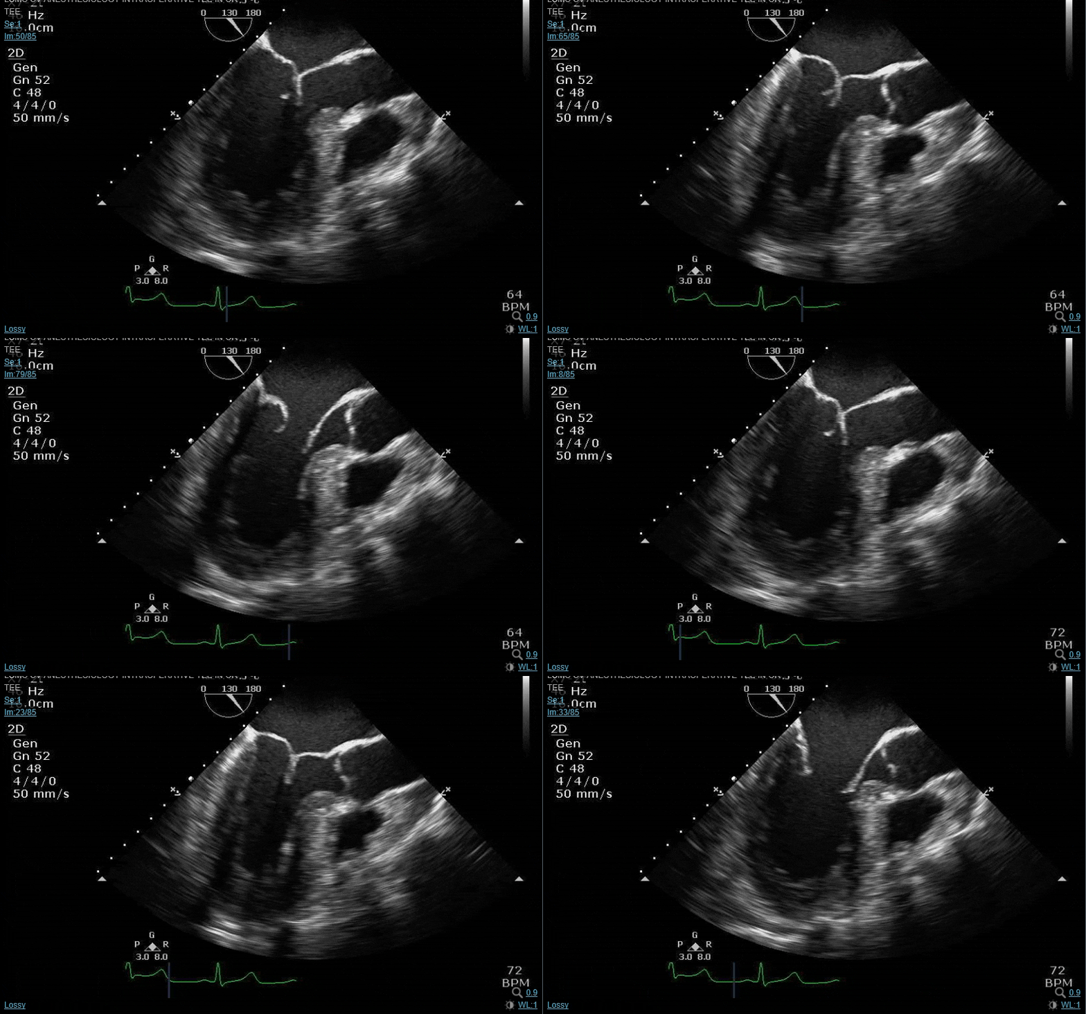
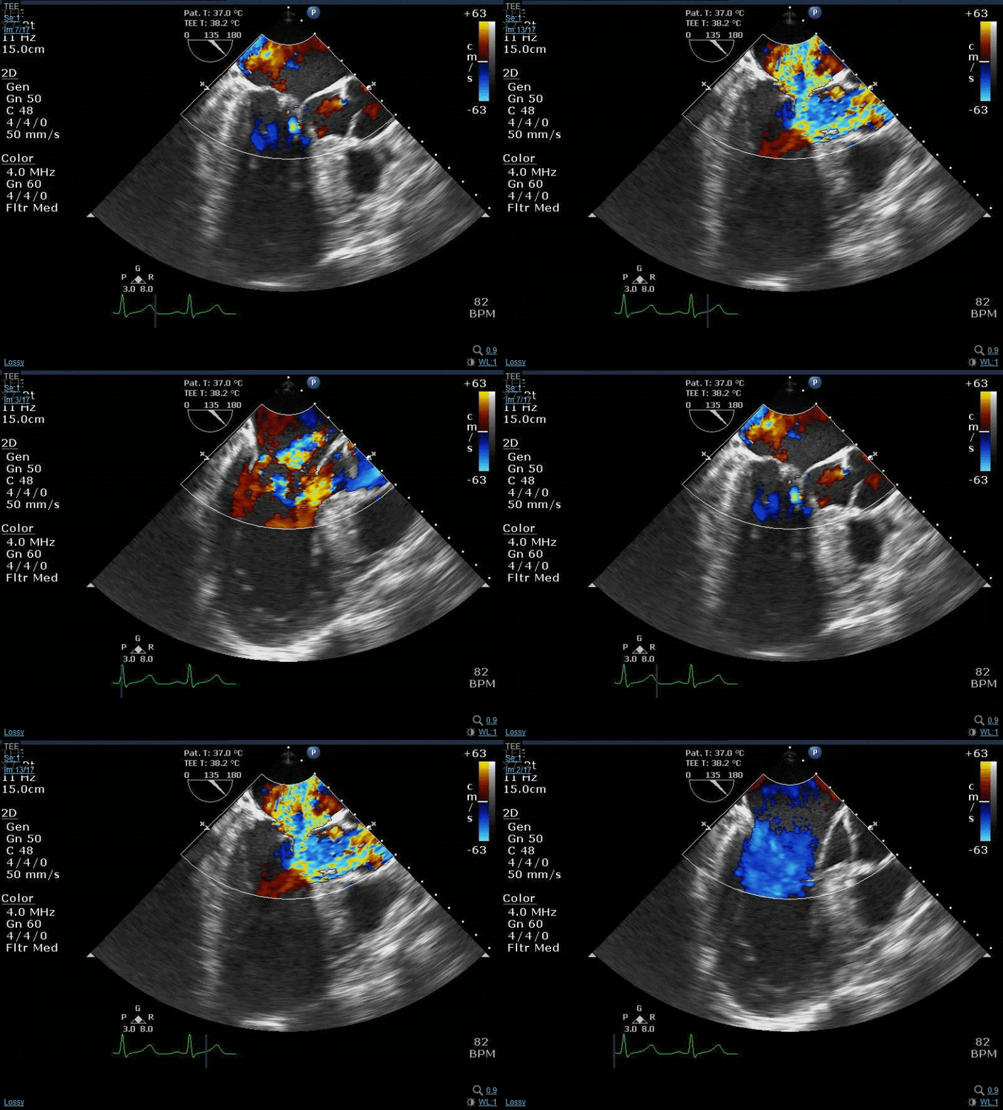
Let's do some echo:

This forum made possible through the generous support of SDN members, donors, and sponsors. Thank you.
Tell this guy not to buy any green bananas...

Other than that there really isnt anything else to say other than the usual. PMS in the groins for a pump, else axil impella, loads of dob, vaso probably epi here too plus probably nitric, straight onto dialysis the second he stops peeing. A good perfusionist that will take plenty volume off him. Mitral Probably repairable with a ring.

We have to look at the tricuspid too though with a right that big even off axis

If anyone sneezes around this guy he's done

Members don't see this ad.
 
Great case Sevo - how did it go?

I wonder would it be better for learning if we got the 2d without color first so we can surmise where the jet should be and what surgical repair technique they could do? We had one guy who used to teach us like that. 3d and color were the icing on the cake, but the pathology was already understood via 2d
 
Another bread and butter case:

50 y/o male admitted to the hospital for progressive SOB. EF 10% on admission. Tuned up for a few weeks b4 surgery and brought to the OR for MVR. You hook up monitors. Resting HR is 130 bpm. After careful induction you obtain the following images.

Comments?
So his EF of 10% on admission, was this prior to developing MR?

I'm envisioning that he's recently infarcted, leading to the severely reduced EF, but during admission the infarct led to papillary rupture hence the multiple flailing chords? And this new MR is likely a major component of his "improved" EF.

Are these reasonable assumptions of the chain of events?
 
Members don't see this ad :)
Yeah...see a flap on the ascending long axis there. E= Ao B= RV C=LV D= LA A =RA.....but I think I see your point...almost looks like the right sided compression is coming from the hematoma? At least impeding RV filling, anyway...

Okay so is the 2nd image the SAX of ascending aorta with the PA going past it? I think that image is the one that was throwing me off so hard.

I also don't really appreciate much effusion on the images. Maybe I'm getting dumber. Please provide big red arrows haha.

It sounds a bit strange, but "be careful about seeing things that you're expecting to see". Understandably with the gain, that thing in the middle of the lumen of the ascending long axis does sorta look like a flap....but it's actually a sidelobe artifact. That echo shot is at the level of the R PA, but when one looks at the CT:

1611454333115.png


There's nothing there except a large intramural hematoma. She did have a kind of flap looking thing on echo and according to the surgeon it was technically a dissection with a discrete flap, but it was not a thin highly mobile piece of intima like one would expect. It was kind of more akin to a big wall adjacent to a penetrating aorta ulcer in the distal ascending that propagated hematoma both retrograde and antegrade.


1611454522669.png


1611455691316.png

Yellow arrow: sidelobe.
Blue arrow: ?"flap" anterior to which the retrograde hematoma is propagating. I think we're foreshortened plus the entry is still a bit distal to where this echo image is

As you can see the "flap" is pretty distal, and definitely in risk of being in the TEE blindspot where the distal trachea/left main bronchus interpose and interrupt the echo interface.

The other point here is that there was no pericardial effusion. Before taking a dissection to the OR, always always always look at the CT yourself and also look at the images from any TTE cards might've done pre-op. @Teillard you are correct in identifying the lettered chambers on the CT. You are also correct in that there is significant mass effect on the right sided chambers, and that is going to significantly affect preoperative management and induction of anesthesia wrt discontinuing antihypertensive drugs, volume loading before induction, assessing for airway compromise or developing SVC syndrome etc. Acute aortic syndromes are sometimes wonky creatures- better to have a good idea of what you're getting into before you wheel the pt into the OR.
 
Last edited:
  • Like
Reactions: 1 users
NuKCNLM.gif

1611459816383.png


Describe what color flow doppler findings you expect to see.
 
Last edited:
  • Like
Reactions: 1 user
Describe what color flow doppler findings you expect to see.
I don't know what terminology you all generally use, but I would expect to see color flowing in a northwest direction through the mitral valve. I think that's towards the posterior side of the leaflets. Or is that the anterior cause it's towards to lateral wall? I'm so bad at remembering those.
 
I don't know what terminology you all generally use, but I would expect to see color flowing in a northwest direction through the mitral valve. I think that's towards the posterior side of the leaflets. Or is that the anterior cause it's towards to lateral wall? I'm so bad at remembering those.
To help remember just look for what you know in the screen.

So you got the RV and then the AoV/LVOT that you probably recognise there on the right and of the screen

We also know the Aortic Valve is always anterior to the MV so therefore the Anterior Mitral Valve Leaflet (AMVL) is Northeast and the PMVL is Northwest in your terminology.

Now im wondering why you think northwest? P2 is billowing so the jet should be eccentric and anteriorly directed. Is that right? If posterior was restricted then it would be posteriorly directed jet. I cant see posterior restriction
The coaptation is abnormal and P2 is elongated - risk for SAM post repair


What is the Cont W Dopp there?

This is Carpentier type 2 MR and repair tech of choice is quadrangular resection with annuloplasty ring. sliding leaflet plasty technique also used to eradicate risk of SAM
There may be chordal sam already
 
Last edited by a moderator:
To help remember just look for what you know in the screen.

So you got the RV and then the AoV/LVOT that you probably recognise there on the right and of the screen

We also know the Aortic Valve is always anterior to the MV so therefore the Anterior Mitral Valve Leaflet (AMVL) is Northeast and the PMVL is Northwest in your terminology

Now im wondering why you think northwest? P2 is billowing so the jet should be eccentric and anteriorly directed. Is that right?
The coaptation is abnormal and P2 is elongated - risk for SAM post repair


What is the Cont W Dopp there?

That is a CW @ almost 5 m/s and a hefty peak gradient.... PWD of the LOVT likely similar. Sometimes you see a y-shaped CFD.

Great cases @vector2. :cool:
 
  • Like
Reactions: 1 user
D-shaped LV/septal flattening indicative of RV pressure/volume overload (going too fast for my eyeballs to tell if it's more during systole vs diastole). Is that like an RV aneurysm or something? It's ballooned out so far around the LV it's definitely more than your usual crescent shape.


Good call @abolt18 . Accidentally cropped out the ekg, but LV flattening occurs during late systole indicating RV pressure overload. @Vector hit the nail on the head in that both pressure and volume overload were indeed present. The distraction on the 3D pictures was definitely the mitral valve. The ever so common p2 fail with an anterrior conda jet with a nice 1+cm gap is always severe plus a little extra. But that was the distraction.
Looking at the top right of the 3d clip and you are looking down on a dilated RV. Cardiac surgeon is approaching the head of the bed and you need to talk about the case. Plan wasn't to do anything on right side of the heart.

I’ll creep out a few more clinical caveats out of this case:
 
Last edited:
What is RVSP and PASP? Cardiac surgeon just pulled up to review some images before he scrubs in.
 
Members don't see this ad :)
what are you measuring there? Ias bulging slightky to the left assoc with rv pressure overload
 
  • Like
Reactions: 1 user
To help remember just look for what you know in the screen.

So you got the RV and then the AoV/LVOT that you probably recognise there on the right and of the screen

We also know the Aortic Valve is always anterior to the MV so therefore the Anterior Mitral Valve Leaflet (AMVL) is Northeast and the PMVL is Northwest in your terminology.

Now im wondering why you think northwest? P2 is billowing so the jet should be eccentric and anteriorly directed. Is that right? If posterior was restricted then it would be posteriorly directed jet. I cant see posterior restriction
The coaptation is abnormal and P2 is elongated - risk for SAM post repair


What is the Cont W Dopp there?

This is Carpentier type 2 MR and repair tech of choice is quadrangular resection with annuloplasty ring. sliding leaflet plasty technique also used to eradicate risk of SAM
There may be chordal sam already
Im a bit confused, this was from the MR with Mvp case? So has AS plus MR?
I think you may be missing the big picture here regarding the pathology of the mitral and why there's a 5 m/s CW jet....look closer.
 
You have it dude. AS gradient vs TR gradient = which way is the envelope on tee?
 
Ah. Just wondering why measure RA to the IAS? But i see now it was a general indication not an actual measurement per se
Ya... took it on the fly but the IVC is right there with a cannula in it.
 
I think you may be missing the big picture here regarding the pathology of the mitral and why there's a 5 m/s CW jet....look closer.
torn posterior leaflet? idk

Ah so TR, i was thinking the deep TG looked wierd but couldnt really see at that resolution from a static image
 
torn posterior leaflet? idk

Ah so TR, i was thinking the deep TG looked wierd but couldnt really see at that resolution from a static image
Nope. That CW cursor is directed through the LVOT and the AV and is 5 m/s. You had some suspicion for AS...but look at the aortic valve on the 2d clip and tell me how those AV leaflets look. Aortic stenosis should be obvious even before one does any color or doppler. And then look very closely at the LVOT and AMVL on the 2d clip.....
 
  • Like
Reactions: 1 users
I don't have a good pic of the hepatic veins, but it was very dilated as well with SFR. Had some coags that were messed up due to liver congestion and pump run. Came off on epi/milrinone with a touch of norepi with some products. The nice thing about working with a competent CT surgeon is coming off the first time and not dealing with surgical issues. Currently doing well and had a great result.
 
Ok.
The 'anterior' AoV cusp we initially saw was sclerotic restricted but the rcc moved well. I was thinking mild maybe mod AS not severe...

Now i see the septal hypertrophy! Ok. Didnt do anything with that?

So the mr jet was what direction?
 
I must put up our first case from Friday. It was a hot dang. Euro score 35%. Refused by 3 other places. Doing ok
 
  • Like
Reactions: 1 user
Ok.
The 'anterior' AoV cusp we initially saw was sclerotic restricted but the rcc moved well. I was thinking mild maybe mod AS not severe...

Now i see the septal hypertrophy! Ok. Didnt do anything with that?

So the mr jet was what direction?
You got it with the septal hypertrophy...now keep going. There is no aortic stenosis, not even mild. Look at the LVOT in systole.....
 
You got it with the septal hypertrophy...now keep going. There is no aortic stenosis, not even mild. Look at the LVOT in systole.....
I said sam with mr in my first post, not as.

the risk fx for sam even were listed

I appreciate the coaching but trying to lead ppl to an answer they already had implied you might not have read my first post fully. Great case and discussion tho.
 
I said sam with mr in my first post, not as.

the risk fx for sam even were listed

I appreciate the coaching but trying to lead ppl to an answer they already had implied you might not have read my first post fully. Great case and discussion tho.
Your first post said CHORDAL sam, which is a normal variant that isn’t associated with LVOT obstruction. This is leaflet SAM, a whole different ballgame.

@sevoflurane great cases!

sorry, I must have missed something though: what where you measuring in the RA with that inflow-outflow view? And why did you take the time to trace the TR jet for a VTI when all you needed for PASP was peak velocity?
 
  • Like
Reactions: 1 user
I said sam with mr in my first post, not as.

the risk fx for sam even were listed

I appreciate the coaching but trying to lead ppl to an answer they already had implied you might not have read my first post fully. Great case and discussion tho.
Yeah man, you said chordal SAM and then talked about post-MV repair SAM. This guy had already had full blown native AMVL SAM and an enormous intraventricular gradient from said LVOT obstruction.
 
  • Like
Reactions: 1 user
Settle down ladies 😂
I was still trying to figure out where north west was
 
Your first post said CHORDAL sam, which is a normal variant that isn’t associated with LVOT obstruction. This is leaflet SAM, a whole different ballgame.

@sevoflurane great cases!

sorry, I must have missed something though: what where you measuring in the RA with that inflow-outflow view? And why did you take the time to trace the TR jet for a VTI when all you needed for PASP was peak velocity?

Good job everyone. I just realized I posted the wrong pic. This dude had a giant IVC basically adding 15 mmhg to your calculated RVSP.
 
  • Like
Reactions: 1 user
iLai2Kw.gif


So for this guy:

Normal BiV function
HCM, mostly DUST with a 2.1 thickness
AMVL SAM with LVOTO. Peak gradient > 90 mmHg. The magic number is 50 mmHg for ablation or repair as that is where risk of HF and SCD starts increasingly dramatically.
Fellows should measure C-sept, Aortomitral angle, ALPL ratio. I didn't because I'm lazy
Moderate MR with an anterior, central, and posterior jet components cause he had posterior prolapse too. Typically with MR in SAM, the jet is *posteriorly* directed

1611503253907.png
 
Last edited:
  • Like
Reactions: 1 user
Nice y shaped CFD if you slow it down. 👌🏾
 
  • Like
Reactions: 1 user
Thanks for posting @vector2.
Super fun super educational.
Keep on posting your juicy ones. 🤘🏽
 
  • Like
Reactions: 2 users
It sounds a bit strange, but "be careful about seeing things that you're expecting to see". Understandably with the gain, that thing in the middle of the lumen of the ascending long axis does sorta look like a flap....but it's actually a sidelobe artifact. That echo shot is at the level of the R PA, but when one looks at the CT:

View attachment 328136


There's nothing there except a large intramural hematoma. She did have a kind of flap looking thing on echo and according to the surgeon it was technically a dissection with a discrete flap, but it was not a thin highly mobile piece of intima like one would expect. It was kind of more akin to a big wall adjacent to a penetrating aorta ulcer in the distal ascending that propagated hematoma both retrograde and antegrade.


View attachment 328137

View attachment 328140
Yellow arrow: sidelobe.
Blue arrow: ?"flap" anterior to which the retrograde hematoma is propagating. I think we're foreshortened plus the entry is still a bit distal to where this echo image is

As you can see the "flap" is pretty distal, and definitely in risk of being in the TEE blindspot where the distal trachea/left main bronchus interpose and interrupt the echo interface.

The other point here is that there was no pericardial effusion. Before taking a dissection to the OR, always always always look at the CT yourself and also look at the images from any TTE cards might've done pre-op. @Teillard you are correct in identifying the lettered chambers on the CT. You are also correct in that there is significant mass effect on the right sided chambers, and that is going to significantly affect preoperative management and induction of anesthesia wrt discontinuing antihypertensive drugs, volume loading before induction, assessing for airway compromise or developing SVC syndrome etc. Acute aortic syndromes are sometimes wonky creatures- better to have a good idea of what you're getting into before you wheel the pt into the OR.
This post was a very nice and helpful explanation. This case had me lost for a while but this post brought it together. Thanks. Very interesting. What's the end result for the patient?
 
This post was a very nice and helpful explanation. This case had me lost for a while but this post brought it together. Thanks. Very interesting. What's the end result for the patient?
Did well in the case, came off on just a bit of juice with a mild coagulopathy. Unfortunately she was a pretty bad surgical candidate to begin with. Super fat, prexisting small stroke, dementia and some schizophrenia history. Had prolonged intubation course but was extubated right before trach decision and flew. Went into renal failure too. CRRT and now she's gonna be on IHD. This case was from awhile back and she actually just made floor status today. Going to facility after she's discharged, I imagine.
 
Did well in the case, came off on just a bit of juice with a mild coagulopathy. Unfortunately she was a pretty bad surgical candidate to begin with. Super fat, prexisting small stroke, dementia and some schizophrenia history. Had prolonged intubation course but was extubated right before trach decision and flew. Went into renal failure too. CRRT and now she's gonna be on IHD. This case was from awhile back and she actually just made floor status today. Going to facility after she's discharged, I imagine.
What was the surgical intervention. Just ascending aorta +root? Or hemi-arch? Valve too?
 
What was the surgical intervention. Just ascending aorta +root? Or hemi-arch? Valve too?
Oh yea, duh, forgot I didn’t say. R ax cannulation, circ arrest, Hemi arch, ascending graft, resuspended valve without having to replace it.
 
  • Like
Reactions: 1 user
Another very good (albeit old school) method for evaluating for dynamic changes to SAM, especially when instituting therapeutic maneuvers or if the surgeon has decided to go back on pump to change their repair...is M-mode across the AV leaflets in the ME AV LAX view. The AV should open into a rectangular box shape on M-mode. Coarse fluttering with a bird-beak appearance and early systolic closure is indicative of ongoing SAM. This, along with CFD, presence of a dagger shaped CWD envelope and elevated gradients across the LVOT in the deep TG LAX are how you tell the surgeon if SAM is improving, the same, or getting worse.
 
  • Like
Reactions: 2 users
Another very good (albeit old school) method for evaluating for dynamic changes to SAM, especially when instituting therapeutic maneuvers or if the surgeon has decided to go back on pump to change their repair...is M-mode across the AV leaflets in the ME AV LAX view. The AV should open into a rectangular box shape on M-mode. Coarse fluttering with a bird-beak appearance and early systolic closure is indicative of ongoing SAM. This, along with CFD, presence of a dagger shaped CWD envelope and elevated gradients across the LVOT in the deep TG LAX are how you tell the surgeon if SAM is improving, the same, or getting worse.
I actually went back to PACS to try and find this exact thing from the case I posted but of course I forgot to do it this time. Highlights the importance of having a baseline comprehensive exam for the bread and butter and then a mental checklist of additional things needed for the relatively rarer pathologies that come to the OR.
 
Some examples of what Peterman is talking about

Screenshot_20210124-174715_Chrome.jpg
Screenshot_20210124-174626_Chrome.jpg


M-mode through the AV leaflets is also nice to get a qualitative sense of how good LV function is. M-mode the leaflets of a normal function CABG and compare the visually assessed ejection time to an EF 30% guy and you'll see what I mean.
 
Last edited:
  • Like
Reactions: 1 user
Top