Lost battle with the SIJ

This forum made possible through the generous support of SDN members, donors, and sponsors. Thank you.

NJPAIN

Full Member
10+ Year Member
Joined
Nov 28, 2011
Messages
2,666
Reaction score
1,687
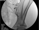
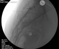
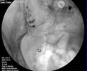
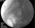
A lost battle with my arch nemesis the SI joint today. Very petite 82 year old with osteoporosis s/p L4-S1 PLIF with transitional anatomy manifesting as lumbarized S1. Old fashioned right side iliac crest bone graft harvest scar. Right buttock pain. Weight bearing LS spine x-ray showing right SIJ and axial MRI image showing only cross sectional view of SIJ. On fluoro straight AP there was what looked to be a long radiolucent “stripe”. The remainder of the sacrum was just a gray ground glass pattern. I tried to access the lower pole and middle. Each time after contrast injection there was a vascular pattern. I tried to oblique the image and separate that anterior and posterior joint surfaces but I could not establish the cortical margins of the joint. I tried the “Benyamin technique” where you tilt the II caudal and add some contralateral oblique to form a lucency where the joint lines cross in the middle. I couldn’t see any lucency but drove the needle where the joint lines appeared to cross. Felt like it entered the joint but all I got was a vascular pattern. After 25 minutes I just gave up. If our IR guy would do it I would send her for CT guidance. He won’t do anything MSK.

I’ve had this happen before in the elderly with osteoporosis. I can’t distinguish the tactile sensation of entry into the joint from entry into the soft bone.

Anyone have suggestions for this kind of situation? Any value to getting a CT and trying again with a better cross sectional view?
1633128611058.jpeg

1633128653333.jpeg

Members don't see this ad.
 
Why not just go with nerve block to the joint and plan on RFA if you can’t get in?
 
  • Like
Reactions: 2 users
LBBB is a possibility. I do the caudal tilt and ipsilateral oblique and if can't line up in the normal view go for the medial line that is the posterior one. you can use a stiffer needle and almost dilate the joint like your were doing an SIJ fusion, but if OP you have to make sure you don't go through the cortex; other option is square off L5-S1 disc space and contralateral oblique to see the upper pole usually superolateral to S1 foramen is the the area for access, if all else fails inject the tendons as that may get some relief as well.
 
  • Like
Reactions: 1 users
Members don't see this ad :)
I bet periarticular injection would work well, after 5 mins of trying to get into the joint and failing this is what I would do.
 
  • Like
Reactions: 16 users
Inject around the joint..
 
  • Like
Reactions: 3 users
Take a close look at an old CT of the area (usually there is one somewhere) and see if there is a good spot to get in.
 
  • Like
Reactions: 1 user
If I didn’t get an absolutely perfect placement with textbook contrast spread on each and every SI joint, including that one…. Well I’d probably just resign.
 
  • Like
  • Haha
Reactions: 6 users
In my opinion, “periarticular” injections for SIJ means you didn’t get in and are justifying your treatment stating that people get relief from periarticular injections. She’s old, her joints are jacked, you tried. Cut your losses..you could always PRP, rfa, send for fusion. My money is on none of it will work for her..Mary Jane/cbd, might not be a bad option for her
 
In my opinion, “periarticular” injections for SIJ means you didn’t get in and are justifying your treatment stating that people get relief from periarticular injections. She’s old, her joints are jacked, you tried. Cut your losses..you could always PRP, rfa, send for fusion. My money is on none of it will work for her..Mary Jane/cbd, might not be a bad option for her
I disagree, there are papers showing periarticular injections work as well in intra articular.
 
  • Like
Reactions: 4 users
In my opinion, “periarticular” injections for SIJ means you didn’t get in and are justifying your treatment stating that people get relief from periarticular injections. She’s old, her joints are jacked, you tried. Cut your losses..you could always PRP, rfa, send for fusion. My money is on none of it will work for her..Mary Jane/cbd, might not be a bad option for her

If anything kicks my a** on a fairly regular basis it’s an SIJ inj. It still gets to me after many years of doing this. This was intended to be a diagnostic injection so I really needed an arthrogram. I guess lateral branch blocks are an option. Though it will be tough to do that properly with such poor landmarks. I guess I need to get beyond the urge for perfection, lay some local down medial to the joint line join and call it a day.

In retrospect I’ll bet that I entered the joint several times relatively superficially but rather than having a hard stop against bone I just pushed the needle straight through the soft bone. If I had access I would do this with CT.
 
  • Like
Reactions: 1 users
If anything kicks my a** on a fairly regular basis it’s an SIJ inj. It still gets to me after many years of doing this. This was intended to be a diagnostic injection so I really needed an arthrogram. I guess lateral branch blocks are an option. Though it will be tough to do that properly with such poor landmarks. I guess I need to get beyond the urge for perfection, lay some local down medial to the joint line join and call it a day.

In retrospect I’ll bet that I entered the joint several times relatively superficially but rather than having a hard stop against bone I just pushed the needle straight through the soft bone. If I had access I would do this with CT.
Why subject this person to that much radiation. Even if you get into the joint then what, steroid injection only lasts so long.
 
Why subject this person to that much radiation. Even if you get into the joint then what, steroid injection only lasts so long
XR of SIJ don't often show the osteophytes where CT scans will pick up on the subtle things blocking your path of entry and allows you know when your in the joint vs in the bone for this OP patient, not unreasonable for special cases like this if available, just like for celiac plexus blocks they can be helpful for distorted anatomy, the pt is 82 not 20, different circumstances requiring using a broad range of tools in the armentarium. With all that said, unless you have access to this CT scan is often not practical
 
XR of SIJ don't often show the osteophytes where CT scans will pick up on the subtle things blocking your path of entry and allows you know when your in the joint vs in the bone for this OP patient, not unreasonable for special cases like this if available, just like for celiac plexus blocks they can be helpful for distorted anatomy, the pt is 82 not 20, different circumstances requiring using a broad range of tools in the armentarium. With all that said, unless you have access to this CT scan is often not practical

sometimes just the tip is enough
 
  • Like
  • Haha
Reactions: 4 users
Members don't see this ad :)
there is usually vascular flow everywhere after a fusion due to collateral formation. These are always tough. Punt and try again on a different day.
 
  • Like
Reactions: 1 user
I usually don't do CT but in this case it can help determine if there is indeed an opening and the obliquity needed at the inferior pole, as looks can be deceiving on XR. Wouldn't worry about radiation at this age.

Something that has helped me a ton is checking a lateral before injecting contrast. If needle tip is not around the anterior margin of the sacrum, don't inject contrast--you're too shallow to be in the joint, and injecting there will muddy your picture. Go back to AP and try again. Since doing this about a year ago I get clean arthrograms most the time.
 
  • Like
Reactions: 1 users
I usually don't do CT but in this case it can help determine if there is indeed an opening and the obliquity needed at the inferior pole, as looks can be deceiving on XR. Wouldn't worry about radiation at this age.

Something that has helped me a ton is checking a lateral before injecting contrast. If needle tip is not around the anterior margin of the sacrum, don't inject contrast--you're too shallow to be in the joint, and injecting there will muddy your picture. Go back to AP and try again. Since doing this about a year ago I get clean arthrograms most the time.

I always check a lateral and agree that when entering the joint at the inferior pole most often the needle tip will be close to ventral border of sacrum if it’s in the joint. Not true at middle of joint.
In this case, every time I injected contrast it was at the ventral border and ever time the pattern was vascular.

How about oblique 5 degrees contra and ipsilateral after needle placement to see if tip remains in the joint. Anyone find that useful and reliable?
 
  • Like
Reactions: 1 user
If anything kicks my a** on a fairly regular basis it’s an SIJ inj. It still gets to me after many years of doing this. This was intended to be a diagnostic injection so I really needed an arthrogram. I guess lateral branch blocks are an option. Though it will be tough to do that properly with such poor landmarks. I guess I need to get beyond the urge for perfection, lay some local down medial to the joint line join and call it a day.

In retrospect I’ll bet that I entered the joint several times relatively superficially but rather than having a hard stop against bone I just pushed the needle straight through the soft bone. If I had access I would do this with CT.
Definitely agree. For such a "simple" injection, it can still be challenging. The SI joint is an irregular three dimensional structure where I feel the alignment on fluoro may not reflect the most posterior aspect of the joint. Even after confidently placing the needle, I now regularly rainbow through the fluoro and not uncommonly the tip is not in the actual joint (though appearing to be in the first view).
 
Definitely agree. For such a "simple" injection, it can still be challenging. The SI joint is an irregular three dimensional structure where I feel the alignment on fluoro may not reflect the most posterior aspect of the joint. Even after confidently placing the needle, I now regularly rainbow through the fluoro and not uncommonly the tip is not in the actual joint (though appearing to be in the first view).

Dumb question, but is the implication here that if in any view the needle appears to be out of the joint, then it is indeed out of the joint? What is your thought process when rainbbowing (great word, going to steal it).
Loving the sij tips. Very frustrating.
 
  • Like
Reactions: 1 users
How about oblique 5 degrees contra and ipsilateral after needle placement to see if tip remains in the joint. Anyone find that useful and reliable?
“Rainbow” under live fluoroscopy, move contra and ipsilateral with C arm. If in joint needle tip will not move at all.
 
  • Like
Reactions: 1 users
Live fluoro for a SI joint. That’s dedication
 
  • Like
Reactions: 5 users
Good new sis case presentation by Tim Maus on SIJ with some helpful tips as always

I start in AP. I prefer to ipsi oblique to separate anterior and posterior joint spaces. I oblique in 2-3 degree increments til inferior joint has very crisp opaque borders and lucent center. Aim at medial joint space. Worth the time to set up well so not dinking around for 5-10 mins

Sometimes I just don’t get that crisp joint opening like this and need to move in small scale increments clo.

Sometimes you really are aiming at posterior joint space or cortical margins that are not at entry point of joint, ie around a curve….. if not entering by look or feel as anticipated then oblique in opposite direction and you’ll generally prove that to yourself.

Joint entry should feel like entering a pencil eraser. If feels like scratching against os or you don’t have firm resistance to initial injection… you’re not in.

Take the time to set it up well. The phrase “can’t polish a turd” holds true on this procedure.
 
  • Like
Reactions: 7 users
It's only a few seconds to quickly confirm position if in doubt. Better than di@king around for 15 minutes.
Every second of live fluoro is 30 radiographs…using live fluoro for a joint injection seems like overkill as opposed to just taking a few images at 3-5 degree increments from the AP view. Also patients do get relief from periarticular injections as well. I actually often deposit a little bit of injectate just outside the joint purposefully on my way out…
 
  • Like
Reactions: 3 users
If anything kicks my a** on a fairly regular basis it’s an SIJ inj. It still gets to me after many years of doing this. This was intended to be a diagnostic injection so I really needed an arthrogram. I guess lateral branch blocks are an option. Though it will be tough to do that properly with such poor landmarks. I guess I need to get beyond the urge for perfection, lay some local down medial to the joint line join and call it a day.

In retrospect I’ll bet that I entered the joint several times relatively superficially but rather than having a hard stop against bone I just pushed the needle straight through the soft bone. If I had access I would do this with CT.
Does anyone do these with ultrasound? Could be a way to visualize a joint opening without using so much radiation.
 
I read an article before that found 5% of the time the lateral joint line is actually the posterior joint. If I can’t get into the medial sometimes I will try for the lateral one.
 
Does anyone do these with ultrasound? Could be a way to visualize a joint opening without using so much radiation.
Think of it this way…. How often do you put the needle in the si joint under fluoro and then still do not get an arthrogram with contrast? How would you verify arthrogram with US? Periarticular? Sure
 
  • Like
Reactions: 1 user
Think of it this way…. How often do you put the needle in the si joint under fluoro and then still do not get an arthrogram with contrast? How would you verify arthrogram with US? Periarticular? Sure

Nazarian does them over there all the time. If you’re good, you’re good.
 
Nazarian does them over there all the time. If you’re good, you’re good.
I am not an ultrasound guru. I know you can confirm intra-articular needle placement with US for SIJ. However, for the gurus- can you definitely confirm arthrogram?

You also can’t bill SIJ code…
 
  • Like
Reactions: 1 user
Every second of live fluoro is 30 radiographs…using live fluoro for a joint injection seems like overkill as opposed to just taking a few images at 3-5 degree increments from the AP view. Also patients do get relief from periarticular injections as well. I actually often deposit a little bit of injectate just outside the joint purposefully on my way out…
That doesn't sound right....I thought it was less. Maybe 10x more than you said.


How much radiation does a person get from medical imaging studies?​

Getting a CT scan gives a patient as much radiation as 100 to 800 chest X-rays.


Getting a nuclear medicine study exposes a patient to as much radiation as 10 to 2,050 chest X-rays.

Getting a fluoroscopic procedure exposes a patient to as much radiation as 250 to 3,500 chest X-rays.

For perspective, a person gets the equivalent of one chest X-ray from normal background radiation in about two and a half days. In 2.7 years, people get as much radiation just from being on the planet as they do from an abdominal CT scan.


Good articles attached. One spine, one not spine.
 

Attachments

  • radiographics.21.4.g01jl271033.pdf
    180.7 KB · Views: 98
  • jkms-31-S55.pdf
    279 KB · Views: 74
  • Like
Reactions: 1 users
So I do a cephalad tilt instead of a caudal tilt as some have mentioned. Then the inferoposterior joint shows up more inferior relative to the anterior joint. I also do the 'rainbow tilt' to make sure both that the joints lines are aligned and, when necessary, to confirm that the needle tip is within the joint. FWIW, I have not necessarily found that periarticular is inferior to arthrogrammed injections. I do always try to at least have some aspect of the joint lines outlined by contrast even if it does not traverse the full length of the joint.
 
  • Hmm
Reactions: 1 user
I am not an ultrasound guru. I know you can confirm intra-articular needle placement with US for SIJ. However, for the gurus- can you definitely confirm arthrogram?

You also can’t bill SIJ code…
I'm no US guru. However, I do SIJ occasionally with US. You cannot confirm arthrogram. You cannot use it to move onto RFN or fusion. It does seem to work for symptomatic relief with CS or PRP though its just as likely periarticular as intraarticular. Billed as TPI with US.
 
I'm no US guru. However, I do SIJ occasionally with US. You cannot confirm arthrogram. You cannot use it to move onto RFN or fusion. It does seem to work for symptomatic relief with CS or PRP though its just as likely periarticular as intraarticular. Billed as TPI with US.
Why not bill as 20610 and ultrasound?
 
I used to use the technique most of you describe - caudal tilt, oblique until inferior joint space is lined up, then aim for that. However, I noticed that on lateral, even though it looked well-aligned and I couldn’t push the needle any further into the joint, the needle position in lateral was often unsatisfactory and the contrast often more along the posterior edge of the joint.
I’ve recently switched to starting with no tilt, 15 degrees oblique, and a target point about 1/3 up the joint. If you look at SI joints on MRI or CT you’ll notice that it broadens out quite significantly around this point. The anterior joint line is more distinct but the posterior joint is usually visible as well, distinguished because it is wider. There is usually a point where they cross and are aligned, which makes a nice target.
You can generally still feel it slip in but it’s not hard like a pencil eraser. Injection flows easily. Contrast usually follows the posterior joint line but will sometimes yield that perfect arthrogram. On lateral, the needle tip can usually be found in the middle of the joint, but you have to be a little careful because you can stick it right through.
 
you can also use outlet and inlet pelvic views to confirm IA as well if hard to appreciate on other imaging
 
I used to use the technique most of you describe - caudal tilt, oblique until inferior joint space is lined up, then aim for that. However, I noticed that on lateral, even though it looked well-aligned and I couldn’t push the needle any further into the joint, the needle position in lateral was often unsatisfactory and the contrast often more along the posterior edge of the joint.
I’ve recently switched to starting with no tilt, 15 degrees oblique, and a target point about 1/3 up the joint. If you look at SI joints on MRI or CT you’ll notice that it broadens out quite significantly around this point. The anterior joint line is more distinct but the posterior joint is usually visible as well, distinguished because it is wider. There is usually a point where they cross and are aligned, which makes a nice target.
You can generally still feel it slip in but it’s not hard like a pencil eraser. Injection flows easily. Contrast usually follows the posterior joint line but will sometimes yield that perfect arthrogram. On lateral, the needle tip can usually be found in the middle of the joint, but you have to be a little careful because you can stick it right through.

Would love to see some fluoro images
 
  • Like
Reactions: 1 users
  • Like
Reactions: 1 user
U enter the middle of the joint? Always taught to enter the inferior part
I was too. Noticed it was difficult and often unsatisfactory spread in joint. Gradually started moving target point up and it got easier. Also usually very easy to inject. I remember a fellow a while back asking for tips because their hands were too weak to inject in the SI joint. I don’t run in to that problem, and it’s not because of giant muscular hands.
 
  • Like
Reactions: 1 user
I was too. Noticed it was difficult and often unsatisfactory spread in joint. Gradually started moving target point up and it got easier. Also usually very easy to inject. I remember a fellow a while back asking for tips because their hands were too weak to inject in the SI joint. I don’t run in to that problem, and it’s not because of giant muscular hands.
Entered the middle part today, got nice joint spread. Was still difficult to inject but I liked the arthogram more. Thanks for the tip!
 
  • Like
Reactions: 1 user
I've been looking some of the papers on the straight AP approach as its been suggested to save time and fluoro.

In this trajectory, wouldn't you be hitting the PSIS of the ilium trying to access the posterior joint line? I have marked in red where the PSIS appear to be. I would think you would need to add in caudal tilt or contralateral oblique to get around the PSIS in this case. What am I missing? These are images from the Taheri 2018 paper.
Screen Shot 2022-11-20 at 6.27.51 PM.png
 
  • Like
Reactions: 1 user
2 different patients. Contrast spread looks a little funny but it’s not intravascular. If you look carefully you can see it’s spreading along the joint lines.
View attachment 344988View attachment 344989
View attachment 344991View attachment 344990
This is an approach I'll take if I cannot get into the inferior pole of the joint. Arthrograms are less consistent from this approach in my hands. It is certainly a good option. You can even enter at the superior pole of the joint if you have to. SIJs can be tough!

One thing you can do (and if you are billing insurance you will loose money on this), is do place the needle in inferior pole under u/s, then switch to fluoro to confirm arthrogram.
 
  • Like
Reactions: 1 users
Meh. Inverted Y view than CLO until inferior limbs overlap. Go medial to lateral. Touch os medial. Back off 1-2 mm and turn tip lateral. Walk in. Done.
 
It is interesting as always how so many well trained pain docs have totally different ways of performing one of our most “basic” procedures

Caudad tilt with 5% obliques as needed then needle into inferior pole. Get a nice little half moon contrast pattern filling inferior out pouching of joint capsule and move on.

Have performed most of the other ways mentioned (except steve’s clo lining up limbs) and for years did a superior tpi as well for free. Never noticed a clinical difference.
 
  • Like
Reactions: 1 users
Top