Name That Pathogen!

This forum made possible through the generous support of SDN members, donors, and sponsors. Thank you.
Wow. An electron micrograph of a bunch of rods. Could be almost anything in microbiology. If it's the gut, it could be any number of gram negative bacteria, perhaps salmonella, shigella, or E. coli. I think I see something that looks like a duodenum in the upper right hand part of the corner though, and the bacteria seem to be growing in some kind of pit (read: ulcer), so if I had to only choose one, I'd put my bet on H. pylori. DDx: Harrison's textbook of medicine.

Members don't see this ad.
 
Last edited:
I don't see any flagella and the bacilli seem pretty straight. That picture is way too small at way too low of a resolution. What's with all the EM anyway? It's not too common we'll be ordering EMs on samples/culture.

That is certainly not staph epi or anything coccoid shaped.
 
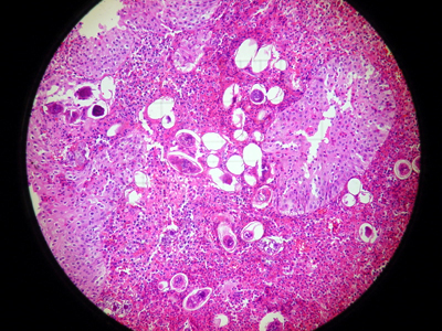
32 y/o male, recent immigrant from the Mediterranean presents to your clinic w/ hematuria. Being the astute clinician that you are, you suspect a particular bug. Some tests are run and finally a biopsy is taken:

Dsc00982.jpg


Bladder, H & E
 
Members don't see this ad :)
32 y/o male, recent immigrant from the Mediterranean presents to your clinic w/ hematuria. Being the astute clinician that you are, you suspect a particular bug. Some tests are run and finally a biopsy is taken:

Dsc00982.jpg


Bladder, H & E

Schistosoma.
 
PT John Doe presents to the ED comatose. Significant other states that they were at the local national park where they had been camping until the PT returned from an absconding 10 minute trip, where he said he was going to "freshen up for the morning" Rapidly thereafter, John Doe began to have diffuse headaches, seizures, and ended up comatose in a few hours. What pathogen is most likely the cause of the PT's symptoms? What Rx is available to treat this pathogen?
 
PT John Doe presents to the ED comatose. Significant other states that they were at the local national park where they had been camping until the PT returned from an absconding 10 minute trip, where he said he was going to "freshen up for the morning" Rapidly thereafter, John Doe began to have diffuse headaches, seizures, and ended up comatose in a few hours. What pathogen is most likely the cause of the PT's symptoms? What Rx is available to treat this pathogen?

That's a very quick onset of neurological symptoms. I'm not sure what could be that quick, but it sounds like some kind of acute meningoencephalitis or encephalititis. Could it be brought on by Naegleria fowleri, tx w/ Amphotericin B?
 
That's a very quick onset of neurological symptoms. I'm not sure what could be that quick, but it sounds like some kind of acute meningoencephalitis or encephalititis. Could it be brought on by Naegleria fowleri, tx w/ Amphotericin B?

:thumbup:
 
A 61 year old New Zealand immigrant presents in anaphylaxis following a blow to the RUQ in a car accident.

CT Imaging of the abdomen shows the following:
wlvw1t.jpg

Echinococcus granulosis.
 
Members don't see this ad :)
A child presents to the clinic with complaints of "strange tingling sensations" in the right lower extremity. Two point discrimination, vibratory sense, and proprioception in the right lower extremity is decreased relative to the left lower extremity. Pain and temperature sensation is also diminished in the right lower extremity. The mother reports that as of the past month, her child has decreased his normal activity, due to complaints of fatigue and progressively worsening diffuse abdominal pain over the last month. The family has returned from a trip overseas from Thailand, where they ate food both offered by the local farings, and on the cruise. Analysis of the child's blood showed the following:

4303021.jpg


What is the infectious pathogen?
 
Last edited:
Lol. Are you guys enjoying yourselves? My cadaver last year had a lot of little cysts that I assume were hydatid cysts. There was also one large one in the pouch of douglas.

I also liked the pernicious anemia case and the hypersegmented PMN as I finished heme 2 weeks ago.
 
Correct! We have yet another new hall of famer:

Hall of Fame
1) themule +2
1) cpants +2
1) Mr hawkings +2
4) Beachblonde +1
4) Whatayear +1
4) Bodonid +1

In an effort to establish some real ranking in the hall of fame, I am going to do a rapid fire round:

Mystery Pathogen #8 (no picture)
...This pathogen is part of a genus that contains 4 known species. It causes severe intestinal epithelium damage, resulting in bloody diarrhea. It also causes local inflammation by recruiting polymorphonucleocytes. Enters through M cells, and is taken up by macrophages. Induces apoptosis of host cell.

Mystery Pathogen #9 (no picture)
...This pathogen evades its degradation via macrophages by blocking phagolysosome formation. As part of a redundant immune evasion repertoire, this pathogen contains a 21 kDa lipoprotein that blocks MHC II expression.

Mystery Pathogen #10 (no picture)
...Transmitted by the kissing bug (triotomines).

Mystery Pathogen #11 (no picture)
...Gram negative bacteria. Causes upper respiratory tract infections and oitis in children. Can also cause meningitis. 1st bacterial genome to be sequenced.

Mystery Pathogen #12 (no picture)
...A patient comes into the hospital after doing missionary work in a West African village 2 weeks prior. They complain of abdominal pain, vomiting, diarrhea, and a sore throat. While doing an exam, you notice facial swelling, and what appears to be conjunctivitis. Labs come back, and one of the first things you notice is your patient has proteinuria. Unsure of what is going on, you go back to the room to get a more detailed history. When you come back, you notice that your patient's gums are starting to bleed.

Please be sure to specify which pathogen you are responding to.

Courtesy of the CDC and my brand new cellular microbiology book :).

human immunodeficiency virus

Which one? HIV-1 or HIV-2?
 
Last edited:
A 72-year old female undergoing chemotherapy for breast cancer presents to your clinic obtunded. She dies soon thereafter. Autopsy reveals this gross image. On closer inspection, biopsy reveals this pathogen.

Which pathogen led to this woman's demise?
 

Attachments

  • Pathogens.pdf
    324.1 KB · Views: 170
HSV-1 encephalitis?
 
HSV-1 encephalitis?

The gross image indicates hemorrhage into the basal ganglia.
Microscopic examination reveals fungal forms.
This is a particularly nasty bug once it makes its way into the CNS!

Keep 'em coming!
 
The gross image indicates hemorrhage into the basal ganglia.
Microscopic examination reveals fungal forms.
This is a particularly nasty bug once it makes its way into the CNS!

Keep 'em coming!

Cerebral aspergillosis (aspergillus spp., maybe a. fumigatus or flavus) would be in my differential because of it's opportunistic nature and the neutropenia secondary to the chemo. Would be especially compelling if the fungal forms were about 15 microns in diameter.
 
Cerebral aspergillosis (aspergillus spp., maybe a. fumigatus or flavus) would be in my differential because of it's opportunistic nature and the neutropenia secondary to the chemo. Would be especially compelling if the fungal forms were about 15 microns in diameter.

You are correct! :)
 
Missed the 2nd picture. I'd say aspergillosis too.
 
Hmm...people w/ silicosis are at higher risk for TB and the presenting S & S seem to corroborate the diagnosis. More rarely, you can get also get actinomyces and aspergillus infections, too. However, I think the organism looks like M. tuberculosis, so that's what I'm going to go with.

Dx: TB (Mycobacterium tuberculosis)

Seconded.
 
A young boy accompanied by his mother presents to your clinic with a low grade fever, lethargy, cough, runny nose, and conjunctivitis. Physical examination reveals <first image>. You send the child home with his mother and instruct them to phone you should his symptoms get worse. Unfortunately, there is a power outage in the city, and several days later as you're watching the evening news, you recognize a photograph of the child you saw in clinic who recently passed away. At autopsy, biopsies are taken and this microscopic finding is made: <second and third images>.

What is the pathognomic finding you recognized on physical examination?
Which pathogen led to this child's demise?
Name the characteristic cellular finding(s) on microscopic examination?
 

Attachments

  • Pathogens2.pdf
    249.8 KB · Views: 182
A young boy accompanied by his mother presents to your clinic with a low grade fever, lethargy, cough, runny nose, and conjunctivitis. Physical examination reveals <first image>. You send the child home with his mother and instruct them to phone you should his symptoms get worse. Unfortunately, there is a power outage in the city, and several days later as you're watching the evening news, you recognize a photograph of the child you saw in clinic who recently passed away. At autopsy, biopsies are taken and this microscopic finding is made: <second and third images>.

What is the pathognomic finding you recognized on physical examination?
Which pathogen led to this child's demise?
Name the characteristic cellular finding(s) on microscopic examination?

Can't open the pdf- adobe stating file is damaged and cannot be repaired... :( might be my pc/my old version of adobe...
Edit: pdf file works just fine.
 
Last edited:
I was able to open it, but couldn't really tell what was going on.

I'm going to say that the kid had strep throat and those were achoff bodies from rheumatic myocarditis.

but that's just pattern recognition here, I couldn't identify anything in the biopsy.
 
Pseudomembrane, metachromatic granules....
C. diphtheriae?
 
wait!

are those koplik spots?

dude's got measles!


(Pretty sure the biopsy would show synctia)

Thought of koplik spots, but didn't see any red/blue/white spots... but I could be wrong.
 
wait!

are those koplik spots?

dude's got measles!


(Pretty sure the biopsy would show synctia)

:thumbup:

The first image is supposed to demonstrate Koplik's spots, the pathognomic finding in rubeola (measles) infection.
The patient developed fatal rubeola pneumonia. The second and third images are biopsies from the patient's lungs. Microscopic examination reveals several Warthin-Finkeldey cells, multinucleate giant cells with eiosinophilic intracytoplasmic and intranuclear inclusions.
 
Hmm...people w/ silicosis are at higher risk for TB and the presenting S & S seem to corroborate the diagnosis. More rarely, you can get also get actinomyces and aspergillus infections, too. However, I think the organism looks like M. tuberculosis, so that's what I'm going to go with.

Dx: TB (Mycobacterium tuberculosis)

Good. Don't forget that the silicosis and TB is likely going to be in the upper lobes vs. asbestosis in the lower lobes.
 
group A strep?
 
Staph aureus impetigo?
 
keep guessing.

A check of the medical records on this this homeless patient reveals he is HIV positive, if that's more of a clue.

Norwegian (or crusted) scabies would be highest on my differential. Sarcoptes scabei gets out of control with the immunocompromised.
 
You, the attending dermatologist, are called down to the ER to consult on this new admission. As you shake hands with the patient, you notice these lesions on his left hand (his right hand was fractured in a scuffle outside the ER): <image one>. With the patient in a sufficiently inebriated state, you proceed to take a biopsy from his skin. This is what you see under microscopic examination: <image two>. To determine how severe this agent is, you plate a petri dish and examine it in the future. This is what you see: <image three>.

Which organism is plaguing this patient's hand?
What is the significance of the petri dish findings?
 

Attachments

  • Pathogens3.pdf
    253.5 KB · Views: 153
You, the attending dermatologist, are called down to the ER to consult on this new admission. As you shake hands with the patient, you notice these lesions on his left hand (his right hand was fractured in a scuffle outside the ER): <image one>. With the patient in a sufficiently inebriated state, you proceed to take a biopsy from his skin. This is what you see under microscopic examination: <image two>. To determine how severe this agent is, you plate a petri dish and examine it in the future. This is what you see: <image three>.

Which organism is plaguing this patient's hand?
What is the significance of the petri dish findings?

birdseed agar is diagnostic for cryptococcus neoformans b/c it is able to metabolize whatever it is in the seed and shows the pigment.

If the dude has cryptococcus in his skin, that's really bad b/c it's usually disseminated from the lungs, right?
 
Top