Pictures of the Week

This forum made possible through the generous support of SDN members, donors, and sponsors. Thank you.
She had true neurological weakness on exam, not pain inhibition.
dorsiflexion is mainly L4, EHL, mainly L5, plantarflexion mainly S1.

if all 3 show progressive weakness, then that is 3 myotomes. definitely no proximal weakness? not many of us check hip abduction or extension
 
dorsiflexion is mainly L4, EHL, mainly L5, plantarflexion mainly S1.

if all 3 show progressive weakness, then that is 3 myotomes. definitely no proximal weakness? not many of us check hip abduction or extension
Did not check hip abduction or extension. She might have had a Trendelenburg gait but that's just based on my own memory so basically useless. EHL was weak, too.
 
I thought diabetic amyotrophic was proximal weakness (?). I think that’s more of a test answer than anything. Never seen it.

Is this patient numb or with sensory deficit?
Classically it is proximal weakness but can affect distal limb. The thing that is confusing is that symptoms started so shortly after "stumbling". Is that the cause of the pain symptoms or was the stumbling because of weakness that maybe wasn't initially perceived and the pain followed? Definitely curious to see what EMG results are.
 
he only posted 1 plane pic - he has an entire MRI that he and the radiologists reviewed. and no surgeon will operate just on basis of perceived weakness without MRI evidence of compression. most of the spine surgeons that i have referred for foot drop defer on surgery.


as noted by others, injections do not change weakness. also, "irritation" of the root will not cause weakness.


Baron - would you consider getting a CT myelogram?

Irritation causes edema, edema causes compression, compression causes decreased blood supply, that causes weakness
 
he only posted 1 plane pic - he has an entire MRI that he and the radiologists reviewed. and no surgeon will operate just on basis of perceived weakness without MRI evidence of compression. most of the spine surgeons that i have referred for foot drop defer on surgery.


as noted by others, injections do not change weakness. also, "irritation" of the root will not cause weakness.


Baron - would you consider getting a CT myelogram?

No it will not show anything, on a good saggital slides you might see an enlarged root in the foremen, but it is very rare
 
yet in this case the rad report is not saying that there is an edematous nerve or significant compression on the nerve to cause weakness.

i am not stating you cant get pain due to inflammatory agents. but theres no significant compression of any sort causing motor weakness in this case that we see on MRI.
 
No it will not show anything, on a good saggital slides you might see an enlarged root in the foremen, but it is very rare
so we get weakness and paralysis without any imaging confirmation?


so patients with normal MRIs and leg weakness dont have other neurologic conditions, they all must have severe spinal nerve compression even with the normal MRIs?
 
Ducttape vs mygalperin…. This is gonna be good! 🍿
 
I thought diabetic amyotrophic was proximal weakness (?). I think that’s more of a test answer than anything. Never seen it.

Is this patient numb or with sensory deficit?
I've seen it a handful of times (maybe 3-4x in the last 7 years). Weirdly, saw one last week. Patient (diabetic) came in with horrible right thigh pain (remembered the exact date the pain started) in an L2/3 distribution. Went to ED. Had MRI and an ESI in the hospital (rare, I know). No better. MRI showed a facet cyst at L4/5 (small) abutting the traversing L5 nerve root on the right side, mild stenosis L3/4. Patient had about 2/5 strength hip flexion/knee extension, distal no issues, hamstring strong. By the time I saw him (3-4 weeks out) pain is 7-8/10 but still densely weak (sensory loss and DTR loss also L2/3ish). EMG/NCS this week confirmed amytrophy. Good case for the resident, but I made less $ off him than the guy in the hospital who did the ESI.
 
View attachment 403214

Mystery diagnosis. 74F, afib w/ previous TIA, diabetic with A1c 6.4%. About 4 weeks ago she stumbled but caught herself and a couple hours later began experiencing severe pain down her left leg, predominantly in her posterior calf. I saw her 2 weeks ago and she had some mild dorsiflexion weakness. Saw her for a follow-up a week later and now she's 3/5 plantarflexion and 4/5 dorsiflexion. Got her admitted for a work-up. MRI brain non-revealing. MRI whole spine with mild degeneration, MRI lumbar spine report below. EMG/NCS will take a couple months due to lack of providers an the area. She's going to see Neurology at some point. Any thoughts on what it could be?

IMPRESSION:
1. Redemonstration of mild disc degeneration with mild spinal canal stenosis
at L2-L3 and L3-L4.
2. Multilevel neuroforaminal narrowing, most advanced to a moderate degree at
L4-L5 and on the left at L5-S1.
Could the fall have caused irritation of sciatic nerve given the l5 and S1 weakness? You mentioned no mass on sacral imaging what about a dedicated MRI pelvis. I agree EMG would help. The A1c is pretty good considering age and don't symptoms usually progress to bilateral so that pushed me away from diabetic amyotrophy.

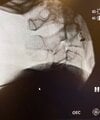
looks epidural but the pics arent great.
Thanks. I was wondering why the pattern looked so wonky when I injected more contrast
 
Could the fall have caused irritation of sciatic nerve given the l5 and S1 weakness? You mentioned no mass on sacral imaging what about a dedicated MRI pelvis. I agree EMG would help. The A1c is pretty good considering age and don't symptoms usually progress to bilateral so that pushed me away from diabetic amyotrophy.


Thanks. I was wondering why the pattern looked so wonky when I injected more contrast
100% of the cases (5-10ish total so not a huge N) of amyotrophy I have seen have been unilateral
 
100% of the cases (5-10ish total so not a huge N) of amyotrophy I have seen have been unilateral
It can be bilateral but usually one side is significantly more affected, and the onset is almost always unilateral. But for most cases, I agree with you that it should be though of as a unilateral disorder.
 
It can be bilateral but usually one side is significantly more affected, and the onset is almost always unilateral. But for most cases, I agree with you that it should be though of as a unilateral disorder.
I don't do EMG or see these. But I was always taught its generally starts unilateral and progressive and more proximal then in at least 50% of cases should become bilateral. Its good to know its unilateral will add to my differential. Does the relatively mild A1c matter for diagnosis (was also taught usually in those with long term suboptimal control and can happen otherwise but rare).
 
Again and again, time is ticking, I assume weakness is getting worse, some people think that whole discussion is funny, but patient is suffering!!! In my first post I said that in a worst scenario injection want help, but will not harm, so what is the problem???
Either do it or sent her to nyc, I will do it for her. Promise to post a procedure pictures. If you afraid of sugar increase, it will temporary.
 
By the way , I just saw the guy with a very similar MRI , just some foraminal narrowing, he walked out of office pain free.
 
Sounds like you need a refund from your weekend course.

This is a professional chat, you comments are not understandable neither pleasant, please keep them for yourself only! Otherwise I might start to consider em an insults, if you have issues , please write directly to me
 
This is a professional chat, you comments are not understandable neither pleasant, please keep them for yourself only! Otherwise I might start to consider em an insults, if you have issues , please write directly to me
TOEFL?
Your thought process is not reassuring . Your plan of care is poor. I am frightened for your patients.
 
so by your comments, you do not see edema of nerve roots on MRI


you also stated that you will not see foraminal stenosis on MRI.


but patients with leg weakness get all better with injections.


i guess 99% of ACGME fellowships are teaching this wrong...
 
so by your comments, you do not see edema of nerve roots on MRI


you also stated that you will not see foraminal stenosis on MRI.


but patients with leg weakness get all better with injections.


i guess 99% of ACGME fellowships are teaching this wrong...

I did not say that you do not see foraminal stenosis on the MRI, that it what doing study for!!!!! I also said that most of the time it is difficult to see root edema on the MRI, and even if you do not see on , that does not eliminate possibility of the root compression. MRI study is secondary to patients complaints and your clinical exam, plus your experience in those cases. With years one should develop a sense of what is going on, MRI will only confirm what you already suspect.
About injection a said that in a worst scenario it will not help. I am not a god, I cannot predict the outcome.
But what do you do when the MRI does not confirm what you had suspected? Shouldn’t you at that point reconsider?
 
Is it possible to get a traction neuritis in this scenario? Would resolve over time.
 
I did not say that you do not see foraminal stenosis on the MRI, that it what doing study for!!!!! I also said that most of the time it is difficult to see root edema on the MRI, and even if you do not see on , that does not eliminate possibility of the root compression. MRI study is secondary to patients complaints and your clinical exam, plus your experience in those cases. With years one should develop a sense of what is going on, MRI will only confirm what you already suspect.
About injection a said that in a worst scenario it will not help. I am not a god, I cannot predict the outcome.
The injections we do still carry risks. The worst case scenario is not that it just doesn’t help. There is the possibility of rare but serious complications. There is a thread on a 60 million dollar case due to a complication from a TFESI.
 
52yo m, 4 weeks of acute onset radicular pain down to the left calf, sharp and burning, 10/10, no pain sitting at rest, increases with ambulation, walks barely 1 block. + left leg weakness. No numbness in the LEs neither. No B/B incontinence. Images from outside facility. Wondering anyone has idea what is the grey material? is it a requested disc or something intrathecal? Thanks.

1747084395587.png
 
Just spitballing here but those look like his nerves in the thecal sac surrounded by epidural lipomatosis. Grade 1 listhesis, no disc herniation. Would like to see the pars and how much fluid is in the facet joints.
1747160647155.png

The left foramen, facet and pars
 
Possibly fractured pars like Baron Samedi mentioned, and a facet joint synovitis/effusion. The L5-S1 foramen looks fairly stenotic to me but still room for the nerve root. Maybe the antelisthesis is increased while standing with, in turn, an increased foraminal stenosis pinching L5? The territory of radicular pain works.
 
52yo m, 4 weeks of acute onset radicular pain down to the left calf, sharp and burning, 10/10, no pain sitting at rest, increases with ambulation, walks barely 1 block. + left leg weakness. No numbness in the LEs neither. No B/B incontinence. Images from outside facility. Wondering anyone has idea what is the grey material? is it a requested disc or something intrathecal? Thanks.

View attachment 403533
Can you share the T1 and/or FS/STIR sequences for the above slices if you have them?

While I agree it looks like lipomatosis and nerve root crowding, I'm confused by the thickened appearing ventral dura vs PLL.
 
Top