Techniques and Pearls for Thoracic MBB/RFA

This forum made possible through the generous support of SDN members, donors, and sponsors. Thank you.
Medial to lateral? Even Ferrismonk's post above has it lateral to medial. Same as Mister Mxyzptlk's.

18 years in on T-spine RF. 100's to over a thousand done.
Medial to lateral, inferior to superior. Needle tip sitting on upper outer quadrant of pedicle shadow. Active tip as flat on lamina as possible.


InkedT-spine RF_LI.jpg
 
Re: pedicle shadow technique, how common is it to get paraspinal twitch vs traditional approach?
 
Pretty common. Ideally touch mid pedicle shadow, then rotate so active tip is flat on lamina and creep it up and laterally.
Pretty common in my experience as well. I make 2 burns with venom tip going med/lat. cover mid pedicle top to bottom then lateral pedicle.
 
IMG_3930.JPG

Recent lower thoracic RFA I did with Nimbus needles
 
I like using diros trident with perpendicular approach for thoracic.
 
Where are you placing for cooled RF? Pics? Thinking about doing this for a current patient.
Here was one from last week. This technique using the cooled technology gives me very consistent and durable results
 

Attachments

  • 7794E0AA-6B98-4D93-8A6F-8585F46780B2.jpeg
    7794E0AA-6B98-4D93-8A6F-8585F46780B2.jpeg
    179.5 KB · Views: 275
  • C9C9F908-FAF3-455F-9BE1-C2EF0906D4B9.jpeg
    C9C9F908-FAF3-455F-9BE1-C2EF0906D4B9.jpeg
    186.3 KB · Views: 265
With cooled being spherical do you still need that angled trajectory? Just curious.
No but I feel like it still may give a somewhat larger burn (no evidence of this) and it was how I was taught originally with traditional RF. Also I feel like it may be safer since you are directing the needle AWAY from the spine while staying over bone
 
“I’ve lost all faith in thoracic medial branch blocks” - MacVicar, former SIS president, at SIS Jamaica (great place for a conference in February, btw).
Take home points for thoracic MBB/RF- for anything higher than T11 or maybe T10, if it works, we don’t know why it works, since we don’t know what we are targeting/burning.
Lol.

I have refrained from publishing this technique, as beyond the adapted drawings I can not find a primary source showing the nerves over pedicle shadow. Closest I found was superolateral foramen under lamina. I’ll just have to keep doing it this way til it stops consistently working so well…
 
“I’ve lost all faith in thoracic medial branch blocks” - MacVicar, former SIS president, at SIS Jamaica (great place for a conference in February, btw).
Take home points for thoracic MBB/RF- for anything higher than T11 or maybe T10, if it works, we don’t know why it works, since we don’t know what we are targeting/burning.

I’ve lost all faith that the SIS thoracic RFA anatomical maps are accurate.

I’m generally a big SIS guy, but this is the one area is which the dogma is just dogma instead of the practical methods we all use for thoracic RFA
 
Lol.

I have refrained from publishing this technique, as beyond the adapted drawings I can not find a primary source showing the nerves over pedicle shadow. Closest I found was superolateral foramen under lamina. I’ll just have to keep doing it this way til it stops consistently working so well…
How about a case report with images and leave it at that? I've done only a handful thoracic mbb since graduating but felt much more comfortable seeing this technique here rather than what I learned in fellowship which was walking off to who knows where.
 
Good question. 5 or 10mm I think. It’s whatever they recommend for the thoracic region. I forget
If you're using the Avanos cooled system, they come with 2/4/5.5 mm active tips. They recommend the 5.5 for thoracic if I remember right. Medtronic's Accurian is 4 and 5.5 mm active tips.

With the cooled lesion projecting forward off the active tip for about 40-50% of the lesion geometry, I would agree with aiming away from the neuraxis if you aren't using a bone backboard to protect things.
Yes it's Avanos and 5.5mm now that I think of it!
 
Place it correctly and use Nimbus and you might get even better results.
how are you all getting paid for thoracic facet interventions/rfa?

I do believe it exists. However the LCDs clearly state it's only medically necessary in Cervical and lumbar spines.......
 
how are you all getting paid for thoracic facet interventions/rfa?

I do believe it exists. However the LCDs clearly state it's only medically necessary in Cervical and lumbar spines.......

I call it C15-17 MBB/RF
 
how are you all getting paid for thoracic facet interventions/rfa?

I do believe it exists. However the LCDs clearly state it's only medically necessary in Cervical and lumbar spines.......
I quit offering them 3 yrs after denials that began to stack up. Several egregious denials.
 
how are you all getting paid for thoracic facet interventions/rfa?

I do believe it exists. However the LCDs clearly state it's only medically necessary in Cervical and lumbar spines.......

Easily covered by WC and Medicare everywhere.
Beyond that it depends on the commercial plan , but I’ve been able to get it paid by most (not all) insurances.
 
Anyone have any lateral images they care to post. I do the pedicle shadow technique above T11 as well but often have to adjust based on lateral and needle tip ends up more cephalad than I would like when I come back to AP
 
Anyone have any lateral images they care to post. I do the pedicle shadow technique above T11 as well but often have to adjust based on lateral and needle tip ends up more cephalad than I would like when I come back to AP
I will look for some, but just use the lateral to make sure you are not way too superficial off bone or deep in the joint. Square the endplate in AP, then try to leave it alone
 
Had a good one today. Old t12 fracture. 2 techniques in one. Lumbar-like at T11/12 mb. Pedicle shadow t10 mb.

It is just me or is the top needle in the second picture too shallow/posterior? Genuine question, trying to learn as I've had some successful TMBBs but haven't made the jump to RF yet.
 
My images are on the right. Very poor quality fluoro machine but you can see the TP better in real life.
In AP view, I hit bone near the center of the TP and walk off the superolateral corner.
Struggling with getting a good lateral view. I know I'm far from the NF but unable to judge depth on the TP, almost looks like I'm on rib.
 

Attachments

  • t2.jpg
    t2.jpg
    96.7 KB · Views: 251
  • t1.jpg
    t1.jpg
    87.5 KB · Views: 256
18 years in on T-spine RF. 100's to over a thousand done.
Medial to lateral, inferior to superior. Needle tip sitting on upper outer quadrant of pedicle shadow. Active tip as flat on lamina as possible.


View attachment 366265
Nice work, i wonder the difference between your approach and superolateral approach of tp. Is it possible the suprolateral approach is ablating the middle or proximal partion of mb, where your approach is really close to the fj, maybe you are burning part of joint capsule as well LOL. I used superolateral approach of tp, the resuls are minic to the results from john’s work, couple of years relief.
 
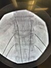
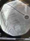
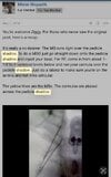
T10 down I place like lumbar. Above that, I start in the middle between the spinous processes below, with an upper outward angulation. If I can’t see the TP, I touch down on pedicle and walk it lateral, then up or down if necessary until I can feel I’m on bone.
Most of my patients are Medicare so no issues with denials. The local Medicaid carrier also covers without issue. Some of the BC/BS plans do not. I probably do it mostly for chronic pain at the site of a healed compression fracture.
View attachment 360460
Call me anesthesia, this is different than anotations done by steve lobel.

I've tried various techniques for thoracic RFA (getting overall good results) but then sometimes failure and wonder what's best
- Pedicle shadow technique where you enter below pedicle and towards it
- or call me anesthesia technique - hitting TP and sliding off to nerve (like SIS diagram), multifidus testing.
 
Not sure if this translates to better burn but I get stronger twitch with pedicle shadow
 
Had a good one today. Old t12 fracture. 2 techniques in one. Lumbar-like at T11/12 mb. Pedicle shadow t10 mb.
Do you think pedicle shadow works because you’re burning the actual joint? Rather than nerve

That technique is similar to how one may access thoracic joint

- how do you chose between pedicle shadow and traditional SIS?
- do you ever land on medial and lateral aspect of TP and bipolar?
 
Why pedicle shadow when the drawing shows them over the TP?

but why male models? Welcome to the forum. Jk

Several excellent discussions here over the years on this. I found some screenshots on my phone from when I was putting everything together a few years ago to publish a technique article on this… Never got around to it. Probably some duplicates in these.
 

Attachments

  • IMG_5535.jpeg
    IMG_5535.jpeg
    120.9 KB · Views: 107
  • IMG_5539.jpeg
    IMG_5539.jpeg
    146.2 KB · Views: 109
  • IMG_5546.jpeg
    IMG_5546.jpeg
    123.1 KB · Views: 94
  • IMG_5548.jpeg
    IMG_5548.jpeg
    79.5 KB · Views: 90
  • IMG_5551.jpeg
    IMG_5551.jpeg
    66.3 KB · Views: 90
  • IMG_5552.jpeg
    IMG_5552.jpeg
    92.6 KB · Views: 86
  • IMG_5554.jpeg
    IMG_5554.jpeg
    98.7 KB · Views: 91
  • IMG_5556.jpeg
    IMG_5556.jpeg
    110.9 KB · Views: 77
  • IMG_5558.jpeg
    IMG_5558.jpeg
    70.9 KB · Views: 89
  • IMG_5536.jpeg
    IMG_5536.jpeg
    81.7 KB · Views: 99
But are you distal or proximal to where the medial branch innervates the joint using that technique? I would say distal which does nothing. Much like the lady who does her cervical mbb’s on the posterior articular pillar.

And if one argues otherwise, this technique still has problems as the probe is placed perpendicular and not parallel to the nerve as you show above. If someone did this in the cervical or lumbar spine, you’d eviscerate em. I’d say you’re living in a glass house
Yet somehow I manage....
20 years doing it this way with success rates = to cervical literature.
Must be lucky.
If following the diagram I posted above, my target appears to be a few mm distal to the lateral branch takeoff. So I am getting almost the entire medial branch from there. Articular branches are not shown... a caveat for us all.
 
Top