Let's do some echo:

This forum made possible through the generous support of SDN members, donors, and sponsors. Thank you.
RV is of paramount importance. If you can't get blood to the LV (via RV ejection), then the LVAD will not work. When you start adding 1-2 l/m to the systemic circulation, that is 1-2 l/m extra flow that the RV suddenly is confronted with. It doesn't have a lot of time to accommodate for such a change in hemodynamics. Therefore, inhaled vasodilators and RV augmentation with volume and inotropes may be necessary. If these maneuvers don't work this may cause the RV to go into failure. One thing to keep in mind is that the RCA (supplies the RV) is anterior. Air tends to go down the RCA when coming off bypass. Furthermore, CPB doesn't protect the RV nearly as well as the LV. This only adds insult to injury when dealing with LVAD placement. Many of the the RVADS that are placed in practice are actually due to an LVAD that overwhelmed the RV leading to RV failure.


The "suckdown" phenomena is usually due to an underfiled LV causing the interventricular septum to get pulled towards the LV free wall. Doing this alters the geometry of the RV. Augmenting the geometry of the RV may equate to poor systolic performance AND tricuspid annulus dilatation leading to acute tricuspid regurgitation.


Here is a modified bicaval view showing this:
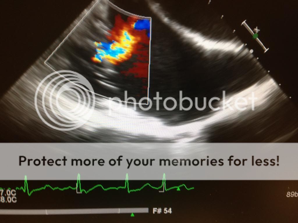
IMG_7335_zps10a137f9.jpg


If you see this coming off bypass you need to think: oh... oh... what can we do to make this better. :nurse:

The answer...? As weird as it sounds, reduce your flow rates on your LVAD and augment volume status. These maneuvers will return the RV to it's regular shape and hopefully increase RV unloading to the LV.

In addition to causing the suckdown phenomena, the inflow canulas can become obstructed. Check for this by using color Doppler and seeing laminar flow. You can also use CWD. Flows should be less than 2.5 m/s. Similarly, the outflow cannula (ascending aorta) should show slightly lower flows. 1-2 m/s.

AI is bad... LVAD worsens AI and therefore you may not achieve increased organ perfusion. You can create a partial closed loop from the ascending aorta back into the LV causing a volume overload problem. You need a competent valve. What are the choices?

1) Repair the valve.
2) Replace the valve and deal with higher morbidity/mortality. Never use mechanical valves. Always bioprosthetic ones.
3) Sounds crazy... but if it's destination therapy or bridge to transplant, you can surgically sew the aortic valve shut so that it doesn't open at all! :eek:


Good stuff.

Love LVAD physiology. I would add one more thing to the management of a suction event- vasopressin. Selectively increasing SVR without increasing PVR will help your RV, and along with decreasing the VAD flow, will help restore septal geometry.

Members don't see this ad.
 
Love LVAD physiology. I would add one more thing to the management of a suction event- vasopressin. Selectively increasing SVR without increasing PVR will help your RV, and along with decreasing the VAD flow, will help restore septal geometry.
Other than vasopressin (svr>pvr) how would you accomplish this?
 
Members don't see this ad :)
I wanna play. Let's kick it up a notch.

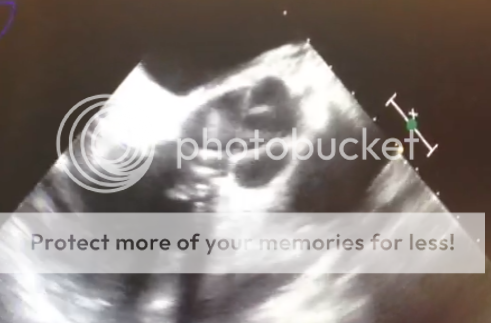
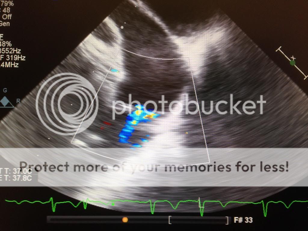
What's going on here?View attachment 181375


Resident here, I'll take a shot.

It looks like an RV inflow-outflow view but the RA looks funny. What I think is the RV and TV also don't look right.

No ASD, no TR jet, some aliasing in the area of the pulmonic valve.

Ebstein's anomaly? Or am I way off?
 
Resident here, I'll take a shot.

It looks like an RV inflow-outflow view but the RA looks funny.
Good for taking a shot. The view you named is correct. I think the RA is normal.
What I think is the RV and TV also don't look right.
This is during diastole as urge mentioned, the TV is in fact normal. What do you think is abnormal about the RV?

No ASD, no TR jet,
True.
some aliasing in the area of the pulmonic valve.
Yeah, what's up with that turbulence over there? Let's focus on that.
Ebstein's anomaly? Or am I way off?
Off, but good try.
 
Common causes of aliasing at a valve are turbulence from stenotic flow or regurgitation. With the timing in diastole, it could be pulmonic regurgitation, but the direction of the jet doesn't appear directed back towards the RV like I would expect.

The RV wall looks a little thick to me but that could just be the image plane. Any more images?
 
When I first looked at the image, I thought Type I/outlet/subpulmonic VSD. Associated with prolapse of the RCC. There is a diastolic rumble heard with a stethoscope, but flow should occur during systole (systolic murmur). So I'm not sure.

Thanks for playing HB. You da man!
 
Do you have a midesophageal LAX view with some color? Maybe the RCC is prolapsing causing AI.
 
Common causes of aliasing at a valve are turbulence from stenotic flow or regurgitation. With the timing in diastole, it could be pulmonic regurgitation, but the direction of the jet doesn't appear directed back towards the RV like I would expect.

Indeed. The flow seems perpendicular to what you'd expect a pulmonic regurg jet to look like. So look at the image again. What does the origin of that jet appear to be?
 
Yeah... I'm sticking to my answer. Type I, subpulmonic VSD with prolapse of the RCC causing AI during diastole. :xf:
 
Very, very close but not quite.

Hint: there is no regurg into the LV. The septum is intact.
 
Members don't see this ad :)
Hmmm... Does this dude have persistent fevers?

How 'bout infective endocarditis of the aortic valve causing a fistula into the pulmonary artery? Prolly reaching at this point: Aortopulmonary fistula. (can also be due to aortic anurysm rupture).

Nice case brah.
 
SOVA rupture into RV/PA? Dang it.... can't let it go.
 
I'm going with a membranous VSD with prolapse of the RCC of the AV. Would love to see a Midesophageal LAX, though
 
I think in a membrenous VSD, the direction of the jet is in the other direction (away from the PV).
 
SOVA rupture into RV/PA? Dang it.... can't let it go.

Winner winner chicken dinner!

Rupture of a Right Sinus of Valsalva Aneurysm, with resultant flow into the RVOT.

Was a fun case. I'll try to rustle up some other images from it tomorrow.

Look at the non color image- you can see the flail wall of the sinus prolapsing into the RV.
 
Winner winner chicken dinner!

Rupture of a Right Sinus of Valsalva Aneurysm, with resultant flow into the RVOT.

Was a fun case. I'll try to rustle up some other images from it tomorrow.

Look at the non color image- you can see the flail wall of the sinus prolapsing into the RV.

Well... Hot diggity dog! :heckyeah:

FWIW, Type I/subpulmonic VSD looks very similar.

Great case and thank you very much for sharing. :thumbup:
 
Dang! I was going to say that the flow looked like it was going from the aorta to the rvot but thought that was a little too ridiculous.
 
  • Like
Reactions: 1 user
K I got another one.

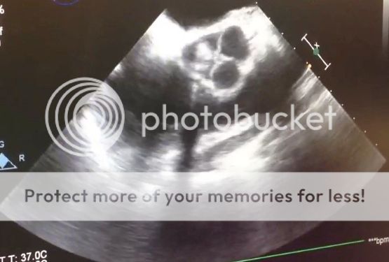
This isn't a completely standard view, and you can't get it in everyone. It's just deep to the aortic arch view positions in the upper esophagus (i.e., still above the blind spot caused by air-filled bronchus). What you see is the main PA, the bifurcation, and the right and left main PAs, left seen better than right. Ascending aorta is seen in short axis abutting the main and right PA.

But what's up with that flow you see in blue, seeming to cross the pulmonic valve? (Don't worry about those two blue splotches in the middle of the main PA, just the single jet that crosses the valve).

echo2.jpg
 
Last edited:
Wild guess.....PDA?

As for SOVA rupture....like everything in medicine, so obvious once you know the answer
 
Tough one Hawaiian Bruin. It isn't a PDA, that would appear near the branch of the main PA into right and left, or further out off the left PA. If that is the pulmonic valve we see, it is closed which leads me to believe the heart is in diastole. I'm just taking a shot in the dark here: getting color flow of mitral inflow showing up in this view, could it be blood flow in a coronary artery, possible aorta-PA fistula. I have no clue, but I love this echo stuff.
 
This view seems similar the the ME AA SAX view. Like Beaker, I think the image is taken in diastole.

With the blue coloring with doppler, my interpretation is the flow is going towards the pulmonic valve during diastole (away from the echo probe). The lighter shade of blue on the more inferior segment near the PV, however, makes me think the flow is faster there (and may be originating there). But that makes no sense if the flow is away from the probe as indicated by the doppler. One structure below the PA that could flow in to the PA at this level would be a pulmonary vein.

Honestly thought, I've got no idea what's going on.

What's the Nyquist limit set at? What omniplane?

I love echo too, trying to get better at it. I'm doing hearts in my PP gig this July, so I've got to.

Thank you for sharing Hawaiian Bruin, I appreciate the challenge.
 
Good shots. A PDA would be in a different anatomic position and would probably have more turbulence, as would an aortopulmonary fistula. But the latter is very, very close.

This is actually a coronary-PA fistula. The flow is more laminar, having made its way through a coronary artery (in this case a diag) and then through a fistulous connection. The fun part of the story is that it was seen preop on cath, but the cardiologist forgot to verbally tell the surgeon about it. The surgeon didn't notice it when reviewing the cath images. But I saw it on echo, told the surgeon, who then fixed it. Would have been a big, big deal otherwise. Why? Coronary steal.

And they say CABG isn't a class 1 indication for echo- this patient would think otherwise!
 
  • Like
Reactions: 1 user
Another good one HB. :thumbup:

Nice view of the main PA with the right and left bifurcation. I find getting the main with the L and R PA's hard to get in the same pic. Purrrty... when you get it though.

IMG_3591_zps7669bd1c.jpg


I tried to get the left to come in today... but no love. :(
 
Last edited:
ME AV SAX view.

Clearly there's a >1cm round lesion on the non-coronary cusp, and it's producing significant echo shadowing. The overall geometry of the valve seems a bit distorted, but that could just be the slice.

Without motion its hard to know if the lesion involves the coaptation surface or if it originates from the cusp.

DDx for Aortic valve mass:
Thrombus
Vegetation
Papillary fibroelastoma.
Weird/rare stuff

I'd get some more images in different planes to get a better sense of what's going on, and some 3d images just for fun.


As an aside, are these posts similar to the PtEExam? I'm taking the Basic in July.
 
Here is a good case for the residents/fellows. I did this case last week, but let's change the scenario a little bit to make it more educational...

37 y/o male presents for I+D of an open tib/fib fracture s/p Fall. When you see the patient in preop, he has normal vital signs and is AAO X 3. Pt states he takes HCTZ/Lisinopril for HTN but denies any other significant past medical/surgical/anesthetic history. Pt is taken back to the OR with standard ASA monitors with starting BP 120/80, HR 80, O2 98% on RA. Pt gets a RSI with Propofol/Sux/Fentanyl/Midazolam and then Sevoflurane/Rocuronium is used for maintainence. 15 minutes after induction, his BP remains low despite continuing methods (fluids, vasopressors, inotropes, etc.) to improve this. Since you have some training in performing rescue TEE, you call for the Echo probe.

1) What do you see? (Note: These are my images but I am having trouble putting videos in, which may give it away. However, the other two findings are pathognomonic for this disease state)
2) What is the C-Sept distance and AL/PL ratio and why is it important?
2) How do you treat this situation?
3) What do you tell the patient post-operatively? What notes do you want to make to future medical providers?
4) What future problems may this patient encounter and what surgical management can be done for this patient?


upload_2014-5-21_17-35-34.png
upload_2014-5-21_17-35-47.png
upload_2014-5-21_17-35-58.png
 
Early peaking "dagger" shapped CWD doppler says it all. M-mode is classic.
Very nice.
 
Just wish I could figure out how to insert the Excelera loop! Oh well, still images for now.
 
Yeah... that would be nice. This is an exceptional example of this pathology. I can't imagine a better representation of what's going on here- except for a vid ;)- in slow motion, the problem becomes very evident and puts to mind the C-sept, AL/PL ratio as well as the aorto-mitral angle. Good stuff sethco.
 
ME AV SAX view.

Clearly there's a >1cm round lesion on the non-coronary cusp, and it's producing significant echo shadowing. The overall geometry of the valve seems a bit distorted, but that could just be the slice.

Without motion its hard to know if the lesion involves the coaptation surface or if it originates from the cusp.

DDx for Aortic valve mass:
Thrombus
Vegetation
Papillary fibroelastoma.
Weird/rare stuff

I'd get some more images in different planes to get a better sense of what's going on, and some 3d images just for fun.


As an aside, are these posts similar to the PtEExam? I'm taking the Basic in July.

Great response. I'll get back to this. For now, sethco's awesomeness trumps my case. His case presentation will likely be on your exam.
 
Here is a good case for the residents/fellows. I did this case last week, but let's change the scenario a little bit to make it more educational...

37 y/o male presents for I+D of an open tib/fib fracture s/p Fall. When you see the patient in preop, he has normal vital signs and is AAO X 3. Pt states he takes HCTZ/Lisinopril for HTN but denies any other significant past medical/surgical/anesthetic history. Pt is taken back to the OR with standard ASA monitors with starting BP 120/80, HR 80, O2 98% on RA. Pt gets a RSI with Propofol/Sux/Fentanyl/Midazolam and then Sevoflurane/Rocuronium is used for maintainence. 15 minutes after induction, his BP remains low despite continuing methods (fluids, vasopressors, inotropes, etc.) to improve this. Since you have some training in performing rescue TEE, you call for the Echo probe.

1) What do you see? (Note: These are my images but I am having trouble putting videos in, which may give it away. However, the other two findings are pathognomonic for this disease state)
2) What is the C-Sept distance and AL/PL ratio and why is it important?
2) How do you treat this situation?
3) What do you tell the patient post-operatively? What notes do you want to make to future medical providers?
4) What future problems may this patient encounter and what surgical management can be done for this patient?

I need to read up on the echo and answer the questions, but this is hypertrophic cardiomypopathy. His preload bottomed out with induction and you are now behind the 8ball on it. He needs fluids and he needs to not be tachycardic like whoa, so he gets beta-blockade intraoperatively (once he's no longer profoundly hypotensive) and he gets a beta blocker for life now. He also gets counseled to avoid strenuous exercise until his next operation. And that should be an alcohol septal ablation.


Edit - I'm guessing the m-mode is giving a beautiful view of SAM.
 
1. Agree with HOCM/Dynamic LVOT obstruction. I did a few myomectomies during my CT months and you've got some great images of classic findings. In the first image you can see an MR jet from the incomplete coaptation of the anterior leaflet. M-mode through the AoV demonstrates premature closure of the valve before the Twave onset. And the classic Dagger shaped CW doppler trace.

2. I remember reading these but had to look them up to refresh the details. These values seem to be more useful for predicting SAM after MVR. C-sept: Distance from MV coaptation point to septum. The smaller the value, the more likely SAM.
Al/Pl: anterior leaflet size divided by posterior leaflet size. The posterior leaflet should be smaller that the anterior, but the closer in size they get (al/pl) ~1, then the more likely the coaptation point is closer to the LVOT and SAM is more likely.

3. Acutely: Slow HR, increase afterload, increase preload, reduce inotropy. give fluids and vasopressors(neo/vaso, not ephedrine). A bit surprising that he's not responding to your current therapy. I'd place and aline and central line for pressor administration. Provided he stabilizes enough, we complete the case (he does have an open fracture after all).

4. If I'm doing the case, the pt gets admitted to the icu postop and has a cardiology consult. He should be made aware of the severity of this situation and since its genetic, he ought to to let his family know.

5. This is one of the causes of sudden death in athletes. They get dehydrated, tachycardic, and arrest from acute obstruciton. Medically can treat with beta-blockers, and can do a surgical myomectomy if symptomatic despite therapy.


Thanks for posting
 
  • Like
Reactions: 1 user
He needs fluids and he needs to not be tachycardic like whoa, so he gets beta-blockade intraoperatively (once he's no longer profoundly hypotensive) and he gets a beta blocker for life now.
Edit - I'm guessing the m-mode is giving a beautiful view of SAM.

So how are you going to correct the hypotension?
 
So how are you going to correct the hypotension?
Norepi or phenylephrine + fluids. Increase preload because these patients are hypersensitive to drops in preload (which is why this patient has been hypotensive since induction), also increase the afterload (partly to prevent tachycardia and the associated worsening of CO with decreased filling time and to decrease venous compliance and thereby increasing preload). Sound reasonable or did I miss something?

Adding to my previous post, I'd also refer this patient to cardiology for consideration of ICD placement.
Phenylephrine yes, norepi no. You don't want any beta agonism.
 
My understanding of the etiology of sudden death in this disorder isn't that they acutely obstruct, but that the myocytes themselves have an abnormally disorganized architecture, which predisposes to microreentry and higher risk of arrhythmia. This is facilitated by the catechol outflow during exercise.
 
Phenylephrine yes, norepi no. You don't want any beta agonism.
I thought at low(er) doses, the beta agonist actions of norepi were nil. I should have been more specific in my post above, but I was hoping to say using the agent in doses that provide for its alpha actions to predominate. Or is even the smallest amount of beta agonism too much?
 
Just a couple points for completion:

  • C-sep distance (Coaptation point of the AL/PL to the interventricular septum) is measured. You want this to be GREATER than 2.5 cm. Otherwise, risk of LVOT obstruction is increased. (measure this post MV repair in patients that may have SAM)
  • AL/PL ratios < 1.3 increase the risk of SAM. Not only is a large AL a problem (>2cm), but a large PL can also be an issue. The venturi effect helps explain part of the problem. Sometimes you need to resect both of them in order to obtain a posteriorly directed inflow pattern to the LV.
  • Aorto-mitral angle is important as well as anterior displacement of the papillary muscles. Inflow blood is usually directed towards the posterior wall. It then comes back up the septum closing the MV. An anteriorly displaced papillary muscle will move the coaptation point towards the LVOT and the direction of blood flow is in the reverse direction (along the septum first). This direction of intraventricular blood flow further pushes mitral valve anteriorly excacerbating the outflow obstruction.
  • AD disorder with variable penetrance and expressivity... i.e. this can run in families.
 
  • Like
Reactions: 1 user
ME AV SAX view.

Clearly there's a >1cm round lesion on the non-coronary cusp, and it's producing significant echo shadowing. The overall geometry of the valve seems a bit distorted, but that could just be the slice.

Without motion its hard to know if the lesion involves the coaptation surface or if it originates from the cusp.

DDx for Aortic valve mass:
Thrombus
Vegetation
Papillary fibroelastoma.
Weird/rare stuff

Very organized way to approach this pic. And you are correct, there is some echo shadowing going on there. This was the point of the picture as it very much resembles a papillary fibroelastoma.

Papillary fibroelastomas can have a stalk on them, but not always.

This one is taken during systole.

91c1f1be-e0ef-4c28-8a1d-372e09000996_zps90a7fe7e.png


4cdc0d38-93ed-42fb-bb7a-4a482cbe4a2b_zpse32f8bb9.jpg
 
Great responses all around. This patient had significant LVOT obstruction with Systolic Anterior Motion of the mitral valve. What you cant really appreciate on the still image was how much aliasing was present in the LVOT, the degree of the anterior mitral valve leaflet being sucked into the LVOT during systole, the degree of hypertrophy of the LV (Can see it if you look close enough at the color wave Doppler image) and the severity of eccentric mitral regurgitant jet directed posteriorly. This was actually a 56 y/o male that we took to the OR for septal myomectomy. He had a peak velocity through the LVOT of 5 m/s (pressure gradient of 100 mmHg) when his systolics got below 120.

The reason I chose to change the presentation of the case was to make this more of a rescue TEE case and less of a planned cardiac surgery, so we could gear this more towards residents/medical students that may not do CT Anesthesia in the future. This case is a classic boards question and with the trend the ABA is going (with emphasizing more TEE), could very well be a TEE question on the written boards. Residents need to know these 20 standard (now 26) TEE images not just for boards, though. Rescue TEE (with or without basic certification) is a very helpful tool in the hemodynamically unstable patient refractory to standard treatment (Class I indication). On your CT rotations, ask your attendings and/or fellows to show you how to obtain some necessary views. You may very well save a patient some day by diagnosing something with rescue echo
 
My understanding of the etiology of sudden death in this disorder isn't that they acutely obstruct, but that the myocytes themselves have an abnormally disorganized architecture, which predisposes to microreentry and higher risk of arrhythmia. This is facilitated by the catechol outflow during exercise.


Thanks for pointing this out, I just presumed that it was acute obstruction.
 
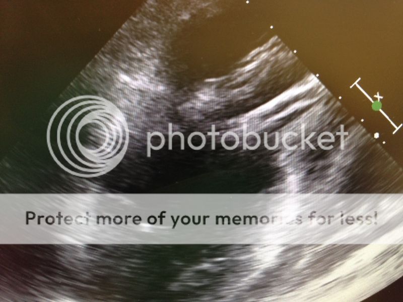
in the image from systole, I see a stalk looking thing, circled in red. The blue circled area appears to be the effective outflow orifice between the cusps.

The second image looks like a closeup, but not sure what to make of it.

Any ME AoV LAX views?
 

Attachments

  • image.jpg
    image.jpg
    63.2 KB · Views: 73
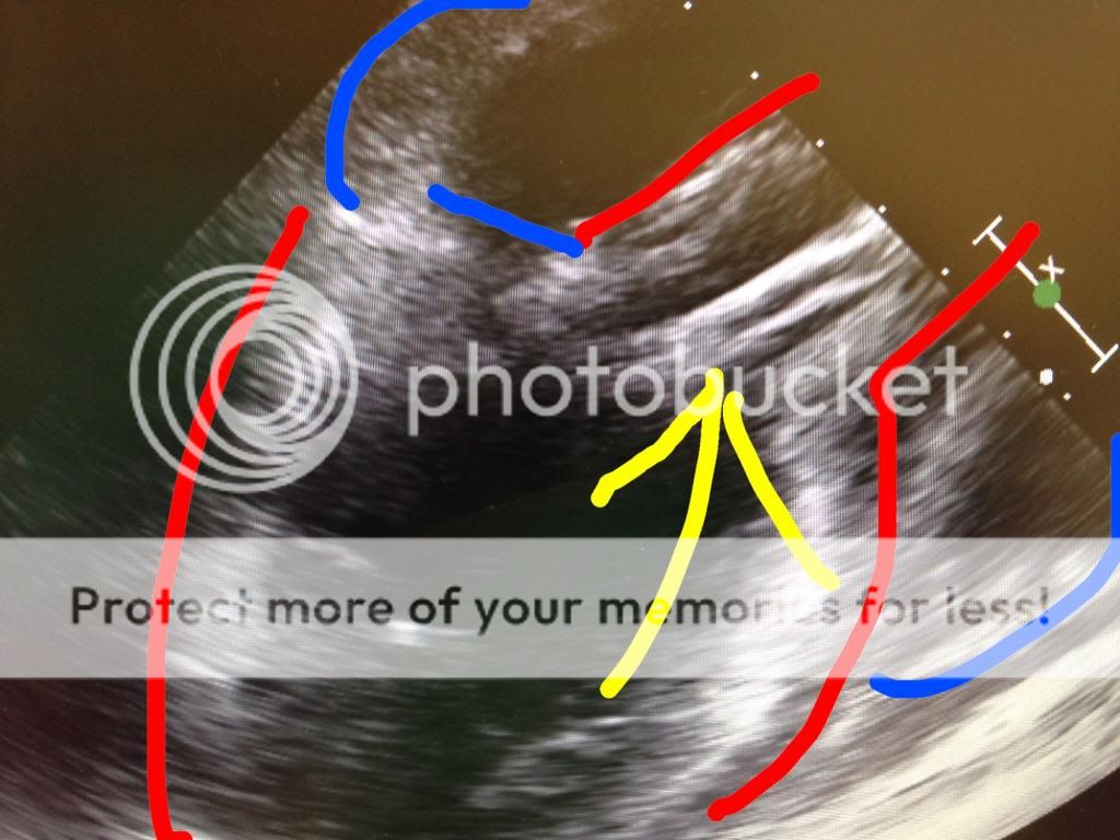
Pretty crappy echo pics, but this may help:

A23DAAC2-DCFB-48BE-AF3D-B8034B465AA1_zpsjtheqoou.jpg


Wow... Love technology. Poor edit... but done at the Denver airport in a couple minutes (on my phone). Wacha think now?

:cigar:
 
Last edited:
Thanks, that helps. Looks like ME AoV LAX shot maybe from early systole.

The structure indicated by the arrow sign looks to be in the LVOT/aorta traversing the aortic valve. It appears to have two echogenic walls and hollow in the middle. Doesn't appear to be artifact. It appears manmade, based on how homogenous the appearance is. The end in the ventricle is poorly defined and hard to comment on. I don't see any identifiable parts of the valvular apparatus either. A catheter of some sort?

The more I stare at it, the more confused I get, especially trying to reconcile it with the SAX views.

If it's not a catheter, then I'm stumped
 
Top