
- Joined
- Aug 15, 2003
- Messages
- 28,059
- Reaction score
- 438
Not sure how long I will keep doing case of the week, but anyway, here is another one
This is an autopsy case, a 71 year old man with a 15 year history of a neurologic disorder resembling parkinson's (a movement disorder). He died of an acute coronary thrombosis.
First: MRI
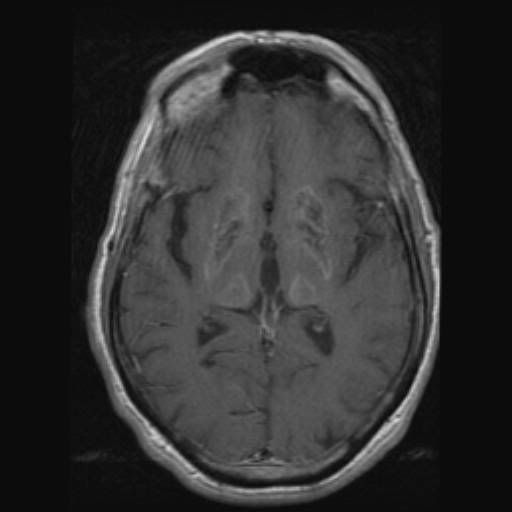

The MRI (and CT, which we don't have) shows an abnormal signal intensity, the same with and without contrast, in all of the basal ganglia, cerebellar dentate, and periventricular white matter. THought to represent mineralization with or without ischemic damage.
This is an autopsy case, a 71 year old man with a 15 year history of a neurologic disorder resembling parkinson's (a movement disorder). He died of an acute coronary thrombosis.
First: MRI
The MRI (and CT, which we don't have) shows an abnormal signal intensity, the same with and without contrast, in all of the basal ganglia, cerebellar dentate, and periventricular white matter. THought to represent mineralization with or without ischemic damage.


")